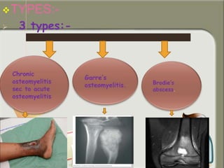
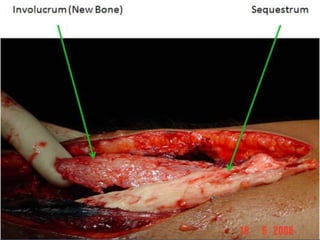
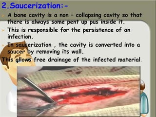
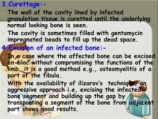
BY:-
SRAVANTHI
BPT III
YEAR.
CARE
INSTITUTE
OF
MEDICAL
SCIENCES.
INTRODUCTION:-
 The term chronic osteomyelitis is
used for chronic pyogenic
osteomyelitis.
INCIDENCE:-
 Occurs less in developed countries and
more in developing countries.
ETIOLOGY:-
 Tuberculosis , fungal infections etc.
TYPES:-
 3 types:-
PATHOLOGY:-
 Acute osteomyelitis commonly leads to
chronic osteomyelitis because of any one
or more of the following reasons:-
a)Delayed and inadequate
treatment:-
 It’s the commonest cause for the
persistence of osteomyelitis.
 Delay causes spread of pus within the
medullary cavity and subperiosteally.
 This results in death of a part of a bone
(sequestrum formation).
 Destruction of cancellous bone leads to
formation of cavities within the bone.
 Such non-collapsing bone cavities and
sequestra are responsible for persistent
infection.
b) Type and virulence of organism:-
 Despite early , adequate treatment of acute
osteomyelitis, the body defense mechanism
may not be able to control the damaging
influence of a highly virulent organism, and
the infection persists.
c)Reduced host resistance:-
 Malnutrition compromises the body's
defense mechanisms, thus letting the
infection persist.
 When the infection persists because
of above reasons, the host bone
responds by generating more and
more subperiosteal new bone.
 This results in a thickening of the
bone.
 The subperiosteal bone is deposited in a
very irregular fashion so that the
osteomyelitic bone has irregular surface.
 The continues discharge of pus results in
the formation of a sinus.
 With time , the wall of the sinus gets
fibrosed and the sinus becomes fixed to
the bone
 With the time, the wall of the sinus gets
fibrosed and sinus becomes fixed to the
bone.
 SEQUESTRUM:-
-It’s a piece of dead bone
surrounded by infected granulation tissue trying
to eat the sequestrum away.
-It appears pale and has
smooth inner and rough outer surface because
the latter is being constantly eroded by the
surrounding granulation tissue.
 INVOLUCRUM:-
-It’s a dense sclerotic bone overlying a
sequestrum .
-There may be some holes in
the involucrum for pus to drain out.
-These holes are called
cloacae.
-The bony cavities are lined
by infected granulation tissue.
DIAGNOSIS:-
 The diagnosis is suspected clinically.
 Its confirmed radio logically by its
characteristic features.
 Disease begins in childhood.
 Lower end of Femur is commonest site.
PRESENTING COMPLAINTS:-
 Chronic discharging is the commonest
presenting
symptom.
 Often sinus heal for short periods, only to
reappear with each acute exacerbation.
 The quality of discharge varies from sero -
purulent to thick pus.
 There may be a history of small bone
fragments from the sinus.
 Pain - minimal but may become aggravated
during acutecondition.
 Generalized symptoms of infection such as
fever etc are present during acute condition.
EXAMINATION:-
 Chronic discharging sinus:-
-sinus is fixed to underlying bone.
-There may be sprouting granulation
tissue at its opening, indicating a sequestrum
within the bone.
-The sequestrum may be visible at
the mouth of the sinus itself.
-The sinus may be surrounded by
healed puckered scars, indicating previous healed
sinuses.
 Thickened irregular bone:-
- This can be
appreciated on comparing the girth of
the affected bone with that of the
bone on the normal side
 Tenderness on deep palpation:-
-Usually mild
 Adjacent joint:-
-Stiff, either due
to excessive soft tissue scarring
around the joint or because of
associated arthritis of the joint.
 INVESTIGATIONS:-
(i) RADIOLOGICAL
EXAMINATION:-
The features seen in chronic osteomyelitis
are:-
 thickening and irregularity of the cortices.
 Patchy sclerosis giving rise to honey
combed appearance.
 Bone cavity:- This is seen as an area of
rarefaction surrounded by sclerosis.
 Sequestrum:-this appears denser than the
surrounding normal bone because the
decalcification which occurs in normal bone does
not o occur here
 The granulation tissue surrounding the
sequestrum gives rise to a radiolucent zone
around it.
 A sequestrum may be visible in soft tissues.
 Involucrum and cloacae may be visible.
(II) BLOOD:-
 A blood examination is usually of no help.
 ESR – mildly elevated
 Total blood counts are increased during acute
exacerbation only.
(III)PUS:-
 Pus culture may grow the causative organism,
 This is useful for the control of the acute stage or
may help in selecting the pre-operative antibiotics
as and when operation is performed.
 DIFFERENTIAL DIAGNOSIS:-
 A history of bone piece from a sinus is used for
diagnosis of chronic osteomyelitis.
other differential diagnosis are:-
1.) Tuberculosis Osteomyelitis:-
 The discharge is often thin and watery .
 A tubercular sinus may show its characteristic
featured like undetermined margins and bluish
surrounding skin.
 Tubercular osteomyelitis is often multi focal .
 The patient may be suffering from or may have
suffered from pulmonary tuberculosis.
2) Soft-tissue infection:-
 A long standing soft tissue infection with a long standing
discharging sinus may mimic osteomyelitis.
 The absence of any radiological changes in the bone would
help in diagnosis.
3) Ewing’s sarcoma:-
 A child with this disease presents with a rather sudden
onset of pain and swelling mostly in the diaphysis.
 Radiological appearance often resembles that of
osteomyelitis
 A biopsy will settle the diagnosis.
TREATMENT:-
o Principles of treatment:-
o Its treatment primarily is surgery.
o Antibiotics are useful only during acute exacerbations and during the
post-operative period
o Aims:-
(i) The removal of dead bone.
(ii)The elimination of dead space.
(iii)The removal of infected granulation tissue and
sinuses.
o Operative measures:-
1. Sequestrectomy:-
 Removal of sequestrum.
 If it lies within the medullary cavity , a window is made
in the overlying involucrum and sequestrum removed.
 One must wait for adequate involucrum formation before
performing Sequestrectomy.
2.Saucerization:-
 A bone cavity is a non – collapsing cavity so that
there is always some pent up pus inside it.
 This is responsible for the persistence of an
infection.
 In saucerization , the cavity is converted into a
saucer by removing its wall.
This allows free drainage of the infected material.
3.Curettage:-
 The wall of the cavity lined by infected
granulation tissue is curetted until the underlying
normal looking bone is seen.
 The cavity is sometimes filled with gentamycin
impregnated beads to fill up the dead space.
4.Excision of an infected bone:-
 In a case where the affected bone can be excised
en-bloc without compromising the functions of the
limb, it is a good method e.g., osteomyelitis of a
part of the fibula.
 With the availability of ilizarov’s technique, an
aggressive approach i.e, excising the infected
bone segment and building up the gap by
transporting a segment of the bone from adjacent
part shows good results.
5.Amputation:-
 It may very rarely be preferred in a case
with a long standing discharging sinus
especially if the sinus undergoes a
malignant change.
 In most cases, a combination of these
procedures is required.
 After surgery the wound is closed over a
continuous suction irrigation system.
 This system has an inlet tube going to the
medullary cavity and an outlet tube
bringing the irrigation fluid out.
 A slow suction is applied to the outlet
tube.
 The irrigation fluid consists of antibiotics
and a detergent.
 The medullary canal is irrigated in this
way for 4-7 days
COMPLICATIONS:-
1.An acute exacerbation or flare up of the
infection occurs commonly.
 It subsides with a a period of rest, and
antibiotics-either broad spectrum or based on
a pus culture and sensitivity report.
2. Growth abnormalities:
 The osteomyelitis may cause growth
disturbance at the adjacent growth plate by:-
 shortening when the growth plate is damaged.
 lengthening because of the increased
vascularity of the growth plate due to the
nearby osteomyelitis.
 Deformities may appear if a part of the growth
plate is damaged and the remaining keeps
growing.
3) A pathological fracture may occur through a
weakened area of the bone. Treatment is by
conservative methods.
4)A joint stiffness may occur because of
scarring of the soft tissues or a secondary
infection of the joint.
5)Sinus tract malignancy is a rare complication.
It occurs many years after the onset of
osteomyelitis. It is usually a squamous cell
carcinoma. The patient may need amputation.
6)Amyloidosis as with all other long standing
suppuratios, this is a late complication of
osteomyelitis.
 PROGNOSIS:-
 To cure bone infection is very difficult.
 Operative intervention may be useful if there is an
obvious factor responsible for persistence of the
infection. E.g.., sequestrum, cavity etc.
GARRE’S OSTEOMYELITIS:-
 This is a sclerosing non suppurative chronic
osteomyelitis.
 It may begin with acute local pain, pyrexia and
swelling.
 The pyrexia and pain subside but the fusiform
osseous enlargement persists.
 There is tenderness on deep palpation.
 There is no discharging sinus.
 The shafts of the femur or tibia are most
commonly affected.
 The importance of Garre's osteomyelitis lies
in differentiating it from bone tumors , which
commonly present with similar features e.g..,
Ewing’s tumor or osteosarcoma.
 Treatment is guarded.
 Acute symptoms subside with rest and broad
spectrum antibiotics.
 Sometimes making a gutter or holes in the
bone bring relief in pain.
BRODIE’S ABSCESS:-
 It’s a special type of osteomyelitis in which
the body defense mechanism have been able
to contain the infection so as to create a
chronic bone abscess containing pus or jelly
like granulation tissue surrounded by a zone
of sclerosis.
 Clinical features:-
 The patient is usually between 11-20 years
of age.
 The common sites are the upper end of the
tibia and lower end of femur.
 It is usually located at the metaphysis.
 A deep boring pain is the predominant
symptom .
 It may become worse at night.
 In some instances it becomes worse on
walking and is relieved by rest.
 Occasionally, there may be a transient
effusion in the adjacent joint during
exacerbation of the symptoms.
 An examination may reveal tenderness and
thickening of bone.
 Radiological features:-
 The radiological picture is diagnostic.
 It shows a circular or oval lucent area
surrounded by a zone of sclerosis.
 The rest of the bone is normal.
 Treatment is by operation.
 Surgical evacuation and curettage is
performed under antibiotic over.
 If the cavity is large, it is packed with
cancellous bone chips.
SALMONELLA OSTEOMYELITIS:-
 This occurs during the convalescent phase
after an attack of typhoid fever.
 It is sub acute type of osteomyelitis,
usually occurring in the ulna, tibia or
vertebra.
 Often, multiple bones are affected
sometimes bilaterally symmetrical.
 The predominant radiological feature is a
diaphysial sclerosis.
 The disease occurs more commonly in
children with sickle cell anemia.
Chronic osteomyelitis

Chronic osteomyelitis

  • 1.
  • 2.
    INTRODUCTION:-  The termchronic osteomyelitis is used for chronic pyogenic osteomyelitis. INCIDENCE:-  Occurs less in developed countries and more in developing countries. ETIOLOGY:-  Tuberculosis , fungal infections etc.
  • 3.
  • 4.
    PATHOLOGY:-  Acute osteomyelitiscommonly leads to chronic osteomyelitis because of any one or more of the following reasons:- a)Delayed and inadequate treatment:-  It’s the commonest cause for the persistence of osteomyelitis.  Delay causes spread of pus within the medullary cavity and subperiosteally.
  • 5.
     This resultsin death of a part of a bone (sequestrum formation).  Destruction of cancellous bone leads to formation of cavities within the bone.  Such non-collapsing bone cavities and sequestra are responsible for persistent infection. b) Type and virulence of organism:-  Despite early , adequate treatment of acute osteomyelitis, the body defense mechanism may not be able to control the damaging influence of a highly virulent organism, and the infection persists.
  • 6.
    c)Reduced host resistance:- Malnutrition compromises the body's defense mechanisms, thus letting the infection persist.  When the infection persists because of above reasons, the host bone responds by generating more and more subperiosteal new bone.  This results in a thickening of the bone.
  • 7.
     The subperiostealbone is deposited in a very irregular fashion so that the osteomyelitic bone has irregular surface.  The continues discharge of pus results in the formation of a sinus.  With time , the wall of the sinus gets fibrosed and the sinus becomes fixed to the bone  With the time, the wall of the sinus gets fibrosed and sinus becomes fixed to the bone.
  • 8.
     SEQUESTRUM:- -It’s apiece of dead bone surrounded by infected granulation tissue trying to eat the sequestrum away. -It appears pale and has smooth inner and rough outer surface because the latter is being constantly eroded by the surrounding granulation tissue.  INVOLUCRUM:- -It’s a dense sclerotic bone overlying a sequestrum . -There may be some holes in the involucrum for pus to drain out. -These holes are called cloacae. -The bony cavities are lined by infected granulation tissue.
  • 10.
    DIAGNOSIS:-  The diagnosisis suspected clinically.  Its confirmed radio logically by its characteristic features.  Disease begins in childhood.  Lower end of Femur is commonest site. PRESENTING COMPLAINTS:-  Chronic discharging is the commonest presenting symptom.  Often sinus heal for short periods, only to reappear with each acute exacerbation.
  • 11.
     The qualityof discharge varies from sero - purulent to thick pus.  There may be a history of small bone fragments from the sinus.  Pain - minimal but may become aggravated during acutecondition.  Generalized symptoms of infection such as fever etc are present during acute condition.
  • 12.
    EXAMINATION:-  Chronic dischargingsinus:- -sinus is fixed to underlying bone. -There may be sprouting granulation tissue at its opening, indicating a sequestrum within the bone. -The sequestrum may be visible at the mouth of the sinus itself. -The sinus may be surrounded by healed puckered scars, indicating previous healed sinuses.
  • 13.
     Thickened irregularbone:- - This can be appreciated on comparing the girth of the affected bone with that of the bone on the normal side  Tenderness on deep palpation:- -Usually mild  Adjacent joint:- -Stiff, either due to excessive soft tissue scarring around the joint or because of associated arthritis of the joint.
  • 14.
     INVESTIGATIONS:- (i) RADIOLOGICAL EXAMINATION:- Thefeatures seen in chronic osteomyelitis are:-  thickening and irregularity of the cortices.  Patchy sclerosis giving rise to honey combed appearance.  Bone cavity:- This is seen as an area of rarefaction surrounded by sclerosis.
  • 15.
     Sequestrum:-this appearsdenser than the surrounding normal bone because the decalcification which occurs in normal bone does not o occur here  The granulation tissue surrounding the sequestrum gives rise to a radiolucent zone around it.  A sequestrum may be visible in soft tissues.  Involucrum and cloacae may be visible. (II) BLOOD:-  A blood examination is usually of no help.  ESR – mildly elevated  Total blood counts are increased during acute exacerbation only.
  • 16.
    (III)PUS:-  Pus culturemay grow the causative organism,  This is useful for the control of the acute stage or may help in selecting the pre-operative antibiotics as and when operation is performed.  DIFFERENTIAL DIAGNOSIS:-  A history of bone piece from a sinus is used for diagnosis of chronic osteomyelitis. other differential diagnosis are:- 1.) Tuberculosis Osteomyelitis:-  The discharge is often thin and watery .  A tubercular sinus may show its characteristic featured like undetermined margins and bluish surrounding skin.
  • 17.
     Tubercular osteomyelitisis often multi focal .  The patient may be suffering from or may have suffered from pulmonary tuberculosis. 2) Soft-tissue infection:-  A long standing soft tissue infection with a long standing discharging sinus may mimic osteomyelitis.  The absence of any radiological changes in the bone would help in diagnosis. 3) Ewing’s sarcoma:-  A child with this disease presents with a rather sudden onset of pain and swelling mostly in the diaphysis.  Radiological appearance often resembles that of osteomyelitis  A biopsy will settle the diagnosis.
  • 18.
    TREATMENT:- o Principles oftreatment:- o Its treatment primarily is surgery. o Antibiotics are useful only during acute exacerbations and during the post-operative period o Aims:- (i) The removal of dead bone. (ii)The elimination of dead space. (iii)The removal of infected granulation tissue and sinuses. o Operative measures:- 1. Sequestrectomy:-  Removal of sequestrum.  If it lies within the medullary cavity , a window is made in the overlying involucrum and sequestrum removed.  One must wait for adequate involucrum formation before performing Sequestrectomy.
  • 19.
    2.Saucerization:-  A bonecavity is a non – collapsing cavity so that there is always some pent up pus inside it.  This is responsible for the persistence of an infection.  In saucerization , the cavity is converted into a saucer by removing its wall. This allows free drainage of the infected material.
  • 20.
    3.Curettage:-  The wallof the cavity lined by infected granulation tissue is curetted until the underlying normal looking bone is seen.  The cavity is sometimes filled with gentamycin impregnated beads to fill up the dead space. 4.Excision of an infected bone:-  In a case where the affected bone can be excised en-bloc without compromising the functions of the limb, it is a good method e.g., osteomyelitis of a part of the fibula.  With the availability of ilizarov’s technique, an aggressive approach i.e, excising the infected bone segment and building up the gap by transporting a segment of the bone from adjacent part shows good results.
  • 21.
    5.Amputation:-  It mayvery rarely be preferred in a case with a long standing discharging sinus especially if the sinus undergoes a malignant change.  In most cases, a combination of these procedures is required.  After surgery the wound is closed over a continuous suction irrigation system.  This system has an inlet tube going to the medullary cavity and an outlet tube bringing the irrigation fluid out.
  • 22.
     A slowsuction is applied to the outlet tube.  The irrigation fluid consists of antibiotics and a detergent.  The medullary canal is irrigated in this way for 4-7 days
  • 23.
    COMPLICATIONS:- 1.An acute exacerbationor flare up of the infection occurs commonly.  It subsides with a a period of rest, and antibiotics-either broad spectrum or based on a pus culture and sensitivity report. 2. Growth abnormalities:  The osteomyelitis may cause growth disturbance at the adjacent growth plate by:-  shortening when the growth plate is damaged.  lengthening because of the increased vascularity of the growth plate due to the nearby osteomyelitis.
  • 24.
     Deformities mayappear if a part of the growth plate is damaged and the remaining keeps growing. 3) A pathological fracture may occur through a weakened area of the bone. Treatment is by conservative methods. 4)A joint stiffness may occur because of scarring of the soft tissues or a secondary infection of the joint. 5)Sinus tract malignancy is a rare complication. It occurs many years after the onset of osteomyelitis. It is usually a squamous cell carcinoma. The patient may need amputation. 6)Amyloidosis as with all other long standing suppuratios, this is a late complication of osteomyelitis.
  • 25.
     PROGNOSIS:-  Tocure bone infection is very difficult.  Operative intervention may be useful if there is an obvious factor responsible for persistence of the infection. E.g.., sequestrum, cavity etc. GARRE’S OSTEOMYELITIS:-  This is a sclerosing non suppurative chronic osteomyelitis.  It may begin with acute local pain, pyrexia and swelling.  The pyrexia and pain subside but the fusiform osseous enlargement persists.  There is tenderness on deep palpation.  There is no discharging sinus.  The shafts of the femur or tibia are most commonly affected.
  • 26.
     The importanceof Garre's osteomyelitis lies in differentiating it from bone tumors , which commonly present with similar features e.g.., Ewing’s tumor or osteosarcoma.  Treatment is guarded.  Acute symptoms subside with rest and broad spectrum antibiotics.  Sometimes making a gutter or holes in the bone bring relief in pain. BRODIE’S ABSCESS:-  It’s a special type of osteomyelitis in which the body defense mechanism have been able to contain the infection so as to create a chronic bone abscess containing pus or jelly like granulation tissue surrounded by a zone of sclerosis.
  • 27.
     Clinical features:- The patient is usually between 11-20 years of age.  The common sites are the upper end of the tibia and lower end of femur.  It is usually located at the metaphysis.  A deep boring pain is the predominant symptom .  It may become worse at night.  In some instances it becomes worse on walking and is relieved by rest.  Occasionally, there may be a transient effusion in the adjacent joint during exacerbation of the symptoms.  An examination may reveal tenderness and thickening of bone.
  • 28.
     Radiological features:- The radiological picture is diagnostic.  It shows a circular or oval lucent area surrounded by a zone of sclerosis.  The rest of the bone is normal.  Treatment is by operation.  Surgical evacuation and curettage is performed under antibiotic over.  If the cavity is large, it is packed with cancellous bone chips.
  • 29.
    SALMONELLA OSTEOMYELITIS:-  Thisoccurs during the convalescent phase after an attack of typhoid fever.  It is sub acute type of osteomyelitis, usually occurring in the ulna, tibia or vertebra.  Often, multiple bones are affected sometimes bilaterally symmetrical.  The predominant radiological feature is a diaphysial sclerosis.  The disease occurs more commonly in children with sickle cell anemia.