Osteosarcoma (knee joint)
•Download as PPT, PDF•
10 likes•3,615 views

This is a powerpoint(case presentation) for radiology and imaging resident.There are many animations used inside this presentation so to see all the pictures which are placed layer by layer with the help of animations you simple need to download this presentation first.... Thanx.

Recommended
More Related Content
What's hot
What's hot (20)
Viewers also liked
Viewers also liked (20)
Similar to Osteosarcoma (knee joint)
Similar to Osteosarcoma (knee joint) (20)
More from Dr.Santosh Atreya
More from Dr.Santosh Atreya (12)
Recently uploaded
Recently uploaded (20)
Osteosarcoma (knee joint)
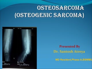
- 1. Presented By Dr. Santosh Atreya MD Resident,Phase-A,BSMMU
- 2. Outline of Presentation What is Osteosarcoma? Characteristics Gross Pathology and Appearance Classification Clinical Presentation Regional Distribution Diagnosis/Radiological Shortly about Parosteal, Periostial osteosarcoma Treatment and Prognosis D/D of Osteosarcoma Difference among Osteosarcoma, Osteomyelitis and Ewing Sarcoma
- 4. Osteo=bone/osteoid tissue. Sarcoma=malignant tumour of connective tissue. An osteosarcoma is the commonest primary malignant bone tumour. They account for 25 % of all primary bone tumors.
- 5. Age: 10-25 years In older age groups associated with Pagets disease Gender: slight male preponderance exists Incidence
- 6. Location Any bone may be involved, rather more than half located around the knee involving the metadiaphyses of the distal end of femur and proximal end of tibia Infrequently in pelvis, spine Clavicle ribs scapula and small bones of hands and feet -rare 10% arise in diaphysis
- 7. Clinical Presentation Patients usually present with localised pain or swelling particularly around the knee, occasionally accompanied by a soft- tissue mass or swelling. Sometimes, the first symptoms are related to pathological fracture.
- 8. Characteristics It is histologically pleomorphic. Two diagnostic features are – a)It’s ability to produce osteoid tissue without necessarily the development of cartilaginous precursor. b)The presence of abundant alkaline phosphatase histochemically within the tumor cells
- 9. Pleomorphic nature of sarcoma A dominant cell line may modify the appearance. If osteoblasts predominate, tumour bone formation will result. Whereas if cells of cartilage origin are present, extensive calcification may be a presenting feature.
- 10. Fig:1.Osteosarcoma of the tibia and fibula-predominantly osteoblastic.Amorphous calcification/ossification is present in the soft tissues with cortical destruction and a little periosteal new bone formation. Fig:2.Osteosarcoma of the distal femur-predominantly chondroblastic.Note the well- defined soft tissue mass and radiating spiculation of calcification within it.
- 11. Metastasis: It is highly vascular & metastases occurs by hematogenous route to the lung. Any lung lesion arising in a patient with osteosarcoma should be regarded with suspicion. .Later stage metastasis may spread to bone. Skip metastases-5 to 8%. Lymphatic spread is rare. Fig: Osteosarcoma-metastasis in the lungs presents with a pneumothorax.
- 12. Gross pathology Arise from multipotent mesenchymal cells. Mixture of osteoid, fibrous, cartilaginous tissue,necrotic, hemorrhagic,cystic areas, destruction of cortex Arise eccentrically in the medullary cavity with ill defined cortical destruction and soft tissue involvement.
- 13. Gross Appearance Large tumors Gritty & grayish-white in color Hemorrhage Cystic degeneration Cortical destruction Spreads – medullary canal Soft tissue masses present
- 14. Classification A.Primary osteosarcoma B.Secondary osteosarcoma A.Primary osteosarcoma: According to dominant cell line( Histopathology) classified as- a. Osteoblastic b. Chondroblastic c. Fibroblastic d. Anaplastic and e.Telangiectatic Accoarding to site: a.Diaphyseal b.Central c.Multifocal and d.Soft tissue osteosarcoma
- 15. B.Secondary osteosarcoma: Secondary to- I. Paget’s disease(paget’s sarcoma) II. Radiation or ingestion of radioactive material.
- 16. S.N PRIMARY OSTEOSARCOMA SECONDARY OSTEOSARCOMA 1. In young patients (10 - 25 years) Occurs in the elderly 2. 75% < age of 20 Secondary to malignant degeneration of pagets disease,extensive bone infarcts or post-radiotherapy 3. M>F M>F 4. Typically occur in the metaphyseal regions of long bones, and have a striking predilection for the knee (60%) Wider distribution,higher incidence in flat bones, especially the pelvis.
- 17. Diagnosis 1.Laboratory studies: CBC, ESR, CRP, LDH (elevated level indicates poor prognosis) ALP (Highly Osteogenic) Platelet count, Electrolyte levels, Liver function tests, Renal function tests, Urinalysis
- 18. 2.Radiological features: Findings: Typical appearances include: Medullary and cortical bone destruction Wide zone of transition Permeative or moth-eaten appearance Aggressive periosteal reaction Codman triangle Sunburst type lamellated (onion skin) reaction( less frequently seen) soft-tissue mass tumour matrix ossification / calcification variable: reflects a combination of the amount of tumour bone production, calcified matrix, and osteoid
- 19. Cont.. 3. CT Scan: It is the most sensitive means in detecting pulmonary metastasis. CT scanning may be helpful locally when the radiographic appearances are confusing, particularly in areas of complex anatomy. Cross-sectional images provide a clearer information of bone destruction, as well as the extent of any soft tissue mass, than do radiographs.
- 20. Cont.. 4. MRI It is the prime investigation of choice for Osteosarcoma An obvious heterogeneous tumor is demonstrated with surrounded bones and usually a soft tissue mass Intramedullary skip lesion may also be identified
- 21. T1WI soft tissue non-mineralized component : intermediate signal intensity mineralised / ossified components : low signal intensity peri-tumoural oedema : intermediate signal intensity scattered regions of haemorrhage will have variable signal T2WI soft tissue non-mineralized component : high signal intensity mineralised / ossified components : low signal intensity peri-tumoural oedema : high signal intensity
- 22. abnormal signal intensity in the metaphyseal marrow and the soft tissue mass (black arrow). Early tumor extension is shown beyond the growth plate into the epiphysis (white arrows). Coronal T1-weighted MRI.
- 23. STIR suppresses signal from fat, Sensitive to edema and bone pathology Normal marrow and fat: dark Fluid & edema: bright Bone findings: 1. Increased signal in the medullary canal. 2. Irregular pattern in the metaphysis. 3. Ill defined cortical outline. 4. Extension to the epiphysis. 5. High signal around distal femur, suggesting edema and growth into the surrounding tissue. Coronal STIR of the left knee
- 24. Cont.. 5.Biopsy to confirm the diagnosis. Histology confirmed radiological suspicion of osteosarcoma in the distal femur of patient 1. Formation of new, abnormal bone with a coarse lacelike architecture 2. Variable tumor cell size & shape, with hyperchromatic nuclei and mitoses.
- 25. Cont.. 6. Scintigraphy Osteosarcomas typically show increased uptake of radioisotope on bone scans obtained by use of technetium-99m (99m Tc) methylene diphosphonate (MDP). A scan in the early blood-pool (left) and delayed phases (right) demonstrates an extensive abnormality. the activity is more uniform and extensive than the apparent involvement shown on the plain film.
- 26. Cont.. 7. Angiogram Angiogram Determine vascularity of the tumour ,Detect vascular displacement and relationship of vessels to the tumour Telangiectatic osteosarcoma of the distal femur. predominantly radiolucent defect is shown on conventional radiograph. Angiographically is shown to contain large, tortuous, pathological vessels.
- 27. Some confusion in nomenclature relates to osteosarcoma arising in or near the periosteum. They are divided into two groups: a.Parosteal osteosarcoma and b.Periosteal osteosarcoma
- 28. Parosteal Osteosarcoma Most patient affected in 3rd & 4th decade. Typically dense tumour surrounds a long bone,particularly femur or a tibia. Margin are sharply defined but tend to undulate. The tumour is denser centrally and at the base than peripherally. Characteristically there is a radiolucent zone between the ossified outer margins of the tumour and adjacent host bone. Usually, the tumour appears to be attached to the cortex by a broad pedicle.
- 29. Parosteal osteosarcoma of the proximal humerus. A well-defined mass of dense tumour bone surrounds the humeral shaft. Parosteal osteosarcoma arising from the anterior aspect of the femur shown angiographically to be unremarkable apart from a slight increase in the number of branches going into the tumour.
- 31. Telangiectatic Osteosarcoma (2.5- 12.5%) Lytic tumors consisting of large cystic cavities filled with blood usually diametaphyseal in location. Has been considered more aggressive than classic osteosarcoma, but studies of long-term survival after optimum treatment now indicate that the aggressiveness of telangiectatic osteosarcoma is similar to that of the classic type.
- 32. Frontal radiograph of the distal femur in a patient with telangiectatic osteosarcoma. the radiograph shows mixed medullary sclerosis and lucency, cortical destruction medially, aggressive periosteal changes, and a large soft-tissue mass with peripheral ossification
- 33. Sarcoma in Paget’s Disease Malignant tumours are said to arise in bone affected by Paget's disease in about 1 % of cases. Overall, the skull, pelvis and long bones are typical sites, predilection for the humerus in the later. Men are more commonly affected, even allowing for the increased male incidence of Paget's disease. However, the tumour is very aggressive and the outlook is very poor. Radiologically, the lesion is lytic, mixed or sclerotic.
- 34. Xray of the proximal femur in a patient with Paget disease demonstrates the typical features of cortical thickening, osseous expansion, and trabecular coarsening. In addition, irregular bone lucency and cortical destruction are shown in the medial aspect of the shaft; Coronal T1WIof the same patient showing -the tumor is shown in the proximal shaft of the right femur (white arrow), with cortical destruction and a large soft-tissue component (black arrow).
- 35. Treatment and Prognosis Treatment options for classic osteosarcoma Surgery alone: 20% cure rate. Surgery & chemotherapy: 60-80% cure rate. Radical surgical treatment • Wide surgical resection • Limb salvage (used in 80-90% of all cases) Bone replaced with a bone allograft or a prosthesis. • Amputation Currently, the 5-year survival rate after adequate therapy is approximately 60 - 80% .
- 36. Differential Diagnosis Osteomyelitis Other tumours : metastatic lesion to bone Malignant round cell tumours (Ewing sarcoma)
- 37. Age Age is the most important clue in differentiating possible bone tumors. Osteosarcoma-Between 10 &25 yrs Ewing’s sarcoma-5 to 30 yrs. Location within the skeleton The location of a bone lesion within the skeleton can be a clue in differential diagnosis. • Osteomyelitis-femur, tibia, humerus, fibula, radius • Osteosarcoma-femur • Ewing's sarcoma-iliac bone, fibula, rib, tibia, humerus,pelvis.
- 38. Site and Location Osteosarcoma Osteomyelitis Ewing’s sarcoma Site Metaphyseal Metaphysis Diaphysis Location juxtacortical centric Eccentric juxtacortical centric
- 39. Periosteal reaction & Zone of transition osteosarcoma Osteomyelitis Ewing’s sarcoma Periosteal reaction Sunburst spiculation Formation of involucrum,se questra Onion peel lameller type zone of transition Wide zone of transition Wide zone of transition Wide zone of transition
- 40. Ewing's sarcoma. well-defined soft-tissue mass. Advanced osteomvelitis involving the whole of the right tibia and lowervoend of fibula. Note sequestrum in tibia and further sequestrum being extruded from the fibula. Osteosarcoma of the distal femur-. well- defined soft-tissue mass and radiating spiculation of calcification within it.
- 41. Thank You