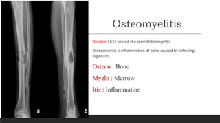
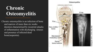
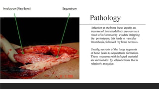
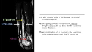
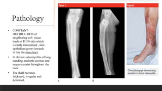
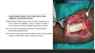
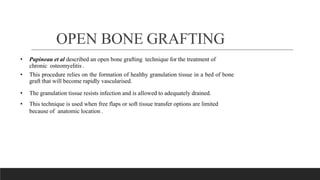
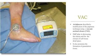
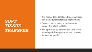
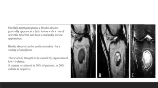
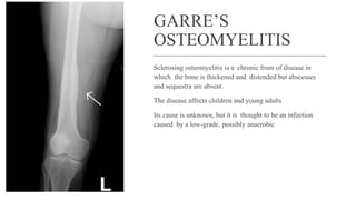
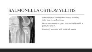
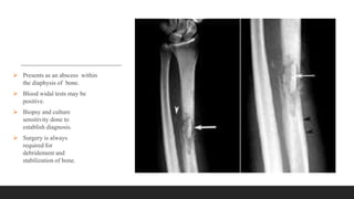
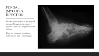
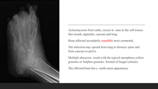
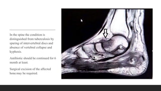
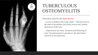
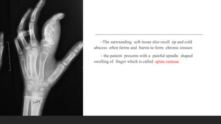
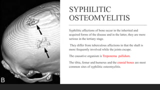
SlideShare a Scribd company logo
1 of 57
Chronic
Osteomyelitis
DR. DIWAKAR PRATAP
ASSISTANT PROFESSOR
GMC-HALDWANI
Osteomyelitis
Nelaton 1834 coined the term Osteomyelitis
Osteomyelitis is inflammation of bone caused by infecting
organism.
Osteon : Bone
Myelo : Marrow
Itis : Inflammation
Classification
A) Based on duration
Acute Osteomyelitis < 2 weeks
Subacute Osteomyelitis 2-6 weeks
Chronic Osteomyelitis > 6 weeks
B) Based on Mechanism
Direct Innolucation Open wound, post surgery
Hematogenous Bacteremia
Chronic
Osteomyelitis
Chronic osteomyelitis is an infection of bone
and marrow of more than six weeks
duration characterised by recurrent attacks
of inflammation with discharging sinuses
and presence of infected dead
bone(sequestra).
BACTERIOLOGY• Staph. aureus(most common)
• Staph. epidermidis
• Strep. Pyogenes
• Haemophilus influenzae (between 6 months. to 4 yrs.)
• Salmonella (in sickle cell anemia)
• Pseudomonas (intravenous drug users)
Predisposing factors
Inadequate treatment of acute osteomyelitis
An acute becomes chronic due to one of the following reasons:
 Improper drainage of the pus in acute osteomyelitis
 Formation of an undrained non collapsible cavity in the bone
 Presence of sequestra
 Presence of foreign bodies in case of osteomyelitis following open injuries
 Compound fracture
 Direct invasion from adjacent soft tissues, such as from diabetic ulcers
 Around an infected implant or prosthesis
Pathology
Infection at the bone locus creates an
increase of intramedullary pressure as a
result of inflammatory exudate stripping
the periosteum; this leads to vascular
thrombosis, followed by bone necrosis.
Usually, necrosis of the large segments
of bone leads to sequestrum formation.
These sequestra with infected material
are surrounded by sclerotic bone that is
relatively avascular.
The haversian canals are blocked
with scar tissue, and the bone is
surrounded by thickened
periosteum and scarred muscle
Antibiotics cannot penetrate
these relatively avascular
tissues and are hence
ineffective in clearing the
infection.
New bone formation occurs at the same time (involucrum)
around the dead bone
Multiple openings appear in this involucrum, (cloacae)
through which exudates and debris from the sequestrum
pass via the sinuses.
The periosteal reaction acts to circumscribe the sequestrum,
producing a thick sheet of new bone or involucrum.
Sequestrum:
It is a piece of dead bone separated from
healthy bone. The area of dead bone gets
demarcated by granulation tissue and
gradually separates and forms a loose piece
of sequestrum.
Different
types of
SEQUESTRA
TYPE DISEASE
TUBULAR PYOGENIC
RING EXTERNAL
FIXATORS
BLACK ACTINOMYCOSIS
CORALLIFORM PERTHE’S DISEASE
COKE TUBERCULOSIS
SANDY TUBERCULOSIS
FEATHERY SYPHILIS
Pathology
• CONSTANT
DESTRUCTION of
neighboring soft tissue
leads to THIN skin which
is easily traumatised , skin
epithelium grows inwards
to line the sinus tract.
• In chronic osteomyelitis of long
standing, multiple cavities and
sequestra exist throughout the
bone
• The shaft becomes
thickened, irregular and
deformed.
CLINICAL
• During the period of in-activity no
symptoms are present.
• The bone is mis-shapen and the shin
is dusky, thin, scarred and poorly
nourished.
• A break in the skin causes an
ulceration that is slow to heal.
• Muscles are scarred and cause
contractures of the adjacent joints.
CLINICAL
Pain is aching type
and usually worsens
in the night.
The overlying soft
tissues become
swollen ,
edematous , warm ,
reddened and
tender.
As the infection
progresses a sinus is
formed and is
drained indefinitely.
Spontaneous
closure of the sinus
and subsidence of
infection often
occur following
explusion of large
fragment.
CLINICAL
Recurrent flare ups occurs indefinitely
over a period of months and years . A
sinus may drain continously.
Recurrent toxemia over a long period
will causes amyloidosis.
DIAGNOSIS
• The diagnosis is based on
Clinical , Laboratory and
Imaging studies.
• The “GOLD STANDARD” is to obtain a biopsy specimen for histological and
microbiological evaluation of the infected bone.
• Physical examination should be focused on integrity of skin and soft
tissue .
• Determination of area of tenderness.
• Assessing bone stability.
• And evaluation of neuro vascular status of the limb
LABORATORY
• Lab studies generally are nonspecific and
give no indication for severity of the
infection.
• ESR and C- Reactive protein are elevated in most
patients.
• But WBC’S elevated in only 35%.
Multiple imaging technique are available to
evaluate chronic osteomyelitis ,however no
technique can absolutely confirm or exclude
presence of osteomyelitis.
• Imaging should be done to confirm
the diagnosis and prepare for
surgery.
• Initial plain radiographs to be
performed it yields valuable info .
• Signs of cortical destruction and
periosteal reaction strongly suggest
the diagnosis of osteomyelitis.
• Sinography can be preformed if a sinus track is present and can be valuable
adjunct to surgical planning.
• Isotopic bone scanning is more useful in acute osteomyelitis than chronic
osteomyelitis.
• CT provides excellent definition of cortical bone and a fair evaluation of
the surrounding soft tissues and is especially useful in identifying
sequestra.
• MRI provides a fairly accurate measure of pathological insult to bone and
soft tissue, so it is superior to CT in soft tissue evaluation.
• MRI may reveal a well defined rim of high signal intensity surrounding
the focus of active disease (RIM SIGN).
TREATMENT
• Requires a multi faceted approach.
In addition to antibiotic and surgical debridement and reconstruction.
• 1st objective is removal of dead bones(sequestrum).
• 2nd objective is to find a method of obliterating any dead space left after
debridement.
• 3rd objective is to obtain soft tissue coverage of exposed bone which is a part of
the objective of the obliterating dead space.
• Surgery for osteomyelitis consists of sequestrectomy and
resection of scarred and infected bone and soft tissue.
• Ring External fixators are generally used for soft tissue and
dead space management after radical debridement.
• The GOAL of surgery is to eradicate infection by achieving a
viable and vascular environment.
• Extensive debridement creates a large dead space – this is
treated with ANTIBIOTIC POLYMETHYL METH
ACRYLATE (PMMA) beads that fills the dead space and
prevents recurrences.
• The duration of post
operative antibiotics is
controversial .
• Traditionally, a 6 week
course of intravenous
antibiotics is prescribed after
surgical debridement.
• The methods to eliminate the
dead space are –
1. Bone grafting with
primary and secondary
closure.
2. Use of PMMA as a
temporary filler of dead
space.
3. Local muscle
flaps and
skin grafting
with or
without bone
grafting.
4. Microvascular transfer of
muscle, osseous flaps.
5. The use of bone transport
(ILIZAROV
TECHNIQUE).
• SEQUESTRECTOMY AND CURETTAGE FOR
CHRONIC OSTEOMYELITIS
SEQUESTRECTOMY means removal of the sequestrum, if
it lies within the medullary cavity, a window is made in
the overlying involucrum and the sequestrum removed .
One must wait for adequate involucrum formation before
performing sequestrectomy.
Sinus tracks can be injected with methylene blue 24 hours before
surgery to make them easier to locate and excise.
OPEN BONE GRAFTING
• Papineau et al described an open bone grafting technique for the treatment of
chronic osteomyelitis .
• This procedure relies on the formation of healthy granulation tissue in a bed of bone
graft that will become rapidly vascularised.
• The granulation tissue resists infection and is allowed to adequately drained.
• This technique is used when free flaps or soft tissue transfer options are limited
because of anatomic location .
VAC
• Archdeacon described a
modification of the papineau
technique using a vaccum
assisted closure (VAC).
• VAC helps in decreasing
the edema and for the
closure of soft tissue
dead space.
• It also promotes the
formation of granulation
tissue.
POLYMETHYLMETHACRYLATE
ANTIBIOTIC BEAD CHAINS (BONE CEMENT)
• Studies have shown that the local concentrations achieved
are 200 times more than intravenous.
• High concentration can be achieved by primary closure of
the wound.
• Short term (10 days), long term(80days) , permanent
implantation of PMMA beads is possible.
BIODEGRADABLE ANTIBIOTIC
DELIVERY SYSTEM
• It offers a significant advantage over PMMA in that a second procedure is not required to remove
the implant.
• It is useful when bone stability is not an issue and soft tissue coverage is adequate.
• Many manufacturers produce a variety of bioabsorbable substrates(calcium sulfate or calcium
phosphate)that can be mixed with antibiotics like vancomycin and tobramycin).
SOFT
TISSUE
TRANSFER
• It is mainly done to fill dead space which is
left behind after extensive debridement.
• Success rate reported in the literature
ranges from 66% to 100%.
• For eg chronic osteomyelitis of tibia a local
muscle graft from gastrocnemius or soleus
is used for transfer.
ILIZAROV
TECHNIQUE
• This technique allows radical resection of the infected bone
• A corticotomy is performed through the normal bone proximal and
distal to the area of the disease.
• Disadvantage is – long time to achieve solid union and high
chances of infections.
• The treatment of segmental defects of up to 13cms can be
achieved.
ADJUNCTIVE
THERAPIES
• Hyperbaric Oxygen is not reliably effective but is used as more
traditional methods of treatment.
• Bone morphogenic proteins (BMPs) and even Platelet Rich Plasmas
(PRPs) have been advocated as it can accelerate or enhance
osteogenesis.
COMPLICATIONS
An acute exacerbation of the infections occurs commonly.
Growth Abnormalities :
• shortening –if growth plate is damaged.
• Lengthening – coz of increased vasularity of the growth plate due to near by osteomyelitis.
Pathological fracture .
Joint stiffness – may occur because of scarring of soft tissues around the joint.
Sinus tract malignancy – rare complication (squamous cell carcinoma)
Muscle contracture.
Epithelioma.
Amyloidosis.
PRIMARY SUBACUTE
OSTEOMYELITIS
Brodie abscess
Garres’osteomyelitis
Salmonella osteomyelitis
BRODIE ABSCESS
A Brodie abscess is a localized form of subacute
osteomyelitis that occurs most often in the long
bones of the lower extremities of young adults.
In adults the metaphyseal- epiphyseal area is
involved.
Intermittent pain of long duration is the presenting
complaint, along with local tenderness over the
affected area.
On plain roentgenograms a Brodie abscess
generally appears as a lytic lesion with a rim of
sclerotic bone but can have a markedly varied
appearance.
Brodie abscess can be easily mistaken for a
variety of neoplasm.
The lesion is thought to be caused by organisms of
low virulence.
S. aureus is cultured in 50% of patients, in 20%
culture is negative.
This condition often requires an open biopsy with curettage to make the diagnosis.
The wound should be closed loosely over a drain.
Following Biopsy and curettage, antibiotics are given for 3 weeks duration.
GARRE’S
OSTEOMYELITIS
Sclerosing osteomyelitis is a chronic from of disease in
which the bone is thickened and distended but abscesses
and sequestra are absent.
The disease affects children and young adults
Its cause is unknown, but it is thought to be an infection
caused by a low-grade, possibly anaerobic
Patients report intermittent pain of moderate intensity and
usually of long duration.
Swelling and tenderness over the affected bone may be
found
Roentgenograms show an expanded bone with generalized
sclerosis
The ESR usually is slightly elevated Biopsy shows only
chronic, low-grade, nonspecific inflammation.
A secondary lesion at a distant site can occur years after onset. No
treatment has been predictably helpful, but fenestration of the
sclerotic bone and antibiotics are advisable
The condition must be distinguished from osteoid osteoma and
Paget’s disease.
SALMONELLA OSTEOMYELITIS
Subacute type of osteomyelitis usually occurring
in the ulna, ribs and vertebrae
Occurs some months or years after attack of typhoid or
paratyphoid fever.
Commonly associated with sickle cell anemia
 Presents as an abscess within
the diaphysis of bone.
 Blood widal tests may be
positive.
 Biopsy and culture
sensitivity done to
establish diagnosis.
 Surgery is always
required for
debridement and
stabilization of bone.
FUNGAL
(MYCOTIC)
INFECTION
Mycotic osteomyelitis is the general
term used to describe a group of
diseases caused by fungal infections
of bone.
There are two main organisms-
Actinomyces and Maduramyces.
Actinomycosis from cattle, occurs in man in the soft tissues
like mouth, appendix, caecum and lung.
Bone affected secondarily, mandible most commonly.
The infection may spread from lung to thoracic spine and
from caecum to pelvis.
Multiple abscesses result with the typical amorphous yellow
granules or Sulphur granules formed of fungal colonies.
The affected bone has a moth-eaten appearance.
In the spine the condition is
distinguished from tuberculosis by
sparing of intervertebral discs and
absence of vertebral collapse and
kyphosis.
Antibiotic should be continued for 6
month at least.
Surgical excision of the affected
bone may be required.
TUBERCULOUS
OSTEOMYELITIS
Tuberculous dactylitis aka Spina Ventosa
~ occurs in children and young adults ~ infection starts in
the shaft of the phalanx and causes erosion and gradual
destruction of the bone
~ subperiosteal new bone formation and thickening of
bone this phenomenon is peculiar to the tuberculous
infection in the long bones.
~The surrounding soft tissue also swell up and cold
abscess often forms and bursts to form chronic sinuses
~ the patient presents with a painful spindle shaped
swelling of finger which is called spina ventosa
SYPHILITIC
OSTEOMYELITIS
Syphilitic affections of bone occur in the inherited and
acquired forms of the disease and in the latter, they are more
serious in the tertiary stage.
They differ from tuberculous affections in that the shaft is
more frequently involved while the joints escape.
The causative organism is Treponema pallidum.
The tibia, femur and humerus and the cranial bones are most
common sites of syphilitic osteomyelitis.
Manifestations –
Pain – this may vary from slight dull ache to most excruciating
pains. There are no local abnormalities on clinical exam. And a
diagnosis of neuralgia is often made.
Periostitis – frequently occurs and affects multiple long bones.
1. Diffuse osteoperiostitis – this is a chronic inflammation
affecting the whole bone or the greater portion of it inside
the periosteal envelope. X-ray shows double outline which is
very characteristic. A second sheath of compact bone
surrounds the original compact layer but an intervening
space exists which may be filled with granulation tissue.
2. Syphilitic osteochondritis –
children with inherited syphilis
show an irregularity of the
epiphyseal line. This irregularity is
due to transformation of cartilage
into bone. There is thickening of
epiphysis and pain on passive
movement.
3.Gummatous Osteomyelitis –
Gumma can occur on surface of the
bone or within it. The surface
gumma resembles an ordinary
periosteal nod except that its
speedily softens at its centre.
A gumma is in the interior of long
bone is a serious condition as it is is
taken for a malignant tumour.
4. Syphilitic dactylitis – the importance
of syphilis of the phalanges lies in the
fact that it may be is taken for
tuberculosis. But there is little
tendency to break down and ulcerate as
in tuberculosis.
The condition is usually painless.
Antibiotics are usually ineffective.

More Related Content

Similar to Chronic Osteomyelitis, Bone infection slides

osteomyelitis ppt.pptx
osteomyelitis ppt.pptxosteomyelitis ppt.pptx
osteomyelitis ppt.pptxaasrithakotha2
 
osteomyelitisbydr-171123063448.pptx
osteomyelitisbydr-171123063448.pptxosteomyelitisbydr-171123063448.pptx
osteomyelitisbydr-171123063448.pptxAmerManzoorPak
 
L- shaped Corticotomy - osteomyelitis
L- shaped Corticotomy - osteomyelitis L- shaped Corticotomy - osteomyelitis
L- shaped Corticotomy - osteomyelitis Ponnilavan Ponz
 
chronicosteomyelitis Powerpt power point
chronicosteomyelitis Powerpt power pointchronicosteomyelitis Powerpt power point
chronicosteomyelitis Powerpt power pointVaisHali822687
 
Implant related periodontal disease
Implant related periodontal diseaseImplant related periodontal disease
Implant related periodontal diseaseNavneet Randhawa
 
Masquelet technique ppt
Masquelet technique pptMasquelet technique ppt
Masquelet technique pptApoorv Garg
 
Osteomyelitis and osteoradionecrosis of jaws
Osteomyelitis and osteoradionecrosis of jawsOsteomyelitis and osteoradionecrosis of jaws
Osteomyelitis and osteoradionecrosis of jawsShibani Sarangi
 
Chronic osteomyelitis
Chronic osteomyelitisChronic osteomyelitis
Chronic osteomyelitisorthoprince
 
Bone and joint infections
Bone and joint infectionsBone and joint infections
Bone and joint infectionsLekhraj Bairwa
 

Similar to Chronic Osteomyelitis, Bone infection slides (20)

Osteomyelitis
OsteomyelitisOsteomyelitis
Osteomyelitis
 
osteomyelitis.pptx
osteomyelitis.pptxosteomyelitis.pptx
osteomyelitis.pptx
 
osteomyelitis.pptx
osteomyelitis.pptxosteomyelitis.pptx
osteomyelitis.pptx
 
osteomyelitis ppt.pptx
osteomyelitis ppt.pptxosteomyelitis ppt.pptx
osteomyelitis ppt.pptx
 
osteomyelitisbydr-171123063448.pptx
osteomyelitisbydr-171123063448.pptxosteomyelitisbydr-171123063448.pptx
osteomyelitisbydr-171123063448.pptx
 
chronic OM.pptx
chronic OM.pptxchronic OM.pptx
chronic OM.pptx
 
L- shaped Corticotomy - osteomyelitis
L- shaped Corticotomy - osteomyelitis L- shaped Corticotomy - osteomyelitis
L- shaped Corticotomy - osteomyelitis
 
chronicosteomyelitis Powerpt power point
chronicosteomyelitis Powerpt power pointchronicosteomyelitis Powerpt power point
chronicosteomyelitis Powerpt power point
 
Implant related periodontal disease
Implant related periodontal diseaseImplant related periodontal disease
Implant related periodontal disease
 
Chronic osteomyelitis
Chronic osteomyelitisChronic osteomyelitis
Chronic osteomyelitis
 
Masquelet technique ppt
Masquelet technique pptMasquelet technique ppt
Masquelet technique ppt
 
bone fracture.pptx
bone fracture.pptxbone fracture.pptx
bone fracture.pptx
 
bone fracture.pptx
bone fracture.pptxbone fracture.pptx
bone fracture.pptx
 
Osteomyelitis OF JAWS
 Osteomyelitis OF JAWS Osteomyelitis OF JAWS
Osteomyelitis OF JAWS
 
Osteomyelitis and osteoradionecrosis of jaws
Osteomyelitis and osteoradionecrosis of jawsOsteomyelitis and osteoradionecrosis of jaws
Osteomyelitis and osteoradionecrosis of jaws
 
Avascular necrosis
Avascular necrosis Avascular necrosis
Avascular necrosis
 
Chronic osteomyelitis
Chronic osteomyelitisChronic osteomyelitis
Chronic osteomyelitis
 
Osteomyelitis Case Report Session
Osteomyelitis Case Report SessionOsteomyelitis Case Report Session
Osteomyelitis Case Report Session
 
Osteomyelitis
OsteomyelitisOsteomyelitis
Osteomyelitis
 
Bone and joint infections
Bone and joint infectionsBone and joint infections
Bone and joint infections
 

Recently uploaded

Pharmacognosy Flower 3. Compositae 2023.pdf
Pharmacognosy Flower 3. Compositae 2023.pdfPharmacognosy Flower 3. Compositae 2023.pdf
Pharmacognosy Flower 3. Compositae 2023.pdfMahmoud M. Sallam
 
call girls in Kamla Market (DELHI) 🔝 >༒9953330565🔝 genuine Escort Service 🔝✔️✔️
call girls in Kamla Market (DELHI) 🔝 >༒9953330565🔝 genuine Escort Service 🔝✔️✔️call girls in Kamla Market (DELHI) 🔝 >༒9953330565🔝 genuine Escort Service 🔝✔️✔️
call girls in Kamla Market (DELHI) 🔝 >༒9953330565🔝 genuine Escort Service 🔝✔️✔️9953056974 Low Rate Call Girls In Saket, Delhi NCR
 
MARGINALIZATION (Different learners in Marginalized Group
MARGINALIZATION (Different learners in Marginalized GroupMARGINALIZATION (Different learners in Marginalized Group
MARGINALIZATION (Different learners in Marginalized GroupJonathanParaisoCruz
 
ECONOMIC CONTEXT - PAPER 1 Q3: NEWSPAPERS.pptx
ECONOMIC CONTEXT - PAPER 1 Q3: NEWSPAPERS.pptxECONOMIC CONTEXT - PAPER 1 Q3: NEWSPAPERS.pptx
ECONOMIC CONTEXT - PAPER 1 Q3: NEWSPAPERS.pptxiammrhaywood
 
Interactive Powerpoint_How to Master effective communication
Interactive Powerpoint_How to Master effective communicationInteractive Powerpoint_How to Master effective communication
Interactive Powerpoint_How to Master effective communicationnomboosow
 
भारत-रोम व्यापार.pptx, Indo-Roman Trade,
भारत-रोम व्यापार.pptx, Indo-Roman Trade,भारत-रोम व्यापार.pptx, Indo-Roman Trade,
भारत-रोम व्यापार.pptx, Indo-Roman Trade,Virag Sontakke
 
Framing an Appropriate Research Question 6b9b26d93da94caf993c038d9efcdedb.pdf
Framing an Appropriate Research Question 6b9b26d93da94caf993c038d9efcdedb.pdfFraming an Appropriate Research Question 6b9b26d93da94caf993c038d9efcdedb.pdf
Framing an Appropriate Research Question 6b9b26d93da94caf993c038d9efcdedb.pdfUjwalaBharambe
 
Capitol Tech U Doctoral Presentation - April 2024.pptx
Capitol Tech U Doctoral Presentation - April 2024.pptxCapitol Tech U Doctoral Presentation - April 2024.pptx
Capitol Tech U Doctoral Presentation - April 2024.pptxCapitolTechU
 
Introduction to ArtificiaI Intelligence in Higher Education
Introduction to ArtificiaI Intelligence in Higher EducationIntroduction to ArtificiaI Intelligence in Higher Education
Introduction to ArtificiaI Intelligence in Higher Educationpboyjonauth
 
Earth Day Presentation wow hello nice great
Earth Day Presentation wow hello nice greatEarth Day Presentation wow hello nice great
Earth Day Presentation wow hello nice greatYousafMalik24
 
Final demo Grade 9 for demo Plan dessert.pptx
Final demo Grade 9 for demo Plan dessert.pptxFinal demo Grade 9 for demo Plan dessert.pptx
Final demo Grade 9 for demo Plan dessert.pptxAvyJaneVismanos
 
DATA STRUCTURE AND ALGORITHM for beginners
DATA STRUCTURE AND ALGORITHM for beginnersDATA STRUCTURE AND ALGORITHM for beginners
DATA STRUCTURE AND ALGORITHM for beginnersSabitha Banu
 
Proudly South Africa powerpoint Thorisha.pptx
Proudly South Africa powerpoint Thorisha.pptxProudly South Africa powerpoint Thorisha.pptx
Proudly South Africa powerpoint Thorisha.pptxthorishapillay1
 
Employee wellbeing at the workplace.pptx
Employee wellbeing at the workplace.pptxEmployee wellbeing at the workplace.pptx
Employee wellbeing at the workplace.pptxNirmalaLoungPoorunde1
 
Painted Grey Ware.pptx, PGW Culture of India
Painted Grey Ware.pptx, PGW Culture of IndiaPainted Grey Ware.pptx, PGW Culture of India
Painted Grey Ware.pptx, PGW Culture of IndiaVirag Sontakke
 
History Class XII Ch. 3 Kinship, Caste and Class (1).pptx
History Class XII Ch. 3 Kinship, Caste and Class (1).pptxHistory Class XII Ch. 3 Kinship, Caste and Class (1).pptx
History Class XII Ch. 3 Kinship, Caste and Class (1).pptxsocialsciencegdgrohi
 
“Oh GOSH! Reflecting on Hackteria's Collaborative Practices in a Global Do-It...
“Oh GOSH! Reflecting on Hackteria's Collaborative Practices in a Global Do-It...“Oh GOSH! Reflecting on Hackteria's Collaborative Practices in a Global Do-It...
“Oh GOSH! Reflecting on Hackteria's Collaborative Practices in a Global Do-It...Marc Dusseiller Dusjagr
 
KSHARA STURA .pptx---KSHARA KARMA THERAPY (CAUSTIC THERAPY)————IMP.OF KSHARA ...
KSHARA STURA .pptx---KSHARA KARMA THERAPY (CAUSTIC THERAPY)————IMP.OF KSHARA ...KSHARA STURA .pptx---KSHARA KARMA THERAPY (CAUSTIC THERAPY)————IMP.OF KSHARA ...
KSHARA STURA .pptx---KSHARA KARMA THERAPY (CAUSTIC THERAPY)————IMP.OF KSHARA ...M56BOOKSTORE PRODUCT/SERVICE
 
POINT- BIOCHEMISTRY SEM 2 ENZYMES UNIT 5.pptx
POINT- BIOCHEMISTRY SEM 2 ENZYMES UNIT 5.pptxPOINT- BIOCHEMISTRY SEM 2 ENZYMES UNIT 5.pptx
POINT- BIOCHEMISTRY SEM 2 ENZYMES UNIT 5.pptxSayali Powar
 

Recently uploaded (20)

Pharmacognosy Flower 3. Compositae 2023.pdf
Pharmacognosy Flower 3. Compositae 2023.pdfPharmacognosy Flower 3. Compositae 2023.pdf
Pharmacognosy Flower 3. Compositae 2023.pdf
 
call girls in Kamla Market (DELHI) 🔝 >༒9953330565🔝 genuine Escort Service 🔝✔️✔️
call girls in Kamla Market (DELHI) 🔝 >༒9953330565🔝 genuine Escort Service 🔝✔️✔️call girls in Kamla Market (DELHI) 🔝 >༒9953330565🔝 genuine Escort Service 🔝✔️✔️
call girls in Kamla Market (DELHI) 🔝 >༒9953330565🔝 genuine Escort Service 🔝✔️✔️
 
MARGINALIZATION (Different learners in Marginalized Group
MARGINALIZATION (Different learners in Marginalized GroupMARGINALIZATION (Different learners in Marginalized Group
MARGINALIZATION (Different learners in Marginalized Group
 
ECONOMIC CONTEXT - PAPER 1 Q3: NEWSPAPERS.pptx
ECONOMIC CONTEXT - PAPER 1 Q3: NEWSPAPERS.pptxECONOMIC CONTEXT - PAPER 1 Q3: NEWSPAPERS.pptx
ECONOMIC CONTEXT - PAPER 1 Q3: NEWSPAPERS.pptx
 
Interactive Powerpoint_How to Master effective communication
Interactive Powerpoint_How to Master effective communicationInteractive Powerpoint_How to Master effective communication
Interactive Powerpoint_How to Master effective communication
 
भारत-रोम व्यापार.pptx, Indo-Roman Trade,
भारत-रोम व्यापार.pptx, Indo-Roman Trade,भारत-रोम व्यापार.pptx, Indo-Roman Trade,
भारत-रोम व्यापार.pptx, Indo-Roman Trade,
 
Framing an Appropriate Research Question 6b9b26d93da94caf993c038d9efcdedb.pdf
Framing an Appropriate Research Question 6b9b26d93da94caf993c038d9efcdedb.pdfFraming an Appropriate Research Question 6b9b26d93da94caf993c038d9efcdedb.pdf
Framing an Appropriate Research Question 6b9b26d93da94caf993c038d9efcdedb.pdf
 
Capitol Tech U Doctoral Presentation - April 2024.pptx
Capitol Tech U Doctoral Presentation - April 2024.pptxCapitol Tech U Doctoral Presentation - April 2024.pptx
Capitol Tech U Doctoral Presentation - April 2024.pptx
 
Introduction to ArtificiaI Intelligence in Higher Education
Introduction to ArtificiaI Intelligence in Higher EducationIntroduction to ArtificiaI Intelligence in Higher Education
Introduction to ArtificiaI Intelligence in Higher Education
 
Earth Day Presentation wow hello nice great
Earth Day Presentation wow hello nice greatEarth Day Presentation wow hello nice great
Earth Day Presentation wow hello nice great
 
Final demo Grade 9 for demo Plan dessert.pptx
Final demo Grade 9 for demo Plan dessert.pptxFinal demo Grade 9 for demo Plan dessert.pptx
Final demo Grade 9 for demo Plan dessert.pptx
 
DATA STRUCTURE AND ALGORITHM for beginners
DATA STRUCTURE AND ALGORITHM for beginnersDATA STRUCTURE AND ALGORITHM for beginners
DATA STRUCTURE AND ALGORITHM for beginners
 
Proudly South Africa powerpoint Thorisha.pptx
Proudly South Africa powerpoint Thorisha.pptxProudly South Africa powerpoint Thorisha.pptx
Proudly South Africa powerpoint Thorisha.pptx
 
Employee wellbeing at the workplace.pptx
Employee wellbeing at the workplace.pptxEmployee wellbeing at the workplace.pptx
Employee wellbeing at the workplace.pptx
 
Painted Grey Ware.pptx, PGW Culture of India
Painted Grey Ware.pptx, PGW Culture of IndiaPainted Grey Ware.pptx, PGW Culture of India
Painted Grey Ware.pptx, PGW Culture of India
 
OS-operating systems- ch04 (Threads) ...
OS-operating systems- ch04 (Threads) ...OS-operating systems- ch04 (Threads) ...
OS-operating systems- ch04 (Threads) ...
 
History Class XII Ch. 3 Kinship, Caste and Class (1).pptx
History Class XII Ch. 3 Kinship, Caste and Class (1).pptxHistory Class XII Ch. 3 Kinship, Caste and Class (1).pptx
History Class XII Ch. 3 Kinship, Caste and Class (1).pptx
 
“Oh GOSH! Reflecting on Hackteria's Collaborative Practices in a Global Do-It...
“Oh GOSH! Reflecting on Hackteria's Collaborative Practices in a Global Do-It...“Oh GOSH! Reflecting on Hackteria's Collaborative Practices in a Global Do-It...
“Oh GOSH! Reflecting on Hackteria's Collaborative Practices in a Global Do-It...
 
KSHARA STURA .pptx---KSHARA KARMA THERAPY (CAUSTIC THERAPY)————IMP.OF KSHARA ...
KSHARA STURA .pptx---KSHARA KARMA THERAPY (CAUSTIC THERAPY)————IMP.OF KSHARA ...KSHARA STURA .pptx---KSHARA KARMA THERAPY (CAUSTIC THERAPY)————IMP.OF KSHARA ...
KSHARA STURA .pptx---KSHARA KARMA THERAPY (CAUSTIC THERAPY)————IMP.OF KSHARA ...
 
POINT- BIOCHEMISTRY SEM 2 ENZYMES UNIT 5.pptx
POINT- BIOCHEMISTRY SEM 2 ENZYMES UNIT 5.pptxPOINT- BIOCHEMISTRY SEM 2 ENZYMES UNIT 5.pptx
POINT- BIOCHEMISTRY SEM 2 ENZYMES UNIT 5.pptx
 

Chronic Osteomyelitis, Bone infection slides

  • 2. Osteomyelitis Nelaton 1834 coined the term Osteomyelitis Osteomyelitis is inflammation of bone caused by infecting organism. Osteon : Bone Myelo : Marrow Itis : Inflammation
  • 3. Classification A) Based on duration Acute Osteomyelitis < 2 weeks Subacute Osteomyelitis 2-6 weeks Chronic Osteomyelitis > 6 weeks
  • 4. B) Based on Mechanism Direct Innolucation Open wound, post surgery Hematogenous Bacteremia
  • 5. Chronic Osteomyelitis Chronic osteomyelitis is an infection of bone and marrow of more than six weeks duration characterised by recurrent attacks of inflammation with discharging sinuses and presence of infected dead bone(sequestra).
  • 6. BACTERIOLOGY• Staph. aureus(most common) • Staph. epidermidis • Strep. Pyogenes • Haemophilus influenzae (between 6 months. to 4 yrs.) • Salmonella (in sickle cell anemia) • Pseudomonas (intravenous drug users)
  • 7. Predisposing factors Inadequate treatment of acute osteomyelitis An acute becomes chronic due to one of the following reasons:  Improper drainage of the pus in acute osteomyelitis  Formation of an undrained non collapsible cavity in the bone  Presence of sequestra  Presence of foreign bodies in case of osteomyelitis following open injuries  Compound fracture  Direct invasion from adjacent soft tissues, such as from diabetic ulcers  Around an infected implant or prosthesis
  • 8. Pathology Infection at the bone locus creates an increase of intramedullary pressure as a result of inflammatory exudate stripping the periosteum; this leads to vascular thrombosis, followed by bone necrosis. Usually, necrosis of the large segments of bone leads to sequestrum formation. These sequestra with infected material are surrounded by sclerotic bone that is relatively avascular.
  • 9. The haversian canals are blocked with scar tissue, and the bone is surrounded by thickened periosteum and scarred muscle Antibiotics cannot penetrate these relatively avascular tissues and are hence ineffective in clearing the infection.
  • 10. New bone formation occurs at the same time (involucrum) around the dead bone Multiple openings appear in this involucrum, (cloacae) through which exudates and debris from the sequestrum pass via the sinuses. The periosteal reaction acts to circumscribe the sequestrum, producing a thick sheet of new bone or involucrum.
  • 11. Sequestrum: It is a piece of dead bone separated from healthy bone. The area of dead bone gets demarcated by granulation tissue and gradually separates and forms a loose piece of sequestrum.
  • 12. Different types of SEQUESTRA TYPE DISEASE TUBULAR PYOGENIC RING EXTERNAL FIXATORS BLACK ACTINOMYCOSIS CORALLIFORM PERTHE’S DISEASE COKE TUBERCULOSIS SANDY TUBERCULOSIS FEATHERY SYPHILIS
  • 13. Pathology • CONSTANT DESTRUCTION of neighboring soft tissue leads to THIN skin which is easily traumatised , skin epithelium grows inwards to line the sinus tract. • In chronic osteomyelitis of long standing, multiple cavities and sequestra exist throughout the bone • The shaft becomes thickened, irregular and deformed.
  • 14.
  • 15.
  • 16. CLINICAL • During the period of in-activity no symptoms are present. • The bone is mis-shapen and the shin is dusky, thin, scarred and poorly nourished. • A break in the skin causes an ulceration that is slow to heal. • Muscles are scarred and cause contractures of the adjacent joints.
  • 17. CLINICAL Pain is aching type and usually worsens in the night. The overlying soft tissues become swollen , edematous , warm , reddened and tender. As the infection progresses a sinus is formed and is drained indefinitely. Spontaneous closure of the sinus and subsidence of infection often occur following explusion of large fragment.
  • 18. CLINICAL Recurrent flare ups occurs indefinitely over a period of months and years . A sinus may drain continously. Recurrent toxemia over a long period will causes amyloidosis.
  • 19. DIAGNOSIS • The diagnosis is based on Clinical , Laboratory and Imaging studies. • The “GOLD STANDARD” is to obtain a biopsy specimen for histological and microbiological evaluation of the infected bone.
  • 20. • Physical examination should be focused on integrity of skin and soft tissue . • Determination of area of tenderness. • Assessing bone stability. • And evaluation of neuro vascular status of the limb
  • 21. LABORATORY • Lab studies generally are nonspecific and give no indication for severity of the infection. • ESR and C- Reactive protein are elevated in most patients. • But WBC’S elevated in only 35%.
  • 22. Multiple imaging technique are available to evaluate chronic osteomyelitis ,however no technique can absolutely confirm or exclude presence of osteomyelitis. • Imaging should be done to confirm the diagnosis and prepare for surgery. • Initial plain radiographs to be performed it yields valuable info . • Signs of cortical destruction and periosteal reaction strongly suggest the diagnosis of osteomyelitis.
  • 23. • Sinography can be preformed if a sinus track is present and can be valuable adjunct to surgical planning. • Isotopic bone scanning is more useful in acute osteomyelitis than chronic osteomyelitis. • CT provides excellent definition of cortical bone and a fair evaluation of the surrounding soft tissues and is especially useful in identifying sequestra. • MRI provides a fairly accurate measure of pathological insult to bone and soft tissue, so it is superior to CT in soft tissue evaluation. • MRI may reveal a well defined rim of high signal intensity surrounding the focus of active disease (RIM SIGN).
  • 24. TREATMENT • Requires a multi faceted approach. In addition to antibiotic and surgical debridement and reconstruction. • 1st objective is removal of dead bones(sequestrum). • 2nd objective is to find a method of obliterating any dead space left after debridement. • 3rd objective is to obtain soft tissue coverage of exposed bone which is a part of the objective of the obliterating dead space.
  • 25. • Surgery for osteomyelitis consists of sequestrectomy and resection of scarred and infected bone and soft tissue. • Ring External fixators are generally used for soft tissue and dead space management after radical debridement. • The GOAL of surgery is to eradicate infection by achieving a viable and vascular environment. • Extensive debridement creates a large dead space – this is treated with ANTIBIOTIC POLYMETHYL METH ACRYLATE (PMMA) beads that fills the dead space and prevents recurrences.
  • 26. • The duration of post operative antibiotics is controversial . • Traditionally, a 6 week course of intravenous antibiotics is prescribed after surgical debridement.
  • 27. • The methods to eliminate the dead space are – 1. Bone grafting with primary and secondary closure. 2. Use of PMMA as a temporary filler of dead space. 3. Local muscle flaps and skin grafting with or without bone grafting. 4. Microvascular transfer of muscle, osseous flaps. 5. The use of bone transport (ILIZAROV TECHNIQUE).
  • 28. • SEQUESTRECTOMY AND CURETTAGE FOR CHRONIC OSTEOMYELITIS SEQUESTRECTOMY means removal of the sequestrum, if it lies within the medullary cavity, a window is made in the overlying involucrum and the sequestrum removed . One must wait for adequate involucrum formation before performing sequestrectomy. Sinus tracks can be injected with methylene blue 24 hours before surgery to make them easier to locate and excise.
  • 29. OPEN BONE GRAFTING • Papineau et al described an open bone grafting technique for the treatment of chronic osteomyelitis . • This procedure relies on the formation of healthy granulation tissue in a bed of bone graft that will become rapidly vascularised. • The granulation tissue resists infection and is allowed to adequately drained. • This technique is used when free flaps or soft tissue transfer options are limited because of anatomic location .
  • 30. VAC • Archdeacon described a modification of the papineau technique using a vaccum assisted closure (VAC). • VAC helps in decreasing the edema and for the closure of soft tissue dead space. • It also promotes the formation of granulation tissue.
  • 31. POLYMETHYLMETHACRYLATE ANTIBIOTIC BEAD CHAINS (BONE CEMENT) • Studies have shown that the local concentrations achieved are 200 times more than intravenous. • High concentration can be achieved by primary closure of the wound. • Short term (10 days), long term(80days) , permanent implantation of PMMA beads is possible.
  • 32. BIODEGRADABLE ANTIBIOTIC DELIVERY SYSTEM • It offers a significant advantage over PMMA in that a second procedure is not required to remove the implant. • It is useful when bone stability is not an issue and soft tissue coverage is adequate. • Many manufacturers produce a variety of bioabsorbable substrates(calcium sulfate or calcium phosphate)that can be mixed with antibiotics like vancomycin and tobramycin).
  • 33.
  • 34. SOFT TISSUE TRANSFER • It is mainly done to fill dead space which is left behind after extensive debridement. • Success rate reported in the literature ranges from 66% to 100%. • For eg chronic osteomyelitis of tibia a local muscle graft from gastrocnemius or soleus is used for transfer.
  • 35. ILIZAROV TECHNIQUE • This technique allows radical resection of the infected bone • A corticotomy is performed through the normal bone proximal and distal to the area of the disease. • Disadvantage is – long time to achieve solid union and high chances of infections. • The treatment of segmental defects of up to 13cms can be achieved.
  • 36. ADJUNCTIVE THERAPIES • Hyperbaric Oxygen is not reliably effective but is used as more traditional methods of treatment. • Bone morphogenic proteins (BMPs) and even Platelet Rich Plasmas (PRPs) have been advocated as it can accelerate or enhance osteogenesis.
  • 37. COMPLICATIONS An acute exacerbation of the infections occurs commonly. Growth Abnormalities : • shortening –if growth plate is damaged. • Lengthening – coz of increased vasularity of the growth plate due to near by osteomyelitis. Pathological fracture . Joint stiffness – may occur because of scarring of soft tissues around the joint. Sinus tract malignancy – rare complication (squamous cell carcinoma) Muscle contracture. Epithelioma. Amyloidosis.
  • 39. BRODIE ABSCESS A Brodie abscess is a localized form of subacute osteomyelitis that occurs most often in the long bones of the lower extremities of young adults. In adults the metaphyseal- epiphyseal area is involved. Intermittent pain of long duration is the presenting complaint, along with local tenderness over the affected area.
  • 40. On plain roentgenograms a Brodie abscess generally appears as a lytic lesion with a rim of sclerotic bone but can have a markedly varied appearance. Brodie abscess can be easily mistaken for a variety of neoplasm. The lesion is thought to be caused by organisms of low virulence. S. aureus is cultured in 50% of patients, in 20% culture is negative.
  • 41. This condition often requires an open biopsy with curettage to make the diagnosis. The wound should be closed loosely over a drain. Following Biopsy and curettage, antibiotics are given for 3 weeks duration.
  • 42. GARRE’S OSTEOMYELITIS Sclerosing osteomyelitis is a chronic from of disease in which the bone is thickened and distended but abscesses and sequestra are absent. The disease affects children and young adults Its cause is unknown, but it is thought to be an infection caused by a low-grade, possibly anaerobic
  • 43. Patients report intermittent pain of moderate intensity and usually of long duration. Swelling and tenderness over the affected bone may be found Roentgenograms show an expanded bone with generalized sclerosis The ESR usually is slightly elevated Biopsy shows only chronic, low-grade, nonspecific inflammation.
  • 44. A secondary lesion at a distant site can occur years after onset. No treatment has been predictably helpful, but fenestration of the sclerotic bone and antibiotics are advisable The condition must be distinguished from osteoid osteoma and Paget’s disease.
  • 45. SALMONELLA OSTEOMYELITIS Subacute type of osteomyelitis usually occurring in the ulna, ribs and vertebrae Occurs some months or years after attack of typhoid or paratyphoid fever. Commonly associated with sickle cell anemia
  • 46.  Presents as an abscess within the diaphysis of bone.  Blood widal tests may be positive.  Biopsy and culture sensitivity done to establish diagnosis.  Surgery is always required for debridement and stabilization of bone.
  • 47. FUNGAL (MYCOTIC) INFECTION Mycotic osteomyelitis is the general term used to describe a group of diseases caused by fungal infections of bone. There are two main organisms- Actinomyces and Maduramyces.
  • 48. Actinomycosis from cattle, occurs in man in the soft tissues like mouth, appendix, caecum and lung. Bone affected secondarily, mandible most commonly. The infection may spread from lung to thoracic spine and from caecum to pelvis. Multiple abscesses result with the typical amorphous yellow granules or Sulphur granules formed of fungal colonies. The affected bone has a moth-eaten appearance.
  • 49. In the spine the condition is distinguished from tuberculosis by sparing of intervertebral discs and absence of vertebral collapse and kyphosis. Antibiotic should be continued for 6 month at least. Surgical excision of the affected bone may be required.
  • 50. TUBERCULOUS OSTEOMYELITIS Tuberculous dactylitis aka Spina Ventosa ~ occurs in children and young adults ~ infection starts in the shaft of the phalanx and causes erosion and gradual destruction of the bone ~ subperiosteal new bone formation and thickening of bone this phenomenon is peculiar to the tuberculous infection in the long bones.
  • 51. ~The surrounding soft tissue also swell up and cold abscess often forms and bursts to form chronic sinuses ~ the patient presents with a painful spindle shaped swelling of finger which is called spina ventosa
  • 52. SYPHILITIC OSTEOMYELITIS Syphilitic affections of bone occur in the inherited and acquired forms of the disease and in the latter, they are more serious in the tertiary stage. They differ from tuberculous affections in that the shaft is more frequently involved while the joints escape. The causative organism is Treponema pallidum. The tibia, femur and humerus and the cranial bones are most common sites of syphilitic osteomyelitis.
  • 53. Manifestations – Pain – this may vary from slight dull ache to most excruciating pains. There are no local abnormalities on clinical exam. And a diagnosis of neuralgia is often made. Periostitis – frequently occurs and affects multiple long bones.
  • 54. 1. Diffuse osteoperiostitis – this is a chronic inflammation affecting the whole bone or the greater portion of it inside the periosteal envelope. X-ray shows double outline which is very characteristic. A second sheath of compact bone surrounds the original compact layer but an intervening space exists which may be filled with granulation tissue.
  • 55. 2. Syphilitic osteochondritis – children with inherited syphilis show an irregularity of the epiphyseal line. This irregularity is due to transformation of cartilage into bone. There is thickening of epiphysis and pain on passive movement.
  • 56. 3.Gummatous Osteomyelitis – Gumma can occur on surface of the bone or within it. The surface gumma resembles an ordinary periosteal nod except that its speedily softens at its centre. A gumma is in the interior of long bone is a serious condition as it is is taken for a malignant tumour.
  • 57. 4. Syphilitic dactylitis – the importance of syphilis of the phalanges lies in the fact that it may be is taken for tuberculosis. But there is little tendency to break down and ulcerate as in tuberculosis. The condition is usually painless. Antibiotics are usually ineffective.