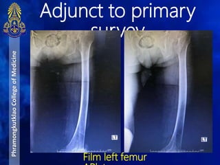
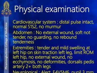
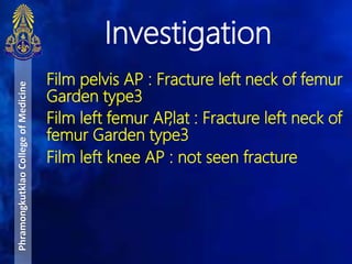
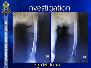
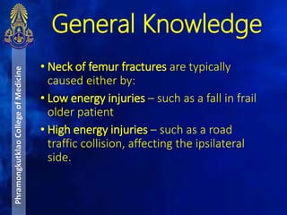
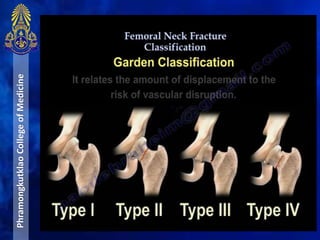
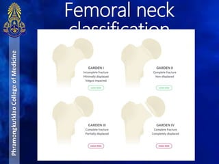
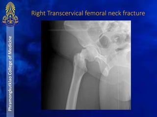
Orthopedic case
BY
THANA MEKSONG
PRIYAPAT MANITYAGUL
TANKHUN CHITTASENEE
MEDICAL CADET OF PHRAMONGKUTKLAO COLLEGE OF MEDICINE
DEPARTMENT OF ORTHOPEDIC
MAHARAT NAKHON RATCHASIMA HOSPITAL
Case
• A 81 year-old Thai female patient
• No known underlying disease
• Chief complaint : ปวดสะโพกซ้าย 3
วันPTA
PhramongkutklaoCollegeofMedicine
A : Can speak, can flex and extend C-
spine, C-spine not tender
B : trachea in midline, RR 20/min, no
subcutaneous emphysema, lungs clear,
equal breath sound both lungs, CCT
negative
C : BP 125/68 mmHg, PR 72 bpm,
tender and mild swelling at left hip on
skin traction left leg, no external wound,
no deformities, no ecchymosis, dorsalis
pedis artery 2+ both legs
Primary Survey in
MNRH
PhramongkutklaoCollegeofMedicine
Primary Survey in
MNRH
PhramongkutklaoCollegeofMedicine
D : E4V5M6, pupil 3 mm react to light
both eyes
E : BT 36.5oC, tender and mild swelling
at left hip on skin traction left leg, no
deformities, limit ROM left hip, dorsalis
pedis artery 2+ both legs, sensory
intact
Primary Survey in
MNRH
PhramongkutklaoCollegeofMedicine
SpO2 98% RA
DTX 98 mg%
CXR AP supine :
Not seen rib fracture, not seen
pneumohemothorax, no
pneumopericardium, CT ratio 0.5, not
seen subcutaneous emphysema
Film pelvis AP : Fracture left neck of
femur Garden type3
Film left femur AP,lat : Fracture left neck
Adjunct to primary
survey
PhramongkutklaoCollegeofMedicine
Adjunct to primary
survey
PhramongkutklaoCollegeofMedicine
CXR AP
Adjunct to primary
survey
PhramongkutklaoCollegeofMedicine
Film Pelvis
Adjunct to primary
survey
PhramongkutklaoCollegeofMedicine
Film left femur
A : No food or drug allergy
M : No underlying disease
P : No previous surgery
L : Last meal at 8 A.M.
E : 3 days prior to MNRH ขณะที่ผู้ป่ วยก้าว
ขึ้นบนเตียงใต้ถุนบ้าน ผู้ป่ วยสะดุดล้ม ก้น
กระแทกพื้น ปวดสะโพกซ้าย ลุกเดินไม่ได้
ขยับแล้วมีอาการปวด ไม่มีศีรษะกระแทก ไม่
สลบ
วันนี้อาการปวดไม่ดีขึ้น ไปรพช. filmพบ
Secondary survey in
MNRH
PhramongkutklaoCollegeofMedicine
Vital signs : BT 36.5oC, PR 72 bpm, RR
20/min, BP 125/68 mmHg
General appearance : An elderly Thai
woman, good consciousness
Head and Maxillofacial : No wound, no
deformity
Cervical spine and Neck : No wound, no
tenderness along the midline
Respiratory system : Equal breath sound
both lungs, no tracheal shift, CCT negative,
Physical examination
PhramongkutklaoCollegeofMedicine
Cardiovascular system : distal pulse intact,
normal S1S2, no murmur
Abdomen : No external wound, soft not
tender, no guarding, no rebound
tenderness
Extremities : tender and mild swelling at
left hip on skin traction left leg, limit ROM
left hip, no external wound, no
ecchymosis, no deformities, dorsalis pedis
artery 2+ both legs
Physical examination
PhramongkutklaoCollegeofMedicine
Film pelvis AP : Fracture left neck of femur
Garden type3
Film left femur AP,lat : Fracture left neck of
femur Garden type3
Film left knee AP : not seen fracture
Investigation
PhramongkutklaoCollegeofMedicine
Film Pelvis
Investigation
PhramongkutklaoCollegeofMedicine
Film left femur
Investigation
PhramongkutklaoCollegeofMedicine
Film left
Investigation
Closed fracture left neck of femur
Garden type3
Diagnosis
PhramongkutklaoCollegeofMedicine
Pain control
Skin traction 2 kg left leg
Set OR for Lt. bipolar hemiarthroplasty
Management
PhramongkutklaoCollegeofMedicine
General Knowledge
• Neck of femur fractures are typically
caused either by:
• Low energy injuries – such as a fall in frail
older patient
• High energy injuries – such as a road
traffic collision, affecting the ipsilateral
side.
PhramongkutklaoCollegeofMedicine
BASIC ANATOMYPhramongkutklaoCollegeofMedicine
1. Reticular vessels
main blood supply.
Originates from an
extra-capsular arterial
ring, supplied by medial
and lateral circumflex
vessels (profunda
femoris A.). Reinforced
by the superior and
inferior gluteal arteries
(internal iliac A.)
2. Foveal artery
3. Metaphyseal vessels
PhramongkutklaoCollegeofMedicine
• Intracapsular – either subcapital (through
the junction of the head and neck) or
basocervical fracture (through the base of
femoral neck)
• Extracapsular – either intertrochanteric
(between the two trochanters) or
subtrochanteric (<5cm distal to the lesser
trochanter)
classification
PhramongkutklaoCollegeofMedicine
Intertrochanter
subcapital
PhramongkutklaoCollegeofMedicine
•Subcapital: femoral head/neck junction
•Transcervical: mid portion of femoral neck
•Basocervical: base of femoral neck
PhramongkutklaoCollegeofMedicine
Femoral neck
classification
PhramongkutklaoCollegeofMedicine
Simplified garden classification
• Nondisplaced : Included garden type I, II
• Displaced : Included garden type III,IV
Pauwels Classification
Based on vertical orientation of fracture line
• Type 1: < 30 deg from horizontal
• Type 2:30 to 50 deg from horizontal
• Type 3:> 50 deg from horizontal (most unstable
with highest risk of nonunion and AVN)
PhramongkutklaoCollegeofMedicine
Mechanism of injury
PhramongkutklaoCollegeofMedicine
•Falls in the elderly
•Significant trauma (e.g. motor vehicle collisions) in
younger patients
• History of a recent fall or trauma
• Significant pain (in the groin, over the hip, in
the thigh, or the knee)
• Inability to weight bear.
• History of chronic metabolic problems such as
osteoporosis or renal failure
Clinical feature
PhramongkutklaoCollegeofMedicine
• shortened leg and externally rotated
• pain on pin-rolling the leg and axial loading
• Unable to straight leg raise.
• Distal neurovascular deficits are rare in isolated
neck of femur fractures
Clinical feature
PhramongkutklaoCollegeofMedicine
• Plain radiograph of hip ( AP with
maximal internal rotation, lateral )
• Normal trabecular pattern, defect in
cortex, shortening or angulation of
femoral neck
• Normal angle between femoral
neck and shaft is 45 degrees
• Normal angle between medial
femoral shaft and trabecular lines
running through the shaft to
femoral head is 160-170 degrees
Investigation
PhramongkutklaoCollegeofMedicine
• Line between anatomical
long axis and femoral shift
Plain radiograph
PhramongkutklaoCollegeofMedicine
•Shenton’s line disruption: loss of contour
between normally continuous line from medial
edge of femoral neck and inferior edge of the
superior pubic ramus
•Lesser trochanter is more prominent due to
external rotation of femur
•Femur often positioned in flexion and external
rotation (due to unopposed iliopsoas)
Plain radiograph
PhramongkutklaoCollegeofMedicine
•Asymmetry of lateral femoral neck/head
•Sclerosis in fracture plane
•Smudgy sclerosis from impaction
•Bone trabeculae angulated
•Non-displaced fractures may be subtle on x-ray
PhramongkutklaoCollegeofMedicine
PhramongkutklaoCollegeofMedicine Left Subcapital femoral neck fracture
Left basicervical femoral fracturePhramongkutklaoCollegeofMedicine
PhramongkutklaoCollegeofMedicine
Right Transcervical femoral neck fracture
ManagementPhramongkutklaoCollegeofMedicine Fracture Type Surgical Option Summary
Subcapital* Hip
hemiarthroplasty
Replacement of the femoral head
and neck via a femoral component
fixed in the proximal femur
Intertrochanteric
and Basocervical*
Dynamic hip
screw
Consists of a lag screw into the neck,
a sideplate, and cortical screws. The
lag screw is able to slide through the
sideplate, allowing for compression
and primary healing of the bone.
Non-displaced
intra-capsular
Cannulated hip
screws
Three non-parallel screws in an
inverted triangle formation. Are also
used in valgus-impacted fractures
Subtrochanteric Intramedullary
Femoral Nail
The metal rod is placed through the
medullary cavity of the femur for
stabilization
• Perform early surgery within 24 hours in
patient with medically stable
• As soon as possible in patient with active
comorbid medical illness
• Avoid delaying surgery beyond 72 hours
• Thrombo-prophylaxis should be performed in
patients awaiting surgery
Timing of operative management
PhramongkutklaoCollegeofMedicine
Reserved for debilitated patients
Stable, impacted patient
Patients over 70 years old with poor health
Non-operative management
PhramongkutklaoCollegeofMedicine
Infection
Thromboembolism
Chronic pain
Osteonecrosis ( Avascular necrosis )
Nonunion
Complications
PhramongkutklaoCollegeofMedicine
Pre & Post-operation
PhramongkutklaoCollegeofMedicine
• Analgesia
• Thromboembolism prophylaxis
• Infection prophylaxis
• Postoperative delirium
• Osteoporosis
• Prevent constipation
• Nutrition
• Prevention of pressure ulcer
• Bladder catheterization
• Blood transfusion
• Rehabilitation
Key Points
PhramongkutklaoCollegeofMedicine
• Neck of femur fractures are associated with a high
one year mortality
• They will present as an acutely painful hip that is
shortened and externally rotated.
• Treatment of neck of femur fracture is primarily
surgical. The specific procedure required will
depend on the classification of the fracture.
• Ensure early assessment by ortho-geriatricians
alongside physiotherapists and occupational
therapists.
THANK YOU
PhramongkutklaoCollegeofMedicine

.Ortho.

Editor's Notes

  • #21 The femoral head’s blood supply is primarily uni-directional, most arriving from the medial femoral circumflex artery* (arising from the deep femoral artery). The medial femoral circumflex artery lies directly on the femoral neck and is thus vulnerable to damage in a fracture. The blood supply to the proximal end of the femur is divided into 3 major groups. The first is the extracapsular arterial ring located at the base of the femoral neck. The second is the ascending cervical branches of the arterial ring on the surface of the femoral neck. The third is the arteries of the ligamentum teres. A large branch of the medial femoral circumflex artery forms the extracapsular arterial ring posteriorly and anteriorly by a branch from the lateral femoral circumflex artery (see images shown below). The ascending cervical branches 1ascend on the surface on the femoral neck anteriorly along the intertrochanteric line. Posteriorly, the cervical branches run under the synovial reflection toward the rim of the articular cartilage, which demarcates the femoral neck from its head. The lateral vessels are the most vulnerable to injury in femoral neck fractures. A second ring of vessels is formed as the ascending cervical vessels approach the articular margin of the femoral head. From this second ring of vessels, the epiphyseal arteries are formed. The lateral epiphyseal arterial group supplies the lateral weight-bearing portion of the femoral head. The epiphyseal vessels are joined by the inferior metaphyseal vessels and vessels from the ligamentum teres. Femoral neck fractures frequently disrupt the blood supply to the femoral head (see images below). The superior reticular and lateral epiphyseal vessels are the most important sources of this blood supply. Widely displaced intracapsular hip fractures tear the synovium and the surrounding vessels. The progressive disruption of the blood supply can lead to serious clinical conditions and complications, including osteonecrosis and nonunion.