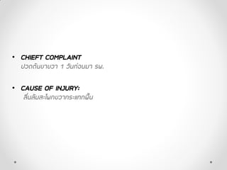
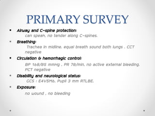
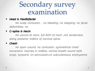
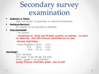
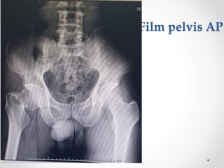
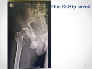
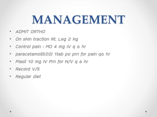
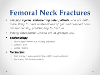
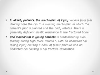
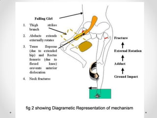
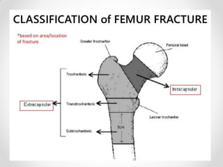
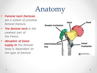
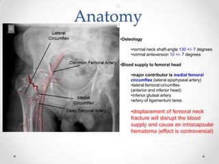
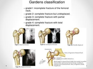
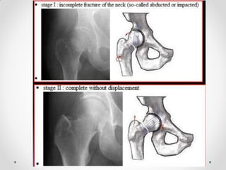
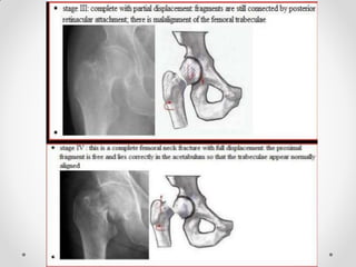
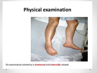
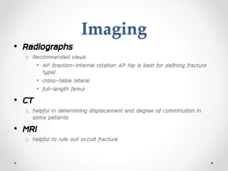
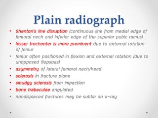
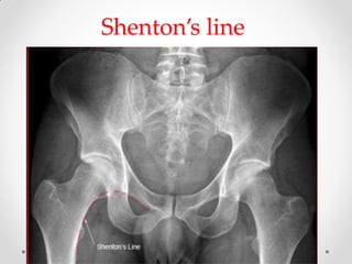
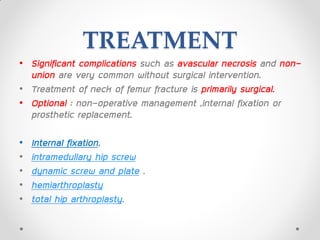
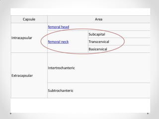
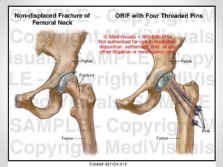
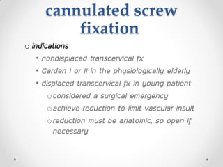
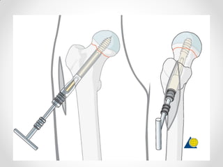
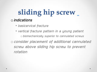
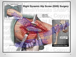
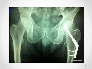
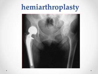
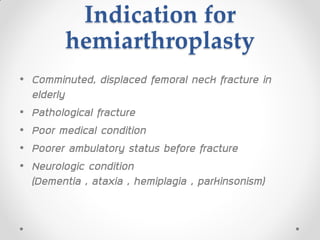
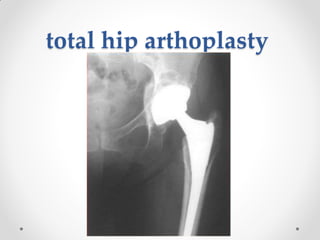
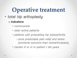
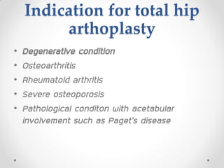
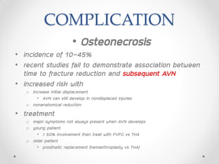
Case conference
extern
PRESENT BY:
EXT. THANAWAT SUPAPONGPICHET
Date 13 February 2019
PATIENT PROFILE
• ผู้ป่วยชายไทยคู่ อายุ 48 ปี , unknown underlying disease
• อาชีพ พนักงาน CP
• ภูมิลาเรา อาเภอ ปักธงชัย จังหวัดนครราชสีมา
• สิทธิ์การรักษาประกันสังคม
• CHIEFT COMPLAINT
ปวดต้นขาขวา 1 วันก่อนมา รพ.
• CAUSE OF INJURY:
ลื่นล้มสะโพกขวากระแทกพื้น
PRIMARY SURVEY
 Airway and C-spine protection:
can speak, no tender along C-spines.
 Breathing:
Trachea in midline. equal breath sound both lungs . CCT
negative
 Circulation & hemorrhagic control:
BP 168/85 mmHg , PR 78/min, no active external bleeding.
PCT negative
 Disability and neurological status:
GCS : E4V5M6, Pupil 3 mm RTLBE.
 Exposure:
no wound , no bleeding
SECONDARY SURVEY
• Allergy: no drug/food allergy
• Medication: no current medication
• Past history: no underlying disease , no alcohol drinking , no
smoking ,
-Previous surgery : ผ่าตัดใส่เหล็กกระดูกแขนขวาหักเนื่องจากอุบัติเหตุ หลายปี
ก่อน
• Last meal: 7.00 A.M. meal&water
• Event/Environment:
1dayPTA(11/2/62 10.00 PM)ผู้ป่วยลื่นล้มสะโพกขวากระแทกพื้น มี
อาการปวดต้นขาขวาและสะโพกขวา ไม่ปวดร้าวไปบริเวณอื่น ปวดมากขึ้นเวลาขยับ
ขา ไม่มีชาบริเวณขาและปลายเท้า ไม่มีขาอ่อนแรง พอเดินลงน้าหนักได้บ้าง ไม่มี
ศีรษะกระทบกระแทก เช้านี้เดินลงน้าหนักไม่ได้ มีอาการเจ็บต้นขาขวามากขึ้น มีบวม
ไม่มีแดงร้อนบริเวณข้อ จึงมา รพ.
Secondary survey
examination
 Head & Maxillofacial:
No scalp contusion, , no bleeding, no stepping, no facial
deformities, no
 C-spine & Neck:
No wound at neck, full ROM of neck ,not tenderness
along posterior midlice of cervical spine.
 Chest:
No open wound, no contusion, symmetrical chest
movement, trachea in midline, normal breath sound both
lungs, tympanic on percussion,no subcutaneous emphysema
Secondary survey
examination
 Abdomen & Pelvis:
Soft, not tender, no guarding, no rebound tenderness
 Perineum/Rectum/:
No wound, no ecchymosis,no bleeding
 Musculoskeletal:
- no wound
- tenderness at Rt.hip and Rt.thigh ,swelling ,no redness , no warm
no deformity , limit ROM Rt.lower extrimities due to pain
-Rt.knee deformities
-Pulse Popliteal a. Rt.2+ Lt.2+
PTA Rt.2+ Lt.2+
DPA Rt. 2+ Lt.2+
Neurologic:
GCS : E4V5M6
CN : pupil 3 mm RTLBE, full EOM
Motor : grade V, all extremities
except Rt.lower extrimities grade I due to pain
Film pelvis AP
Film Rt.Hip lateral
Film Rt.knee AP-lateral
Film CXR
PROVISIONAL DIAGNOSIS
Closed fracture Rt.femoral neck
MANAGEMENT
• ADMIT ORTHO
• On skin traction Rt. Leg 2 kg
• Control pain : MO 4 mg IV q 6 hr
• paracetamol(500) 1tab po prn for pain q6 hr
• Plasil 10 mg IV Prn for N/V q 6 hr
• Record V/S
• Regular diet
Femoral Neck
Fractures
Femoral Neck Fractures
• common injuries sustained by older patients who are both
more likely to have unsteadiness of gait and reduced bone
mineral density, predisposing to fracture.
• Elderly osteopolotic women are at greatest risk.
• Epidemiology
o increasingly common due to aging population
o women > men
o whites > blacks
• Mechanism
o high energy in young patients (e.g. motor vehicle collisions)
o low energy falls in older patients
• In elderly patients, the mechanism of injury various from falls
directly onto the hip to a twisting mechanism in which the
patient’s foot is planted and the body rotates. There is
generally deficient elastic resistance in the fractured bone .
• The mechanism in young patients is predominantly axial
loading during high force trauma 9, with an abducted hip
during injury causing a neck of femur fracture and an
adducted hip causing a hip fracture-dislocation.
Associated injuries
• femoral shaft fractures6-9% associated with femoral neck
fractures
• treat femoral neck first followed by shaft
Anatomy
• Femoral neck fractures
are a subset of proximal
femoral fracture.
• The femoral neck is the
weakest part of
the Femur.
• disruption of blood
supply to the femoral
head is dependent on
the type of fracture
Anatomy
•Osteology
•normal neck shaft-angle 130 +/- 7 degrees
•normal anteversion 10 +/- 7 degrees
•Blood supply to femoral head
•major contributor is medial femoral
circumflex (lateral epiphyseal artery)
•lateral femoral circumflex
(anterior and inferior head)
•inferior gluteal artery
•artery of ligamentum teres
•displacement of femoral neck
fracture will disrupt the blood
supply and cause an intracapsular
hematoma (effect is controversial)
Pathophysiology
• healing potential
ofemoral neck is intracapsular, bathed in
synovial fluid
olacks periosteal layer
ocallus formation limited, which affects
healing
Presentation
• Symptoms
oimpacted and stress fractures
• slight pain in the groin or pain referred
along the medial side of the thigh and
knee
odisplaced fractures
• pain in the entire hip region
Presentation
• Physical exam
o impacted and stress fractures
• no obvious clinical deformity
• minor discomfort with active or passive hip range of
motion, muscle spasms at extremes of motion
• pain with percussion over greater trochanter
o If displaced fractures
• leg in external rotation and abduction, with shortening
• minimal swelling due to intracapsular fracture
Imaging
• Radiographs
o Recommended views
• AP (traction-internal rotation AP hip is best for defining fracture
type)
• cross-table lateral
• full-length femur
• CT
o helpful in determining displacement and degree of comminution in
some patients
• MRI
o helpful to rule out occult fracture
Plain radiograph
• Shenton’s line disruption (continuous line from medial edge of
femoral neck and inferior edge of the superior pubic ramus)
• lesser trochanter is more prominent due to external rotation
of femur
• femur often positioned in flexion and external rotation (due to
unopposed iliopsoas)
• asymmetry of lateral femoral neck/head
• sclerosis in fracture plane
• smudgy sclerosis from impaction
• bone trabeculae angulated
• nondisplaced fractures may be subtle on x-ray
Shenton’s line
TREATMENT
• Significant complications such as avascular necrosis and non-
union are very common without surgical intervention.
• Treatment of neck of femur fracture is primarily surgical.
• Optional : non-operative management ,internal fixation or
prosthetic replacement.
• Internal fixation,
• intramedullary hip screw
• dynamic screw and plate .
• hemiarthroplasty
• total hip arthroplasty.
The Delbet classification
• correlates with the risk of
AVN
• type 1 (transphyseal):
~90% risk of AVN
• type 2 (subcapital):
~50% risk of AVN
• type 3
(basicervical/transcervical):
~25% risk of AVN
• type 4 (intertrochanteric):
~10% risk of AVN
TREATMENT
• internal fixation is recommended for young,
otherwise, fit patients with small risk for AVN.
• While prosthetic replacement is reserved for
fractures with a high risk of AVN and the
elderly.
TREATMENT
• Nonoperative : observation alone
o indications
• may be considered in some patients who are non-
ambulators, have minimal pain, and who are at high
risk for surgical intervention
Operative treatment
•ORIF
o indications
• displaced fractures in young or physiologically
young patients
• ORIF indicated for most pts <65 years of age
cannulated screw
fixation
o indications
• nondisplaced transcervical fx
• Garden I or II in the physiologically elderly
• displaced transcervical fx in young patient
oconsidered a surgical emergency
oachieve reduction to limit vascular insult
oreduction must be anatomic, so open if
necessary
sliding hip screw
oindications
• basicervical fracture
• vertical fracture pattern in a young patient
obiomechanically superior to cannulated screws
oconsider placement of additional cannulated
screw above sliding hip screw to prevent
rotation
hemiarthroplasty
Indication for
hemiarthroplasty
• Comminuted, displaced femoral neck fracture in
elderly
• Pathological fracture
• Poor medical condition
• Poorer ambulatory status before fracture
• Neurologic condition
(Dementia , ataxia , hemiplagia , parkinsonism)
total hip arthoplasty
Operative treatment
• total hip arthoplasty
o indications
• controversial
• older active patients
• patients with preexisting hip osteoarthritis
omore predictable pain relief and better
functional outcome than hemiarthroplasty
• Garden III or IV in patient < 85 years
Indication for total hip
arthoplasty
• Degenerative condition
• Osteoarthritis
• Rheumatoid arthritis
• Severe osteoporosis
• Pathological conditon with acetabular
involvement such as Paget’s disease
COMPLICATION
• Osteonecrosis
• incidence of 10-45%
• recent studies fail to demonstrate association between
time to fracture reduction and subsequent AVN
• increased risk with
o increase initial displacement
• AVN can still develop in nondisplaced injuries
o nonanatomical reduction
• treatment
o major symptoms not always present when AVN develops
o young patient
• > 50% involvement then treat with FVFG vs THA
o older patient
• prosthetic replacement (hemiarthroplasty vs THA)
Nonunion
• Most common complication
• incidence of 5 to 30%
o increased incidence in displaced fractures
o no correlation between age, gender, and rate of nonunion
• varus malreduction most closely correlates with failure of
fixation after reduction and cannulated screw fixation.
• treatment
o valgus intertrochanteric osteotomy
• indicated in patients after femoral neck nonunion
o free vascularized fibula graft (FVFG)
• indicated in young patients with a nonviable femoral head
o arthroplasty
• indicated in older patients or when the femoral head is not viable
o revision ORIF
Dislocation
• higher rate of dislocation with THA (~ 10%)
o about seven times higher than hemiarthroplasty
Failure rates
• high early failure rates in fixation group, which stabilizes after
2 years
o 2-year follow-up (elderly population >70 years) with displaced femoral neck fractures
• 46% with fixation techniques
• 8% with arthroplasty techniques
o 2-to-10 year follow-up
• failure rate approx. 2-4%, respectively
• overall failure rates still higher in fixation vs. arthoplasty at
10-year follow-up
THANK YOU

Case conference extern ortho ed1

  • 1.
    Case conference extern PRESENT BY: EXT.THANAWAT SUPAPONGPICHET Date 13 February 2019
  • 2.
    PATIENT PROFILE • ผู้ป่วยชายไทยคู่อายุ 48 ปี , unknown underlying disease • อาชีพ พนักงาน CP • ภูมิลาเรา อาเภอ ปักธงชัย จังหวัดนครราชสีมา • สิทธิ์การรักษาประกันสังคม
  • 3.
    • CHIEFT COMPLAINT ปวดต้นขาขวา1 วันก่อนมา รพ. • CAUSE OF INJURY: ลื่นล้มสะโพกขวากระแทกพื้น
  • 4.
    PRIMARY SURVEY  Airwayand C-spine protection: can speak, no tender along C-spines.  Breathing: Trachea in midline. equal breath sound both lungs . CCT negative  Circulation & hemorrhagic control: BP 168/85 mmHg , PR 78/min, no active external bleeding. PCT negative  Disability and neurological status: GCS : E4V5M6, Pupil 3 mm RTLBE.  Exposure: no wound , no bleeding
  • 5.
    SECONDARY SURVEY • Allergy:no drug/food allergy • Medication: no current medication • Past history: no underlying disease , no alcohol drinking , no smoking , -Previous surgery : ผ่าตัดใส่เหล็กกระดูกแขนขวาหักเนื่องจากอุบัติเหตุ หลายปี ก่อน • Last meal: 7.00 A.M. meal&water • Event/Environment: 1dayPTA(11/2/62 10.00 PM)ผู้ป่วยลื่นล้มสะโพกขวากระแทกพื้น มี อาการปวดต้นขาขวาและสะโพกขวา ไม่ปวดร้าวไปบริเวณอื่น ปวดมากขึ้นเวลาขยับ ขา ไม่มีชาบริเวณขาและปลายเท้า ไม่มีขาอ่อนแรง พอเดินลงน้าหนักได้บ้าง ไม่มี ศีรษะกระทบกระแทก เช้านี้เดินลงน้าหนักไม่ได้ มีอาการเจ็บต้นขาขวามากขึ้น มีบวม ไม่มีแดงร้อนบริเวณข้อ จึงมา รพ.
  • 6.
    Secondary survey examination  Head& Maxillofacial: No scalp contusion, , no bleeding, no stepping, no facial deformities, no  C-spine & Neck: No wound at neck, full ROM of neck ,not tenderness along posterior midlice of cervical spine.  Chest: No open wound, no contusion, symmetrical chest movement, trachea in midline, normal breath sound both lungs, tympanic on percussion,no subcutaneous emphysema
  • 7.
    Secondary survey examination  Abdomen& Pelvis: Soft, not tender, no guarding, no rebound tenderness  Perineum/Rectum/: No wound, no ecchymosis,no bleeding  Musculoskeletal: - no wound - tenderness at Rt.hip and Rt.thigh ,swelling ,no redness , no warm no deformity , limit ROM Rt.lower extrimities due to pain -Rt.knee deformities -Pulse Popliteal a. Rt.2+ Lt.2+ PTA Rt.2+ Lt.2+ DPA Rt. 2+ Lt.2+ Neurologic: GCS : E4V5M6 CN : pupil 3 mm RTLBE, full EOM Motor : grade V, all extremities except Rt.lower extrimities grade I due to pain
  • 8.
  • 9.
  • 10.
  • 11.
  • 12.
  • 13.
    MANAGEMENT • ADMIT ORTHO •On skin traction Rt. Leg 2 kg • Control pain : MO 4 mg IV q 6 hr • paracetamol(500) 1tab po prn for pain q6 hr • Plasil 10 mg IV Prn for N/V q 6 hr • Record V/S • Regular diet
  • 14.
  • 15.
    Femoral Neck Fractures •common injuries sustained by older patients who are both more likely to have unsteadiness of gait and reduced bone mineral density, predisposing to fracture. • Elderly osteopolotic women are at greatest risk. • Epidemiology o increasingly common due to aging population o women > men o whites > blacks • Mechanism o high energy in young patients (e.g. motor vehicle collisions) o low energy falls in older patients
  • 16.
    • In elderlypatients, the mechanism of injury various from falls directly onto the hip to a twisting mechanism in which the patient’s foot is planted and the body rotates. There is generally deficient elastic resistance in the fractured bone . • The mechanism in young patients is predominantly axial loading during high force trauma 9, with an abducted hip during injury causing a neck of femur fracture and an adducted hip causing a hip fracture-dislocation.
  • 18.
    Associated injuries • femoralshaft fractures6-9% associated with femoral neck fractures • treat femoral neck first followed by shaft
  • 20.
    Anatomy • Femoral neckfractures are a subset of proximal femoral fracture. • The femoral neck is the weakest part of the Femur. • disruption of blood supply to the femoral head is dependent on the type of fracture
  • 21.
    Anatomy •Osteology •normal neck shaft-angle130 +/- 7 degrees •normal anteversion 10 +/- 7 degrees •Blood supply to femoral head •major contributor is medial femoral circumflex (lateral epiphyseal artery) •lateral femoral circumflex (anterior and inferior head) •inferior gluteal artery •artery of ligamentum teres •displacement of femoral neck fracture will disrupt the blood supply and cause an intracapsular hematoma (effect is controversial)
  • 25.
    Pathophysiology • healing potential ofemoralneck is intracapsular, bathed in synovial fluid olacks periosteal layer ocallus formation limited, which affects healing
  • 26.
    Presentation • Symptoms oimpacted andstress fractures • slight pain in the groin or pain referred along the medial side of the thigh and knee odisplaced fractures • pain in the entire hip region
  • 27.
    Presentation • Physical exam oimpacted and stress fractures • no obvious clinical deformity • minor discomfort with active or passive hip range of motion, muscle spasms at extremes of motion • pain with percussion over greater trochanter o If displaced fractures • leg in external rotation and abduction, with shortening • minimal swelling due to intracapsular fracture
  • 29.
    Imaging • Radiographs o Recommendedviews • AP (traction-internal rotation AP hip is best for defining fracture type) • cross-table lateral • full-length femur • CT o helpful in determining displacement and degree of comminution in some patients • MRI o helpful to rule out occult fracture
  • 30.
    Plain radiograph • Shenton’sline disruption (continuous line from medial edge of femoral neck and inferior edge of the superior pubic ramus) • lesser trochanter is more prominent due to external rotation of femur • femur often positioned in flexion and external rotation (due to unopposed iliopsoas) • asymmetry of lateral femoral neck/head • sclerosis in fracture plane • smudgy sclerosis from impaction • bone trabeculae angulated • nondisplaced fractures may be subtle on x-ray
  • 31.
  • 32.
    TREATMENT • Significant complicationssuch as avascular necrosis and non- union are very common without surgical intervention. • Treatment of neck of femur fracture is primarily surgical. • Optional : non-operative management ,internal fixation or prosthetic replacement. • Internal fixation, • intramedullary hip screw • dynamic screw and plate . • hemiarthroplasty • total hip arthroplasty.
  • 34.
    The Delbet classification •correlates with the risk of AVN • type 1 (transphyseal): ~90% risk of AVN • type 2 (subcapital): ~50% risk of AVN • type 3 (basicervical/transcervical): ~25% risk of AVN • type 4 (intertrochanteric): ~10% risk of AVN
  • 35.
    TREATMENT • internal fixationis recommended for young, otherwise, fit patients with small risk for AVN. • While prosthetic replacement is reserved for fractures with a high risk of AVN and the elderly.
  • 36.
    TREATMENT • Nonoperative :observation alone o indications • may be considered in some patients who are non- ambulators, have minimal pain, and who are at high risk for surgical intervention
  • 37.
    Operative treatment •ORIF o indications •displaced fractures in young or physiologically young patients • ORIF indicated for most pts <65 years of age
  • 40.
    cannulated screw fixation o indications •nondisplaced transcervical fx • Garden I or II in the physiologically elderly • displaced transcervical fx in young patient oconsidered a surgical emergency oachieve reduction to limit vascular insult oreduction must be anatomic, so open if necessary
  • 42.
    sliding hip screw oindications •basicervical fracture • vertical fracture pattern in a young patient obiomechanically superior to cannulated screws oconsider placement of additional cannulated screw above sliding hip screw to prevent rotation
  • 45.
  • 46.
    Indication for hemiarthroplasty • Comminuted,displaced femoral neck fracture in elderly • Pathological fracture • Poor medical condition • Poorer ambulatory status before fracture • Neurologic condition (Dementia , ataxia , hemiplagia , parkinsonism)
  • 47.
  • 48.
    Operative treatment • totalhip arthoplasty o indications • controversial • older active patients • patients with preexisting hip osteoarthritis omore predictable pain relief and better functional outcome than hemiarthroplasty • Garden III or IV in patient < 85 years
  • 49.
    Indication for totalhip arthoplasty • Degenerative condition • Osteoarthritis • Rheumatoid arthritis • Severe osteoporosis • Pathological conditon with acetabular involvement such as Paget’s disease
  • 50.
    COMPLICATION • Osteonecrosis • incidenceof 10-45% • recent studies fail to demonstrate association between time to fracture reduction and subsequent AVN • increased risk with o increase initial displacement • AVN can still develop in nondisplaced injuries o nonanatomical reduction • treatment o major symptoms not always present when AVN develops o young patient • > 50% involvement then treat with FVFG vs THA o older patient • prosthetic replacement (hemiarthroplasty vs THA)
  • 51.
    Nonunion • Most commoncomplication • incidence of 5 to 30% o increased incidence in displaced fractures o no correlation between age, gender, and rate of nonunion • varus malreduction most closely correlates with failure of fixation after reduction and cannulated screw fixation. • treatment o valgus intertrochanteric osteotomy • indicated in patients after femoral neck nonunion o free vascularized fibula graft (FVFG) • indicated in young patients with a nonviable femoral head o arthroplasty • indicated in older patients or when the femoral head is not viable o revision ORIF
  • 52.
    Dislocation • higher rateof dislocation with THA (~ 10%) o about seven times higher than hemiarthroplasty
  • 53.
    Failure rates • highearly failure rates in fixation group, which stabilizes after 2 years o 2-year follow-up (elderly population >70 years) with displaced femoral neck fractures • 46% with fixation techniques • 8% with arthroplasty techniques o 2-to-10 year follow-up • failure rate approx. 2-4%, respectively • overall failure rates still higher in fixation vs. arthoplasty at 10-year follow-up
  • 54.