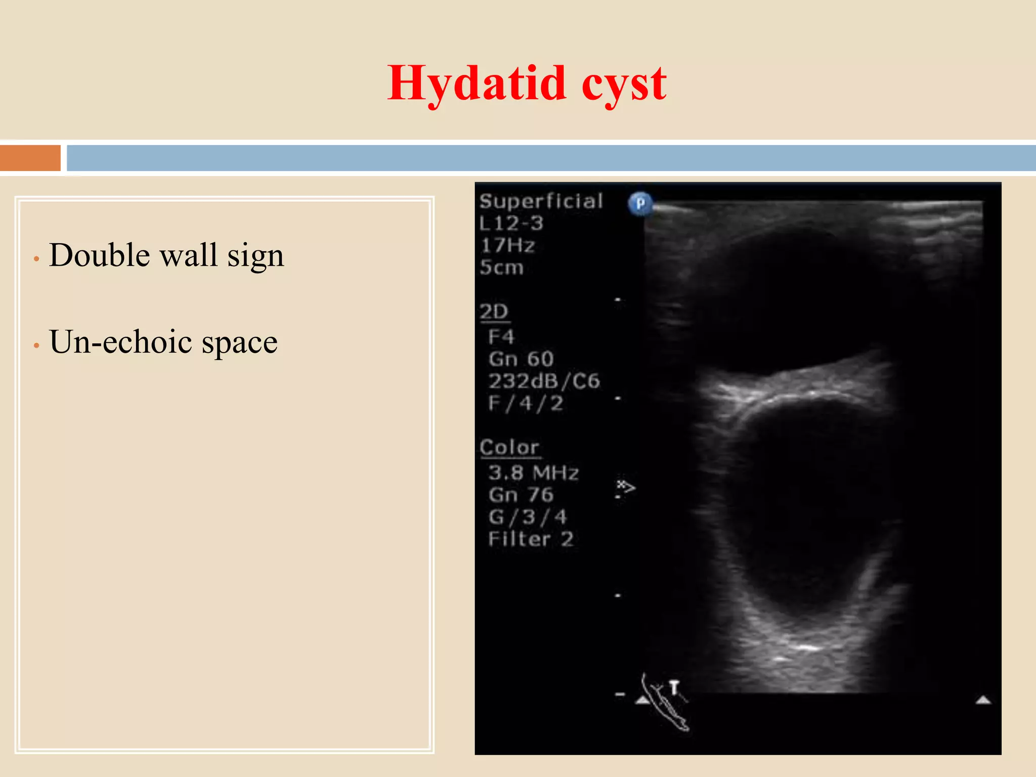
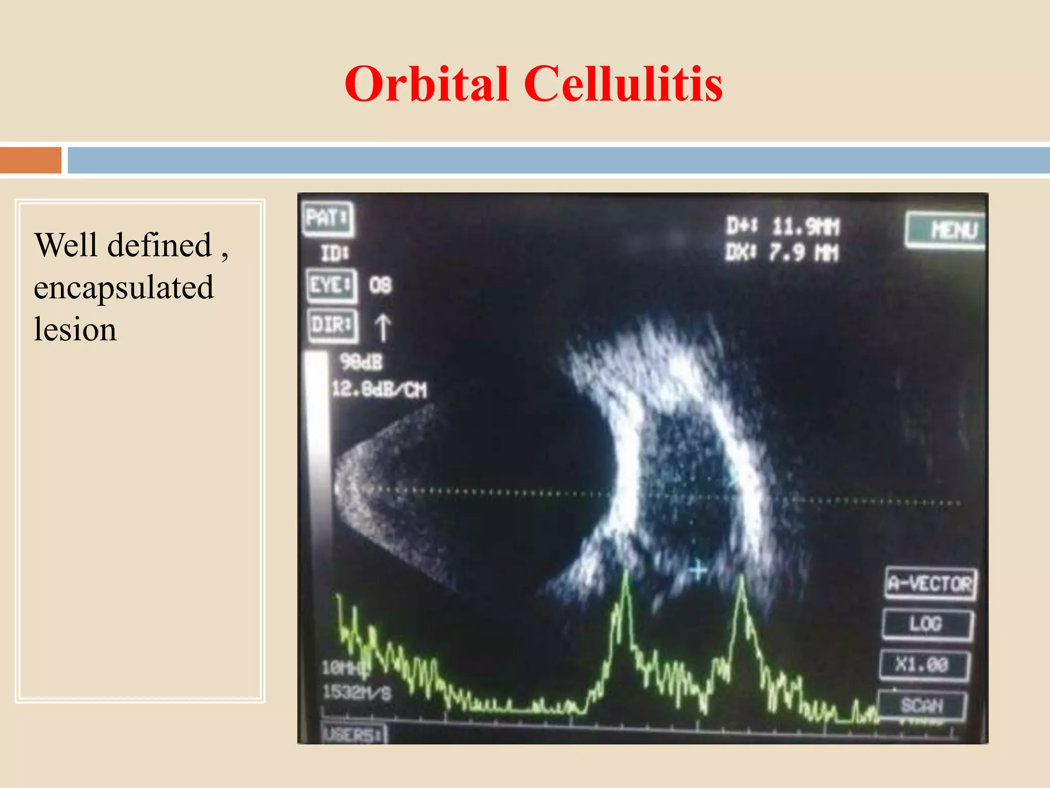
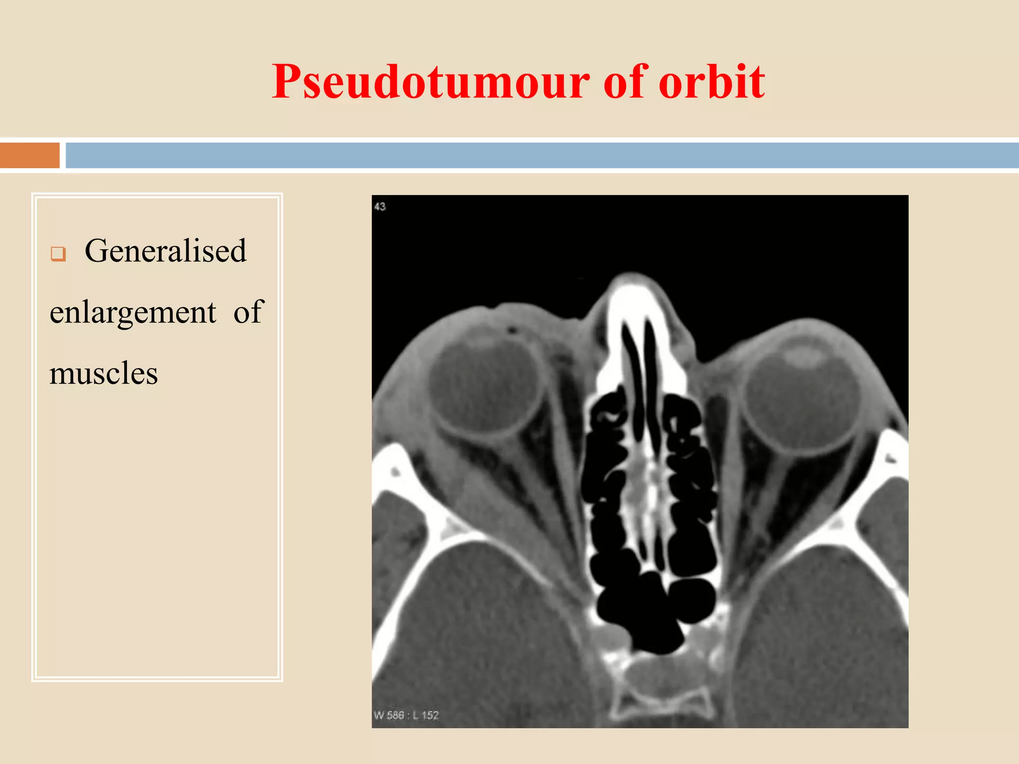
PROPTOSIS :
INVESTIGATION & MANAGEMENT
Dr Ramchandra D Hendge
Proptosis
 Definition :
 Abnormal protrusion of globe
 Displacement of globe relative to orbital rims
 Exophthalmos : abnormal protrusion of eye balls in
endocrine disorders specially in thyroid dysfunction
Classification
 1) Based on direction
 2) Based on severity
 3) Based on presentation
 4) Based on etiology
 5) Based on movement
Etiology
Axial Proptosis- Eccentric/Abaxial Proptosis
Optic nerve tumours Thyroid orbitopathy
Intra conal haemangioma Dermoid cyst
Infections & Inflammations
Neurofibroma
Lacrimal gland tumour
Approach to diagnosis
History Clinical exam Investigations
Inspection Blood Ix
Palpation Imaging
Percussion X-ray
Auscultation USG
Tests CT
MRI
Angiography
Histopathology
FNAC
Biopsy
History
 Onset , duration, progress
 DOV, colour vision defect
 Diplopia
 Discoloration of lids
 Association with straining
 Postural variation
 Oral hygiene
Clinical Examination
 Inspection
 Palpation
 Percussion
 Auscultation
 Clinical Tests
Auscultation
 Globe/temporal region for bruit
Differential Tonometry
 Useful in fibrosis of muscles & lesions compressing globe
 Rise in IOP >6mmHg suggestive of pathology
 Rise in IOP due to mechanical compression of globe
 Gaze- Upward for muscles & in direction of mass in
space occupying lesions
Ruler Test
Exophthalmometry/ Proptometry
1) Clinical
2) Stereo Photographic
3) Radiographic
 Clinical Exophthalmometry
1. Hertel
2. Luedde
3. Naugle
4. Gormaz
Hertel Exophthalmometer
 Measures distance of corneal apex from lateral orbital rim
 Lateral orbital rim serves as reference point
 Difficult to use in lateral wall fracture or surgery
Leudde Exophthalmometer
 Light weight, cheaper, easier
 allows accurate measurement of proptosis by viewing at right
angle
Naugle Exophthalmometer
 Uses fixation points slightly above and below superior &
inferior orbital rims
 Measures difference in proptosis of two eyes rather than
absolute proptosis
 Useful in lateral
orbital wall fracture
or surgery
Gormaz Exophthalmometer
 Measure distance between two lateral orbital
margins
 Require topical anaesthesia
Blood Investigations
 Complete haemogram
 ESR
 FBS/PPBS
 Thyroid function tests
 Serum c-ANCA levels
 Serum ANA levels
Other Investigations
 Sputum for AFB
 Montoux test
X -ray
 Should be the first screening imaging investigation
 Useful specially for bony pathology
 Useful in detecting :
Benign tumours
Calcification( Meningioma, retinoblastoma,
lacrimal gland carcinoma)
Hyperostosis( Meningioma, fibrous dysplasia,
osteoblastic secondaries)
Caldwell view/General view
Structures seen-
Medial wall ,
orbital rim
Water’s view/Orbital view
Structures
seen-
anterior
2/3rd of
orbital
floor
Lateral
Structures
seen-
Orbital
roof
Ultra sonography
 Rapid, non invasive , simple, no radiations
 Compact & Portable
 Can be done in OPD & before surgery by ophthalmologist
 No need to depend upon radiologists
 Gives information about characteristics of lesions
 Size of lesion can be measured
 Principle- Piezo electric effect
 Probes- Sound waves-6-20 MHz
 Lower frequencies-> lower resolution->better penetration
 Higher frequencies-> higher resolution->lesser penetration
 Gives echogenicity based on reflectivity of structure
TYPES
 A scan/Amplitude scan
 B scan/ Brightness scan Trans ocular
 C scan/ Coronal scan Para ocular
 D scan / 3D scan
Hydatid cyst
• Double wall sign
• Un-echoic space
Vascular tumor
• Well
demarcated, intra-
conal lesion with
high internal
reflectivity
Orbital Cellulitis
Well defined ,
encapsulated
lesion
CT Scan
 Is superior to X ray & USG
 Gives shape , size , location & nature of lesion
 Resolution ~0.5 mm
 8 slices required to scan orbit
 Causes radiation exposure but its comparable with orbital X
ray
 HRCT with 1mm sections gives information about optic nerve
tumours
Sections
 Axial section-
shows both globes,
horizontal recti ,
optic nerves, orbital
soft tissue & bony
structures
 Coronal section-
 Anterior- Globe with
recti muscles
 Posterior – all recti
muscles, oblique
muscles, optic nerve &
soft tissue of orbit
Thyroid orbitopathy
Fusiform
dilatation of belly
of recti muscles
Hematoma
Meningioma
 Iso-dense with
optic nerve
 Takes contrast
 Tram track sign
on axial CT
 Non enhancing
dot sign on coronal
CT
Pseudotumour of orbit
 Generalised
enlargement of
muscles
MRI
 Most sensitive modality for soft tissue lesions
 No radiation exposure
 Specially useful in optic nerve lesions, pseudo tumour, orbital
metastasis & tumours having intra cranial expansion
 Metallic implants/ metallic foreign bodies are the only
absolute contraindications
 T1/T2 weighting- refers to methods of measuring the
relaxation times of excited protons after magnetic field is
switch off.
 T1- Fat-Bright
Vitreous- Dark
 T2- Fat-Dark
Vitreous- Bright
Cavernous Hemangioma
 T1- Iso-intese
with muscle, if
thrombosis-hyper
intense
 T2- Hyper intense
with muscle
 Contrast-
Irregular
enhancement with
delayed wash out
Lacrimal gland tumor
Pleo-morphic adenoma-
 T1,T2- Intermediate
intensity
 Contrast- bright
enhancement
Pseudotumor
 T1,T2- Hypo
intense
 Contrast-
Diffuse
enhancement
Angiography
Orbital Venography :
 Useful in diagnosis of orbital varix, cavernous sinus
thrombosis & obstruction of ophthalmic vein by external mass
 Dye is injected in frontal or angular veins & sequential X
rays are taken in AP view.
 not used now a days
Orbital Arteriography :
 Useful in diagnosis of A-V malformations , carotid-cavernous
fistula , aneurysms etc
 Contrast dye is injected in ipsi-lateral common or internal
carotid artery and sequential X rays are taken.
 Contrast medium may cause allergic reactions
 Not used now a days due to CT,MRI & MR Angiography.
Ophthalmic artery Anurysm
Hyper lucent
mass in orbit
Histopathology
 Definitive diagnosis is by histopathology
Biopsy Techniques :
 FNAC
 Core biopsy
 Incisional biopsy
 Excisional biopsy
FNAC
 Minimal invasive
 Rapid diagnosis
 USG/CT guided FNAC
 Accuracy >80%
 Indications :
Lymphoma,melanoma,meningioma
 Scanty material, difficult to evaluate
 Risk of globe perforation,h’age
 Core biopsy –better than FNAC
 Inscisional biopsy- sample obtained under direct visualisation
 Excisional biopsy- Best method for tissue sampling
Both diagnostic &therapeutic
Pathology Techniques
1. Cytology
2. Gross examination
3. Routine histopathology
4. Histochemistry-Sudan black(Fat)
5. Immuno-histochemistry –HMB45 for melanomas, S100 for
schwannomas & neurofibromas
6. Electron microscopy
Management of proptosis
 Local measures
 Medical therapy
 Radiation
 Surgical options
Local measures
 Sun glasses
 Sleep in supine position with head elevated
 Taping of lids at night
 Prisms in diplopia
Medical therapy
 Topical tear substitutes
 Systemic diuretics - minimal role
 Parenteral antibiotics
 Pain killers
 Corticosteroids
Corticosteroids
Indications
 Compressive optic neuropathy
 Prior to orbital decompression
 Pseudo tumor
 Traumatic optic neuropathy
 Hydatid cyst
Corticosteroids - regimen
 60-100mg(1mg/kg) orally prednisolone in divided
doses
 Pulse intravenous therapy of methyl prednisolone
1gm on alternate days for 3-5days
 Local steroid injections have no role
Complications
 Weight gain
 Gastrointestinal irritation
 Reactivation infections (tuberculosis)
 Cataract
 Glaucoma
 Osteoporosis
 Adrenal gland suppression
Immunosuppressive
Indications-
 Refractory cases of thyroid orbitopathy, pseudotumour,
sarcoidosis , hematological malignancies etc
 Commonly used drugs- Cyclosporin, methotrexate,
azathioprine, cyclophosphamide etc
 Anti tumor necrosis factor drugs – infiximab
Radiation Therapy
Indications-
 Pseudotumour
 Lymphoma
 Rhabdomyosarcoma
 Meningioma
 Thyroid orbitopathy
Surgical management
 Orbital decompression
 Strabismus surgery
 Eye lid surgery
Orbital decompression
 Done in stable cases
 Severe exophthalmos
 Exposure keratopathy
 Optic nerve compression
 Cosmetic purpose
Surgical approaches to orbit
(1) Lateral orbitotomy (Krönlein procedure)
(2) Superior decompression via craniotomy(Naffziger procedure)
(3) Medial wall removal and ethmoidectomy
(4) Orbit floor removal
(5) Combined ethmoidectomy
and medial orbit floor
Complications: Orbital Decompression
 Diplopia
 Hyper aesthesia in distribution of infra orbital nerve
 Nasolacrimal duct obstruction
 Cerebrospinal fluid leak
 Frontal lobe hematoma
Strabismus surgery
Indications :
 Done in stable inactive thyroid orbitopathy
 Angle of deviation stable for at least 6–12 months
Goal :
 To minimize diplopia in primary position
Eye lid surgeries
 Mild eyelid retraction- no surgeries
 Main indication is exposure keratopathy
Conclusion
 Proptosis is an important manifestation of a large number of
orbital diseases. Thorough clinical examination coupled with
appropriate investigations clinches the diagnosis and helps in
management.
THANK YOU

Proptosis investigation & management.rdh

  • 1.
    PROPTOSIS : INVESTIGATION &MANAGEMENT Dr Ramchandra D Hendge
  • 2.
    Proptosis  Definition : Abnormal protrusion of globe  Displacement of globe relative to orbital rims  Exophthalmos : abnormal protrusion of eye balls in endocrine disorders specially in thyroid dysfunction
  • 3.
    Classification  1) Basedon direction  2) Based on severity  3) Based on presentation  4) Based on etiology  5) Based on movement
  • 4.
    Etiology Axial Proptosis- Eccentric/AbaxialProptosis Optic nerve tumours Thyroid orbitopathy Intra conal haemangioma Dermoid cyst Infections & Inflammations Neurofibroma Lacrimal gland tumour
  • 5.
    Approach to diagnosis HistoryClinical exam Investigations Inspection Blood Ix Palpation Imaging Percussion X-ray Auscultation USG Tests CT MRI Angiography Histopathology FNAC Biopsy
  • 6.
    History  Onset ,duration, progress  DOV, colour vision defect  Diplopia  Discoloration of lids  Association with straining  Postural variation  Oral hygiene
  • 7.
    Clinical Examination  Inspection Palpation  Percussion  Auscultation  Clinical Tests
  • 8.
  • 9.
    Differential Tonometry  Usefulin fibrosis of muscles & lesions compressing globe  Rise in IOP >6mmHg suggestive of pathology  Rise in IOP due to mechanical compression of globe  Gaze- Upward for muscles & in direction of mass in space occupying lesions
  • 10.
  • 11.
    Exophthalmometry/ Proptometry 1) Clinical 2)Stereo Photographic 3) Radiographic  Clinical Exophthalmometry 1. Hertel 2. Luedde 3. Naugle 4. Gormaz
  • 12.
    Hertel Exophthalmometer  Measuresdistance of corneal apex from lateral orbital rim  Lateral orbital rim serves as reference point  Difficult to use in lateral wall fracture or surgery
  • 14.
    Leudde Exophthalmometer  Lightweight, cheaper, easier  allows accurate measurement of proptosis by viewing at right angle
  • 15.
    Naugle Exophthalmometer  Usesfixation points slightly above and below superior & inferior orbital rims  Measures difference in proptosis of two eyes rather than absolute proptosis  Useful in lateral orbital wall fracture or surgery
  • 17.
    Gormaz Exophthalmometer  Measuredistance between two lateral orbital margins  Require topical anaesthesia
  • 18.
    Blood Investigations  Completehaemogram  ESR  FBS/PPBS  Thyroid function tests  Serum c-ANCA levels  Serum ANA levels
  • 19.
    Other Investigations  Sputumfor AFB  Montoux test
  • 20.
    X -ray  Shouldbe the first screening imaging investigation  Useful specially for bony pathology  Useful in detecting : Benign tumours Calcification( Meningioma, retinoblastoma, lacrimal gland carcinoma) Hyperostosis( Meningioma, fibrous dysplasia, osteoblastic secondaries)
  • 21.
    Caldwell view/General view Structuresseen- Medial wall , orbital rim
  • 22.
  • 23.
  • 25.
    Ultra sonography  Rapid,non invasive , simple, no radiations  Compact & Portable  Can be done in OPD & before surgery by ophthalmologist  No need to depend upon radiologists  Gives information about characteristics of lesions  Size of lesion can be measured
  • 26.
     Principle- Piezoelectric effect  Probes- Sound waves-6-20 MHz  Lower frequencies-> lower resolution->better penetration  Higher frequencies-> higher resolution->lesser penetration  Gives echogenicity based on reflectivity of structure
  • 27.
    TYPES  A scan/Amplitudescan  B scan/ Brightness scan Trans ocular  C scan/ Coronal scan Para ocular  D scan / 3D scan
  • 28.
    Hydatid cyst • Doublewall sign • Un-echoic space
  • 29.
    Vascular tumor • Well demarcated,intra- conal lesion with high internal reflectivity
  • 30.
    Orbital Cellulitis Well defined, encapsulated lesion
  • 31.
    CT Scan  Issuperior to X ray & USG  Gives shape , size , location & nature of lesion  Resolution ~0.5 mm  8 slices required to scan orbit  Causes radiation exposure but its comparable with orbital X ray  HRCT with 1mm sections gives information about optic nerve tumours
  • 32.
    Sections  Axial section- showsboth globes, horizontal recti , optic nerves, orbital soft tissue & bony structures
  • 33.
     Coronal section- Anterior- Globe with recti muscles  Posterior – all recti muscles, oblique muscles, optic nerve & soft tissue of orbit
  • 34.
  • 35.
  • 36.
    Meningioma  Iso-dense with opticnerve  Takes contrast  Tram track sign on axial CT  Non enhancing dot sign on coronal CT
  • 37.
    Pseudotumour of orbit Generalised enlargement of muscles
  • 38.
    MRI  Most sensitivemodality for soft tissue lesions  No radiation exposure  Specially useful in optic nerve lesions, pseudo tumour, orbital metastasis & tumours having intra cranial expansion  Metallic implants/ metallic foreign bodies are the only absolute contraindications
  • 39.
     T1/T2 weighting-refers to methods of measuring the relaxation times of excited protons after magnetic field is switch off.  T1- Fat-Bright Vitreous- Dark  T2- Fat-Dark Vitreous- Bright
  • 40.
    Cavernous Hemangioma  T1-Iso-intese with muscle, if thrombosis-hyper intense  T2- Hyper intense with muscle  Contrast- Irregular enhancement with delayed wash out
  • 41.
    Lacrimal gland tumor Pleo-morphicadenoma-  T1,T2- Intermediate intensity  Contrast- bright enhancement
  • 42.
    Pseudotumor  T1,T2- Hypo intense Contrast- Diffuse enhancement
  • 43.
    Angiography Orbital Venography : Useful in diagnosis of orbital varix, cavernous sinus thrombosis & obstruction of ophthalmic vein by external mass  Dye is injected in frontal or angular veins & sequential X rays are taken in AP view.  not used now a days
  • 45.
    Orbital Arteriography : Useful in diagnosis of A-V malformations , carotid-cavernous fistula , aneurysms etc  Contrast dye is injected in ipsi-lateral common or internal carotid artery and sequential X rays are taken.  Contrast medium may cause allergic reactions  Not used now a days due to CT,MRI & MR Angiography.
  • 46.
    Ophthalmic artery Anurysm Hyperlucent mass in orbit
  • 47.
    Histopathology  Definitive diagnosisis by histopathology Biopsy Techniques :  FNAC  Core biopsy  Incisional biopsy  Excisional biopsy
  • 48.
    FNAC  Minimal invasive Rapid diagnosis  USG/CT guided FNAC  Accuracy >80%  Indications : Lymphoma,melanoma,meningioma  Scanty material, difficult to evaluate  Risk of globe perforation,h’age
  • 49.
     Core biopsy–better than FNAC  Inscisional biopsy- sample obtained under direct visualisation  Excisional biopsy- Best method for tissue sampling Both diagnostic &therapeutic
  • 50.
    Pathology Techniques 1. Cytology 2.Gross examination 3. Routine histopathology 4. Histochemistry-Sudan black(Fat) 5. Immuno-histochemistry –HMB45 for melanomas, S100 for schwannomas & neurofibromas 6. Electron microscopy
  • 51.
    Management of proptosis Local measures  Medical therapy  Radiation  Surgical options
  • 52.
    Local measures  Sunglasses  Sleep in supine position with head elevated  Taping of lids at night  Prisms in diplopia
  • 53.
    Medical therapy  Topicaltear substitutes  Systemic diuretics - minimal role  Parenteral antibiotics  Pain killers  Corticosteroids
  • 54.
    Corticosteroids Indications  Compressive opticneuropathy  Prior to orbital decompression  Pseudo tumor  Traumatic optic neuropathy  Hydatid cyst
  • 55.
    Corticosteroids - regimen 60-100mg(1mg/kg) orally prednisolone in divided doses  Pulse intravenous therapy of methyl prednisolone 1gm on alternate days for 3-5days  Local steroid injections have no role
  • 56.
    Complications  Weight gain Gastrointestinal irritation  Reactivation infections (tuberculosis)  Cataract  Glaucoma  Osteoporosis  Adrenal gland suppression
  • 57.
    Immunosuppressive Indications-  Refractory casesof thyroid orbitopathy, pseudotumour, sarcoidosis , hematological malignancies etc  Commonly used drugs- Cyclosporin, methotrexate, azathioprine, cyclophosphamide etc  Anti tumor necrosis factor drugs – infiximab
  • 58.
    Radiation Therapy Indications-  Pseudotumour Lymphoma  Rhabdomyosarcoma  Meningioma  Thyroid orbitopathy
  • 59.
    Surgical management  Orbitaldecompression  Strabismus surgery  Eye lid surgery
  • 60.
    Orbital decompression  Donein stable cases  Severe exophthalmos  Exposure keratopathy  Optic nerve compression  Cosmetic purpose
  • 61.
    Surgical approaches toorbit (1) Lateral orbitotomy (Krönlein procedure) (2) Superior decompression via craniotomy(Naffziger procedure) (3) Medial wall removal and ethmoidectomy (4) Orbit floor removal (5) Combined ethmoidectomy and medial orbit floor
  • 62.
    Complications: Orbital Decompression Diplopia  Hyper aesthesia in distribution of infra orbital nerve  Nasolacrimal duct obstruction  Cerebrospinal fluid leak  Frontal lobe hematoma
  • 63.
    Strabismus surgery Indications : Done in stable inactive thyroid orbitopathy  Angle of deviation stable for at least 6–12 months Goal :  To minimize diplopia in primary position
  • 64.
    Eye lid surgeries Mild eyelid retraction- no surgeries  Main indication is exposure keratopathy
  • 65.
    Conclusion  Proptosis isan important manifestation of a large number of orbital diseases. Thorough clinical examination coupled with appropriate investigations clinches the diagnosis and helps in management.
  • 66.

Editor's Notes

  • #3 Definition : abnormal protrusion of globe which may be due to retrobulbar lesiond or less commonly a shallow orbit