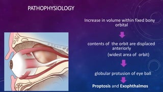
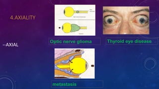
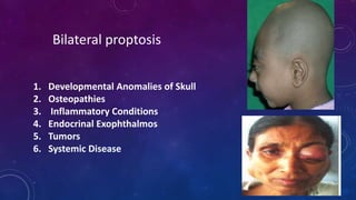
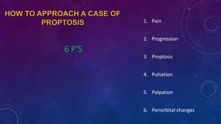
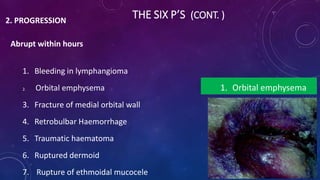
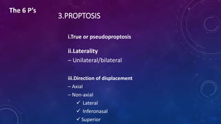
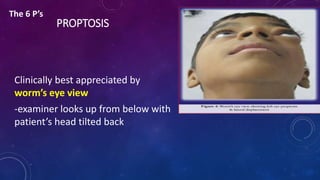
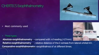
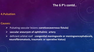
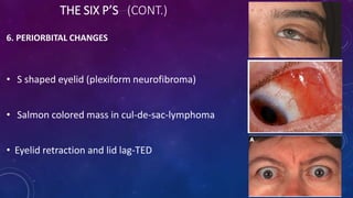
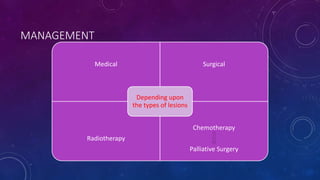
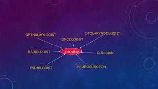
EVALUATION OF
PROPTOSIS
DR. RAKSHYA BASNET
1ST YEAR RESIDENT
NAMS,LEI
LAYOUT
• Introduction
• Clinical anatomy of orbit
• Classification of proptosis
• Evaluation of proptosis on basis of THE 6 P’S
• Investigation
• anterior displacement of globe by >20 mm from lateral orbital rim
• >2mm difference between apex of cornea of two eyes
OTHER TERMS
1.Exophthalmos is a term reserved for proptosis due to endocrine
cause, but it is used interchangeably
2.Exorbitism decrease in volume of orbit : orbital contents to
protrudeforwards
3.Pseudoproptosis
1. Enlarged globe
2. Asymmetric orbital size
3. Asymmetric palpebral fissure
4. Contralateral enophthalmos
CLINICAL ANATOMY
ORBITALWALLRELATIONS
Roof :anterior cranial fossa& frontal sinus above
Medial wall : adjacent nasal cavity, ethmoid&posterior sphenoid sinus
Lateral wall :adjacent to middle cranial ,temporal&pterygopalatine fossa
ORBIT
Below- maxillary sinus
• Relationship of orbit & PNS :By its location &
venous drainage
• orbital venous drainage :devoid of valves –
two way communication between orbit and
sinuses
SPACES IN RELATION TO ORBIT
1. subperiosteal
2. Peripheral/extraconal
3. Central/intraconal
4. Sub tenon’s
5. subarachnoid
1.SUBPERIOSTEAL SPACE
 Common tumors in this space are
Dermoid cyst, epidermoid cyst,mucocele,
subperiosteal abscess, myeloma, osteomatous
tumour, haematoma and fibrous dysplasia
 plain x-rays most useful in this space
• Common tumors in this space:
malignant lymphoma, capillary hemangioma,
intrinsic neoplasm of lacrimal gland,
pseudotumors
• Produces non-axial proptosis
• Tumors in this space are explored by
anterior orbitotomy
2.Peripheral space
• Common tumors include
Cavernous hemangioma of adults,
solitary neurofibroma,
neurilemmomas, nodular orbital
meningiomas, optic nerve gliomas
• Produces axial proptosis
• Tumors in this space explored by
lateral orbitotomy
3.Central space
PATHOPHYSIOLOGY
Increase in volume within fixed bony
orbital
contents of the orbit are displaced
anteriorly
(widest area of orbit)
globular protusion of eye ball
Proptosis and Exophthalmos
patterns of orbital involvement
1. INFLAMMATORY EFFECT: redness, swelling, pain,
heat, and loss of function
2. MASS EFFECT: Displacement with or without signs
of involvement of sensory or neuromuscular sign
3. VASCULAR CHANGE: venous dilation, pulsation,
expansion with straining (Valsalva) and haemorrhage
4. INFILTRATIVE CHANGE: evidence of destruction,
entrapment, or both
CLASSIFICATION
1) Etiological
2) Onset
3) Direction
4) Axiality
5) Laterality
6) Age of onset
CLASSIFICATION
1.Etiology
• Inflammatory
• Infectious
• Vascular
• Neoplastic
• Idiopathic
2.Onset
• Acute-several minutes, several hours, or 1 to 2 days
• Subacute-period of weeks
• Chronic-more insidious onset over several months
3.DIRECTION
• POSITIVE-if the lesion occupies space
and pushes orbital structures away
eg-intraconal schwannoma
• NEGATIVE -if it draws structures toward
eg.orbital metastasis of sclerosing carcinoma
4.AXIALITY
--AXIAL
Thyroid eye diseaseOptic nerve glioma
metastasis
SUPERIOR
1.Maxillary sinus tumours
Inferiomedial
2.Dermoid cyst
3.Lacrimal gland tumour
Non-axial
INFEROLATERAL
•Fronto-ethmoidal mucocele
•abscess
•Sinus carcinomas
•osteomas
5.LATERALITY
• Unilateral Proptosis
1.Congenital
2.Traumatic
3.Inflammatory Lesions
4.Circulatory Disturbances & Vascular Lesions
5.Cysts of orbit:
6.Tumors:
7.Mucocele of PNS:
1. Developmental Anomalies of Skull
2. Osteopathies
3. Inflammatory Conditions
4. Endocrinal Exophthalmos
5. Tumors
6. Systemic Disease
Bilateral proptosis
IN CHILDREN
• Unilateral (most common): orbital cellulites
• Bilateral (most common):leukemia, metastatic
neuroblastoma
6.PROPTOSIS : ACCORDING TO AGE
CAUSES IN INFANT
 congenital lesions like craniosynostosis, cephalocele, micropthalmia
with cyst, teratoma,
 retinoblastoma
 capillary hemangioma
 juvenile xantho granuloma
 metastatic neuroblastoma
CAUSES IN CHILDREN
• Orbital cellulitis
• Dermoid cyst
• Capillary hemangioma
• Optic nerve glioma
• Rhabdomyosarcoma
• Retinoblastoma
• Leukemia
• Lymphangioma
• Metastasis
– Metastatic
neuroblastoma
– Ewing's sarcoma
CAUSES IN ADULTS
• Thyroid Exophthalmos
• Pseudotumor
• Orbital cellulitis
• Trauma
• Meningioma
• Lymphoma
• Histiocytoma
• Cavernous hemangioma
• Osteoma
• Varices
• Carotid-cavernous fistula
• Tumors extending from adjacent areas
– Lacrimal gland
– Sinuses
– lids
• Metastasis
6 P’S
1. Pain
2. Progression
3. Proptosis
4. Pulsation
5. Palpation
6. Periorbital changes
1.PAIN
– Inflammatory disease
– Infectious disease
– Orbital hemorrhage
– Malignant Carcinoma (nasopharyngeal or lacrimal gland)
– Metastatic lesions
The 6 P’s
2.PROGRESSION
THE SIX P’s (Cont. )
THE SIX P’S (CONT. )2. PROGRESSION
Abrupt within hours
1. Bleeding in lymphangioma
2. Orbital emphysema
3. Fracture of medial orbital wall
4. Retrobulbar Haemorrhage
5. Traumatic haematoma
6. Ruptured dermoid
7. Rupture of ethmoidal mucocele
1. Orbital emphysema
Onset occurring over days to
weeks
• Idiopathic orbital inflammatory disease
• Orbital cellulitis
• Thrombophlebitis
• Rhabdomyosarcoma
• Thyroid ophthalmopathy
• Neuroblastoma
• Metastatic tumour
THE SIX P’S (CONT. )
Onset occurring over months to years
• Dermoids
• Benignmixed tumours
• Neurogenic tumours
• Cavernous hemangioma
• Lymphoma
• Fibrous histiocytoma
• Osteoma
Intermittent proptosis
• Orbital varices
• Haemangioma
• Carotid Cavernous fistula
3.PROPTOSIS
i.True or pseudoproptosis
ii.Laterality
– Unilateral/bilateral
iii.Direction of displacement
– Axial
– Non-axial
 Lateral
 Inferonasal
 Superior
The 6 P’s
PROPTOSIS
Clinically best appreciated by
worm’s eye view
-examiner looks up from below with
patient’s head tilted back
The 6 P’s
Nafziger’s Method
• patient sits in front of examiner, head slightly drawn
back & looks downwards
• examiner stands behind patient, looks over
patient’s forehead by bending over patient’s head
• examiner raises patient’s upper lids with his index
fingers from sides
• examiner compares position of apex of cornea on
each side
• patient bends his head forward and cornea should disappear
at same time.
CLINICAL METHODS FOR MEASUREMENT OF PROPTOSIS:
A) PLASTIC RULER:
can measure
proptosis from the
lateral orbital rim to
corneal apex,holding
ruler parallel to ground
B)LUEDDE’S EXOPHTHALMOMETER:
-Transparent plastic mm ruler which is thicker than
normal ruler
- is better than hertel’s if there is facial
asymmetry
C)HERTEL’S Exophthalmometry
• Most commonly used
• Three types
Absolute exophthalmometry - compared with n/lreading (>21mm)
Relative exophthalmometry - relative distance of the2 corneas from lateral orbitalrim.
Comparative exophthalmometry -exophthalmos of at different times.
Steps of hertel’s exophthalmometry
Stepsof Hertel’s exopthalmometer
1. make sure instrument is ready
2. explain patient
3. ask patient to sit in erect position
4. take a seat one arm away from patient and make sure your eyes and
patient’s eyes are at same level
5. ask patient to look at the centre of your forehead
6. place Hertel against lateral walls of patient
7. Measure patient’s left eye with examiner’s right eye
8. move our view so that 2 red lines on prism are are in overlapping
position
9. find the position of corneal apex in millimeter scale in prism
10.record proptosis with base reading
Grading:
• Mild : 21 – 23 mm
• Moderate: 24 – 27 mm
• Severe: 28 mm or more
D)Naugle’s exophthalmometer:
-Useful in fracture patients when lateral canthus has
been displaced
-uses frontal and maxillary bone as references
4.Pulsation
Causes:
 Pulsating vascular lesions (caroticocavernous fistula)
 saccular aneurysm of ophthalmic artery
 deficient orbital roof (congenital meningocele or meningoencephalocele,
neurofibromatosis, traumatic or operative hiatus)
The 6 P’s contd..
Auscultation
• Globe/temporal region for bruit
PULSATION
Without bruit
• Neurofibromatosis
• Meningoencephaloceles
• Encephaloceles
• Result of surgical removal of the orbital roof
With bruit
• Carotid cavernous fistula
• Dural arteriovenous fistula
• Orbital arteriovenous fistula
1. Local Temperature
2. Tenderness
3. orbital margins
4. Retropulsion of globe
5. Regional lymph node
6. If mass palpable note
 Position
 Size,surface, attachnents
 Consistency(hard , rubbery, spongy or soft)
 Compressibility/ Reducibility
5.PALPATION
The 6 P’s contd..
ORBITAL RIM
• Palpation of orbital rim done to note any changes in
contour or dehiscence of orbital wall....
RETROPULSION
• should be estimated by applying equal digital pressure over two eyes,
simultaneously
• best done with examiner’s thumb over closed lids-retroocular
resistance encountered in presence of solid tumors
LYMPH NODES
• regional lymph node preauricular lymph node and
metastatic dz. supraclavicular, and cervical nodes.
THE SIX P’S (CONT.)
6. PERIORBITAL CHANGES
• S shaped eyelid (plexiform neurofibroma)
• Salmon colored mass in cul-de-sac-lymphoma
• Eyelid retraction and lid lag-TED
Ecchymosis of eyelid skin-metastatic neuroblastoma,leukemia,amyloidosis
Eczematous lesions of eyelid-mycosis fungoides
Edematous swelling of lowerlid (meningioma)
-Prominent temple –sphenoid wing
meningioma
-corkscrew conjunctival vessels –arteriovenous
fistula
D. Other tests
1. Transillumination
2. Visual acuity
3. Pupillary reaction
4. Ocular motility
5. Forced duction test
6. Tonometry
1.TRANSILLUMINATION
(fluid / air filled)
2.Visual acuity:
 Loss of vision preceding exophthalmos suggests tumor of optic nerve like glioma in children
 Orbital tumors decrease central acuity by
--pressing on back of eyeball producing changes in refraction or sallman’s macular folds or
optic atropy in late stages
VISION LOSS
• Due to involvement of optic nerve by compression, infiltration, vascular
compromise, inflammation
• Marked proptosis with no visual loss—cavernous hemangioma &
neurilemmoma
• Marked visual loss with mild to moderate proptosis—optic nerve glioma and
optic nerve sheath meningioma
3.FUNDUS
i. Optic disc changes :
- Optic disc oedema
- Optic atrophy
- Optociliary shunt vessels
(optic nerve sheath meningioma,
cavernous haemangioma)
….
ii. Choroidal folds :
- Tumor
- Dysthyroid ophthalmopathy
- inflammatory lesions
iii. Retinal vascular changes
- Venous dilatation & tortuosity
(arteriovenous communication)
- Venous dilatation & disc swelling
(orbital mass)
- Vascular occlusion (optic nerve tumor)
4.PUPILLARY REACTIONS
-Look carefully for RAPD suggestive of optic nerve damage
-Is an indication for plotting visual fields in both eyes
5.OCULAR MOVEMENTS
Limitation of ocular motility due to
 Restrictive myopathy (thyroid opthalmopathy)
 Splinting of optic nerve(optic nerve sheath meningioma)
 Neurological deficit from orbital apex lesions
Differentiation of restrictive from neurological motility defect
6.Forced duction test:
Positive result: difficulty or inability to move globe indicates restrictive problem
Negative result: no resistance will be encountered indicates neurologic problem
7.TONOMETRY
Raised IOP in TED in upward gaze i.e. positional IOP changes
Braley’s sign
Positive result: increase of 6mmHg or more indicates due to muscle restriction
Negative result: < 6mmHg IOP indicates neurological lesion
• Systemic examination
• Café au lait spots
• Skin pigmentation
• Features of hyperthyroidism
• Cutaneous hemangioma elsewhere
• Scalp bony lesions
• Organomegaly or lumps in abdomen
• Otorhinolaryngological examination: paranasal sinus or nasopharyngeal mass
• NERVES; Ocular movements (III, IV, VI),Ptosis (III),Lagophthalmos (VII)
Investigations
LAB.. • Haematological studies (TLC, DLC, ESR, VDRL test)
• Thyroid function test
• Casoni’s test (to rule out hydatid cyst)
• Stool examination: cysts, ova
• Urine: Bence Jones proteins for multiple myeloma
• Serum ACE
• Lysozyme
• Antineutrophil cytoplasmic antibody
RADIOLOGY
1. X-rays
2. Orbital USG
3. CT scan
4. MRI
5. Angiography
Non-invasive
invasive
1. Orbital venography
2. Carotid angiography
IMAGING TECHNIQUE:
(A)Non-invasive techniques:
1.Plain X-rays: Caldwell view, Water’s view, lateral view & Rhese view (for optic foramina).
• Enlargement of orbital cavity & optic foramina, calcification, hyperostosis.
2. Ultrasonography:
• Valuable initial scanning procedure for orbital lesions
• Can usually differentiate between solid, cystic, infiltrative & spongy masses
• Lesions of posterior orbit can’t be viewed
3. Computed Tomography:
- Most valuable for delineating the shape, location , extent and
character of lesions in orbit esp.orbital trauma,bony tumors
- Not only bones but foreign body and soft tissues also
- Contrast for vascularized tumor,orbital abscess
MEASUREMENT OF PROPTOSIS
• By measuring distance from anterior corneal
surface to interzygomatic line
• Distance from the posterior scleral margin to
interzygomatic line.
76
4. Magnetic resonance imaging (MRI):
• Sensitive for detecting differences between normal
& abnormal tissues.
• Better technique for orbitocranial junction or
intracranial,intracanalicular optic nerve
5. CT and MR angiography:
- Arteriovenous malformation
- Aneurysms
- Arteriovenous fistulas
INVASIVE PROCEDURES
Orbital venography: orbital varix suspected
Carotid angiography: pulsating exophthalmos & with bruit or thrill
PATHOLOGY
The diagnosis of an orbital lesion usually requires analysis of
tissue obtained through an orbitotomy.
• FNAC
• incisional biopsy
• Excisional biopsy
• Core biopsy
MANAGEMENT
Medical Surgical
Radiotherapy
Chemotherapy
Palliative Surgery
Depending upon
the types of lesions
OPTHALMOLOGIST OTOLARYNGOLOGIST
RADIOLOGIST
PATHOLOGIST NEUROSURGEON
CLINICIANproptosis
ONCOLOGIST
BIBLIOGRAPHY
• Wolff’s Anatomy of the eye and orbit 7th Edition
• AAO
• BCSC: Orbit Eyelids and Lacrimal System
• Parsons’ Diseases of the eye 22nd edition
• Disease of orbit, jack rootman,2nd edition
•Thank you

proptosis

  • 1.
    EVALUATION OF PROPTOSIS DR. RAKSHYABASNET 1ST YEAR RESIDENT NAMS,LEI
  • 2.
    LAYOUT • Introduction • Clinicalanatomy of orbit • Classification of proptosis • Evaluation of proptosis on basis of THE 6 P’S • Investigation
  • 3.
    • anterior displacementof globe by >20 mm from lateral orbital rim • >2mm difference between apex of cornea of two eyes
  • 4.
    OTHER TERMS 1.Exophthalmos isa term reserved for proptosis due to endocrine cause, but it is used interchangeably 2.Exorbitism decrease in volume of orbit : orbital contents to protrudeforwards
  • 5.
    3.Pseudoproptosis 1. Enlarged globe 2.Asymmetric orbital size 3. Asymmetric palpebral fissure 4. Contralateral enophthalmos
  • 6.
  • 7.
    ORBITALWALLRELATIONS Roof :anterior cranialfossa& frontal sinus above Medial wall : adjacent nasal cavity, ethmoid&posterior sphenoid sinus Lateral wall :adjacent to middle cranial ,temporal&pterygopalatine fossa
  • 8.
    ORBIT Below- maxillary sinus •Relationship of orbit & PNS :By its location & venous drainage • orbital venous drainage :devoid of valves – two way communication between orbit and sinuses
  • 9.
    SPACES IN RELATIONTO ORBIT 1. subperiosteal 2. Peripheral/extraconal 3. Central/intraconal 4. Sub tenon’s 5. subarachnoid
  • 10.
    1.SUBPERIOSTEAL SPACE  Commontumors in this space are Dermoid cyst, epidermoid cyst,mucocele, subperiosteal abscess, myeloma, osteomatous tumour, haematoma and fibrous dysplasia  plain x-rays most useful in this space
  • 11.
    • Common tumorsin this space: malignant lymphoma, capillary hemangioma, intrinsic neoplasm of lacrimal gland, pseudotumors • Produces non-axial proptosis • Tumors in this space are explored by anterior orbitotomy 2.Peripheral space
  • 12.
    • Common tumorsinclude Cavernous hemangioma of adults, solitary neurofibroma, neurilemmomas, nodular orbital meningiomas, optic nerve gliomas • Produces axial proptosis • Tumors in this space explored by lateral orbitotomy 3.Central space
  • 13.
    PATHOPHYSIOLOGY Increase in volumewithin fixed bony orbital contents of the orbit are displaced anteriorly (widest area of orbit) globular protusion of eye ball Proptosis and Exophthalmos
  • 14.
    patterns of orbitalinvolvement 1. INFLAMMATORY EFFECT: redness, swelling, pain, heat, and loss of function 2. MASS EFFECT: Displacement with or without signs of involvement of sensory or neuromuscular sign
  • 15.
    3. VASCULAR CHANGE:venous dilation, pulsation, expansion with straining (Valsalva) and haemorrhage 4. INFILTRATIVE CHANGE: evidence of destruction, entrapment, or both
  • 16.
    CLASSIFICATION 1) Etiological 2) Onset 3)Direction 4) Axiality 5) Laterality 6) Age of onset
  • 17.
  • 18.
    2.Onset • Acute-several minutes,several hours, or 1 to 2 days • Subacute-period of weeks • Chronic-more insidious onset over several months
  • 19.
    3.DIRECTION • POSITIVE-if thelesion occupies space and pushes orbital structures away eg-intraconal schwannoma • NEGATIVE -if it draws structures toward eg.orbital metastasis of sclerosing carcinoma
  • 20.
  • 21.
    SUPERIOR 1.Maxillary sinus tumours Inferiomedial 2.Dermoidcyst 3.Lacrimal gland tumour Non-axial
  • 22.
  • 23.
    5.LATERALITY • Unilateral Proptosis 1.Congenital 2.Traumatic 3.InflammatoryLesions 4.Circulatory Disturbances & Vascular Lesions 5.Cysts of orbit: 6.Tumors: 7.Mucocele of PNS:
  • 24.
    1. Developmental Anomaliesof Skull 2. Osteopathies 3. Inflammatory Conditions 4. Endocrinal Exophthalmos 5. Tumors 6. Systemic Disease Bilateral proptosis
  • 25.
    IN CHILDREN • Unilateral(most common): orbital cellulites • Bilateral (most common):leukemia, metastatic neuroblastoma
  • 26.
    6.PROPTOSIS : ACCORDINGTO AGE CAUSES IN INFANT  congenital lesions like craniosynostosis, cephalocele, micropthalmia with cyst, teratoma,  retinoblastoma  capillary hemangioma  juvenile xantho granuloma  metastatic neuroblastoma
  • 27.
    CAUSES IN CHILDREN •Orbital cellulitis • Dermoid cyst • Capillary hemangioma • Optic nerve glioma • Rhabdomyosarcoma • Retinoblastoma • Leukemia • Lymphangioma • Metastasis – Metastatic neuroblastoma – Ewing's sarcoma
  • 28.
    CAUSES IN ADULTS •Thyroid Exophthalmos • Pseudotumor • Orbital cellulitis • Trauma • Meningioma • Lymphoma • Histiocytoma • Cavernous hemangioma • Osteoma • Varices • Carotid-cavernous fistula • Tumors extending from adjacent areas – Lacrimal gland – Sinuses – lids • Metastasis
  • 29.
    6 P’S 1. Pain 2.Progression 3. Proptosis 4. Pulsation 5. Palpation 6. Periorbital changes
  • 30.
    1.PAIN – Inflammatory disease –Infectious disease – Orbital hemorrhage – Malignant Carcinoma (nasopharyngeal or lacrimal gland) – Metastatic lesions The 6 P’s
  • 31.
  • 32.
    THE SIX P’S(CONT. )2. PROGRESSION Abrupt within hours 1. Bleeding in lymphangioma 2. Orbital emphysema 3. Fracture of medial orbital wall 4. Retrobulbar Haemorrhage 5. Traumatic haematoma 6. Ruptured dermoid 7. Rupture of ethmoidal mucocele 1. Orbital emphysema
  • 33.
    Onset occurring overdays to weeks • Idiopathic orbital inflammatory disease • Orbital cellulitis • Thrombophlebitis • Rhabdomyosarcoma • Thyroid ophthalmopathy • Neuroblastoma • Metastatic tumour
  • 34.
    THE SIX P’S(CONT. ) Onset occurring over months to years • Dermoids • Benignmixed tumours • Neurogenic tumours • Cavernous hemangioma • Lymphoma • Fibrous histiocytoma • Osteoma
  • 35.
    Intermittent proptosis • Orbitalvarices • Haemangioma • Carotid Cavernous fistula
  • 36.
    3.PROPTOSIS i.True or pseudoproptosis ii.Laterality –Unilateral/bilateral iii.Direction of displacement – Axial – Non-axial  Lateral  Inferonasal  Superior The 6 P’s
  • 37.
    PROPTOSIS Clinically best appreciatedby worm’s eye view -examiner looks up from below with patient’s head tilted back The 6 P’s
  • 38.
  • 39.
    • patient sitsin front of examiner, head slightly drawn back & looks downwards • examiner stands behind patient, looks over patient’s forehead by bending over patient’s head • examiner raises patient’s upper lids with his index fingers from sides • examiner compares position of apex of cornea on each side • patient bends his head forward and cornea should disappear at same time.
  • 40.
    CLINICAL METHODS FORMEASUREMENT OF PROPTOSIS: A) PLASTIC RULER: can measure proptosis from the lateral orbital rim to corneal apex,holding ruler parallel to ground
  • 41.
    B)LUEDDE’S EXOPHTHALMOMETER: -Transparent plasticmm ruler which is thicker than normal ruler - is better than hertel’s if there is facial asymmetry
  • 42.
    C)HERTEL’S Exophthalmometry • Mostcommonly used • Three types Absolute exophthalmometry - compared with n/lreading (>21mm) Relative exophthalmometry - relative distance of the2 corneas from lateral orbitalrim. Comparative exophthalmometry -exophthalmos of at different times.
  • 43.
    Steps of hertel’sexophthalmometry
  • 44.
    Stepsof Hertel’s exopthalmometer 1.make sure instrument is ready 2. explain patient 3. ask patient to sit in erect position 4. take a seat one arm away from patient and make sure your eyes and patient’s eyes are at same level 5. ask patient to look at the centre of your forehead 6. place Hertel against lateral walls of patient 7. Measure patient’s left eye with examiner’s right eye 8. move our view so that 2 red lines on prism are are in overlapping position 9. find the position of corneal apex in millimeter scale in prism 10.record proptosis with base reading
  • 45.
    Grading: • Mild :21 – 23 mm • Moderate: 24 – 27 mm • Severe: 28 mm or more
  • 46.
    D)Naugle’s exophthalmometer: -Useful infracture patients when lateral canthus has been displaced -uses frontal and maxillary bone as references
  • 47.
    4.Pulsation Causes:  Pulsating vascularlesions (caroticocavernous fistula)  saccular aneurysm of ophthalmic artery  deficient orbital roof (congenital meningocele or meningoencephalocele, neurofibromatosis, traumatic or operative hiatus) The 6 P’s contd..
  • 48.
  • 49.
    PULSATION Without bruit • Neurofibromatosis •Meningoencephaloceles • Encephaloceles • Result of surgical removal of the orbital roof With bruit • Carotid cavernous fistula • Dural arteriovenous fistula • Orbital arteriovenous fistula
  • 50.
    1. Local Temperature 2.Tenderness 3. orbital margins 4. Retropulsion of globe 5. Regional lymph node 6. If mass palpable note  Position  Size,surface, attachnents  Consistency(hard , rubbery, spongy or soft)  Compressibility/ Reducibility 5.PALPATION The 6 P’s contd..
  • 51.
    ORBITAL RIM • Palpationof orbital rim done to note any changes in contour or dehiscence of orbital wall....
  • 52.
    RETROPULSION • should beestimated by applying equal digital pressure over two eyes, simultaneously • best done with examiner’s thumb over closed lids-retroocular resistance encountered in presence of solid tumors
  • 53.
    LYMPH NODES • regionallymph node preauricular lymph node and metastatic dz. supraclavicular, and cervical nodes.
  • 54.
    THE SIX P’S(CONT.) 6. PERIORBITAL CHANGES • S shaped eyelid (plexiform neurofibroma) • Salmon colored mass in cul-de-sac-lymphoma • Eyelid retraction and lid lag-TED
  • 55.
    Ecchymosis of eyelidskin-metastatic neuroblastoma,leukemia,amyloidosis Eczematous lesions of eyelid-mycosis fungoides Edematous swelling of lowerlid (meningioma)
  • 56.
    -Prominent temple –sphenoidwing meningioma -corkscrew conjunctival vessels –arteriovenous fistula
  • 57.
    D. Other tests 1.Transillumination 2. Visual acuity 3. Pupillary reaction 4. Ocular motility 5. Forced duction test 6. Tonometry
  • 58.
  • 59.
    2.Visual acuity:  Lossof vision preceding exophthalmos suggests tumor of optic nerve like glioma in children  Orbital tumors decrease central acuity by --pressing on back of eyeball producing changes in refraction or sallman’s macular folds or optic atropy in late stages
  • 60.
    VISION LOSS • Dueto involvement of optic nerve by compression, infiltration, vascular compromise, inflammation • Marked proptosis with no visual loss—cavernous hemangioma & neurilemmoma • Marked visual loss with mild to moderate proptosis—optic nerve glioma and optic nerve sheath meningioma
  • 61.
    3.FUNDUS i. Optic discchanges : - Optic disc oedema - Optic atrophy - Optociliary shunt vessels (optic nerve sheath meningioma, cavernous haemangioma)
  • 62.
    …. ii. Choroidal folds: - Tumor - Dysthyroid ophthalmopathy - inflammatory lesions iii. Retinal vascular changes - Venous dilatation & tortuosity (arteriovenous communication) - Venous dilatation & disc swelling (orbital mass) - Vascular occlusion (optic nerve tumor)
  • 63.
    4.PUPILLARY REACTIONS -Look carefullyfor RAPD suggestive of optic nerve damage -Is an indication for plotting visual fields in both eyes
  • 64.
    5.OCULAR MOVEMENTS Limitation ofocular motility due to  Restrictive myopathy (thyroid opthalmopathy)  Splinting of optic nerve(optic nerve sheath meningioma)  Neurological deficit from orbital apex lesions
  • 65.
    Differentiation of restrictivefrom neurological motility defect 6.Forced duction test: Positive result: difficulty or inability to move globe indicates restrictive problem Negative result: no resistance will be encountered indicates neurologic problem
  • 66.
    7.TONOMETRY Raised IOP inTED in upward gaze i.e. positional IOP changes Braley’s sign Positive result: increase of 6mmHg or more indicates due to muscle restriction Negative result: < 6mmHg IOP indicates neurological lesion
  • 67.
    • Systemic examination •Café au lait spots • Skin pigmentation • Features of hyperthyroidism • Cutaneous hemangioma elsewhere • Scalp bony lesions • Organomegaly or lumps in abdomen • Otorhinolaryngological examination: paranasal sinus or nasopharyngeal mass • NERVES; Ocular movements (III, IV, VI),Ptosis (III),Lagophthalmos (VII)
  • 68.
  • 69.
    LAB.. • Haematologicalstudies (TLC, DLC, ESR, VDRL test) • Thyroid function test • Casoni’s test (to rule out hydatid cyst) • Stool examination: cysts, ova • Urine: Bence Jones proteins for multiple myeloma • Serum ACE • Lysozyme • Antineutrophil cytoplasmic antibody
  • 70.
    RADIOLOGY 1. X-rays 2. OrbitalUSG 3. CT scan 4. MRI 5. Angiography Non-invasive invasive 1. Orbital venography 2. Carotid angiography
  • 71.
    IMAGING TECHNIQUE: (A)Non-invasive techniques: 1.PlainX-rays: Caldwell view, Water’s view, lateral view & Rhese view (for optic foramina). • Enlargement of orbital cavity & optic foramina, calcification, hyperostosis.
  • 72.
    2. Ultrasonography: • Valuableinitial scanning procedure for orbital lesions • Can usually differentiate between solid, cystic, infiltrative & spongy masses • Lesions of posterior orbit can’t be viewed
  • 73.
    3. Computed Tomography: -Most valuable for delineating the shape, location , extent and character of lesions in orbit esp.orbital trauma,bony tumors - Not only bones but foreign body and soft tissues also - Contrast for vascularized tumor,orbital abscess
  • 74.
    MEASUREMENT OF PROPTOSIS •By measuring distance from anterior corneal surface to interzygomatic line • Distance from the posterior scleral margin to interzygomatic line. 76
  • 75.
    4. Magnetic resonanceimaging (MRI): • Sensitive for detecting differences between normal & abnormal tissues. • Better technique for orbitocranial junction or intracranial,intracanalicular optic nerve
  • 76.
    5. CT andMR angiography: - Arteriovenous malformation - Aneurysms - Arteriovenous fistulas
  • 77.
    INVASIVE PROCEDURES Orbital venography:orbital varix suspected Carotid angiography: pulsating exophthalmos & with bruit or thrill
  • 78.
    PATHOLOGY The diagnosis ofan orbital lesion usually requires analysis of tissue obtained through an orbitotomy. • FNAC • incisional biopsy • Excisional biopsy • Core biopsy
  • 79.
  • 80.
  • 81.
    BIBLIOGRAPHY • Wolff’s Anatomyof the eye and orbit 7th Edition • AAO • BCSC: Orbit Eyelids and Lacrimal System • Parsons’ Diseases of the eye 22nd edition • Disease of orbit, jack rootman,2nd edition
  • 82.