Orbit (anatomy and diseases) for medical students
•Download as PPTX, PDF•
0 likes•10 views

orbit medical students kafrelsheikh university delta university new mansoura university

Recommended
More Related Content
Similar to Orbit (anatomy and diseases) for medical students
Similar to Orbit (anatomy and diseases) for medical students (20)
More from KafrELShiekh University
More from KafrELShiekh University (20)
Recently uploaded
Recently uploaded (20)
Orbit (anatomy and diseases) for medical students
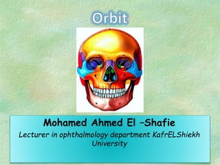
- 1. Mohamed Ahmed El –Shafie Lecturer in ophthalmology department KafrELShiekh University
- 2. Frontal Ethmoid Sphenoid Lacrimal Palatine Maxillary Zygomatic pyramidal or conical in shape consists of an apex, a base and 4 sides: roof, floor, medial wall and lateral wall
- 3. Roof of the Orbit: Contains fossa for the lacrimal gland frontal bone lesser wing of the sphenoid Defect: Pulsatile proptosis
- 5. Lateral wall of the Orbit zygomatic bone greater wing of the sphenoid Inferiorly – inf orbital fissure Medially – sup orbital fissure • Only orbital wall not related to paranasal sinus.
- 6. Medial Wall: The thinnest orbital wall. ethmoid, lacrimal, maxillary and sphenoid bones • Related to : Sphenoid and ethmoid sinus The commonest cause of orbital cellulits is ethmoiditis sinusitis.
- 7. Floor of the Orbit: Forms the roof of the maxillary sinus. maxillary, zygomatic bones palatine • Relatively weak, so Site of blow out fracture • Roof of maxillary sinus below, so maxillary carcinoma invade orbit and displace globe upward.
- 8. FRACTURES OF THE ORBITAL FLOOR • Clinical features • Periocular changes: ecchymosis, edema, subcutaneous emphysema • Enophthalmos • Infraorbital nerve anesthesia • Diplopia
- 11. Contents of orbit Eye ball Orbital fat Connective tissue system: Periorbita Orbital septum Tenon’s capsule Blood vessels Nerves Extraocular muscles
- 12. Orbital septum: Interconnecting / circumferential radial webs of fascial system support and transmit forces in trauma Compressive optic neuropathy following trauma
- 13. ORBITA L DISEASE S Inflammatory: Thyroid eye disease Idiopathic orbital inflammatory disease Tumors and cysts Infection trauma
- 15. Main Clinical Manifestation 1. Eyelid retraction 2. Soft Tissue involvement 3. Proptosis 4. Optic Neuropathy 5. Restrictive Myopathy General manifestations - Raised basal metabolic rate. - Tachycardia. - Tremors. - Loss of weight
- 16. 1-Eyelid Retraction: Muller's muscle overaction (sympathetic overstimulation). Darlymple's Sign: Staring look with upper lid retraction Von Grefe's Sign: Upper lid lag behind the globe on looking down. Stellwage's sign: Infrequent blinking and imperfect closure of lid during the act.
- 17. • 2-Soft Tissue Involvement (infiltration) • 1. Conjunctival Injection • 2. Chemosis • 3. Eyelid swelling • 4. Kerato-conjunctival Sicca
- 18. • • • •
- 19. 4-OPTIC NEUROPATHY • DIRECT COMPRESSION BY RECTI 5-Restrictive Myopathy IR>MR>SR>LR
- 20. CT SCAN • EOM HYPERTROPHY WITH TENDON SPARING
- 22. PRESEPTAL CELLULITIS Infection confined to the eyelids and periorbital tissues anterior to the orbital septum Globe is uninvolved: Pupillary reflexes, visual acuity, EOM’s are NORMAL No chemosis no pain No proptosis
- 23. ORBITAL CELLULITIS • Commonest cause is ethmoiditis
- 26. Investigation: Radiology of paranasal sinuses. • Treatment 1- Medical - Systemic broad spectrum antibiotics , and non steroidal anti-inflammatories. - Warm compresses. - Treatment of the cause. 2- Surgical: Drainage of any suppuration ( pointing ).
- 27. Dermoid Cyst Benign cystic teratoma well-encapsulated lined by stratified squamous & contain dermal appendages
- 30. • The most common cause of proptosis in adults is: A- Dysthyroid orbitopathy B-Orbital cellulitis C-Orbital tumors D-None of the above
- 31. Unilateral proptosis may be seen in all of the following except : a. Cavernous sinus thrombosis b. Oribital tumors c. Retrobulbar hemorrtage following tramua d. Hyphema
- 32. • The commonest sign of Graves’ disease: A- Lid retraction B-Conjunctival chemosis C-Diplopia D-Exophthalmos
- 33. Evisceration is: A. Excision of the entire eyeball. B. Excision of all the inner contents of the eyeball including the uveal tissue. C. Photocoagulation of the retina. D. Removal of orbit contents.
- 34. Lagophthalmos can occur in all of the following except; a. 7th cranial nerve paralysis b. 5th cranial nerve paralysis c. Thyrotoxic exophthalmos d. Symblepharon