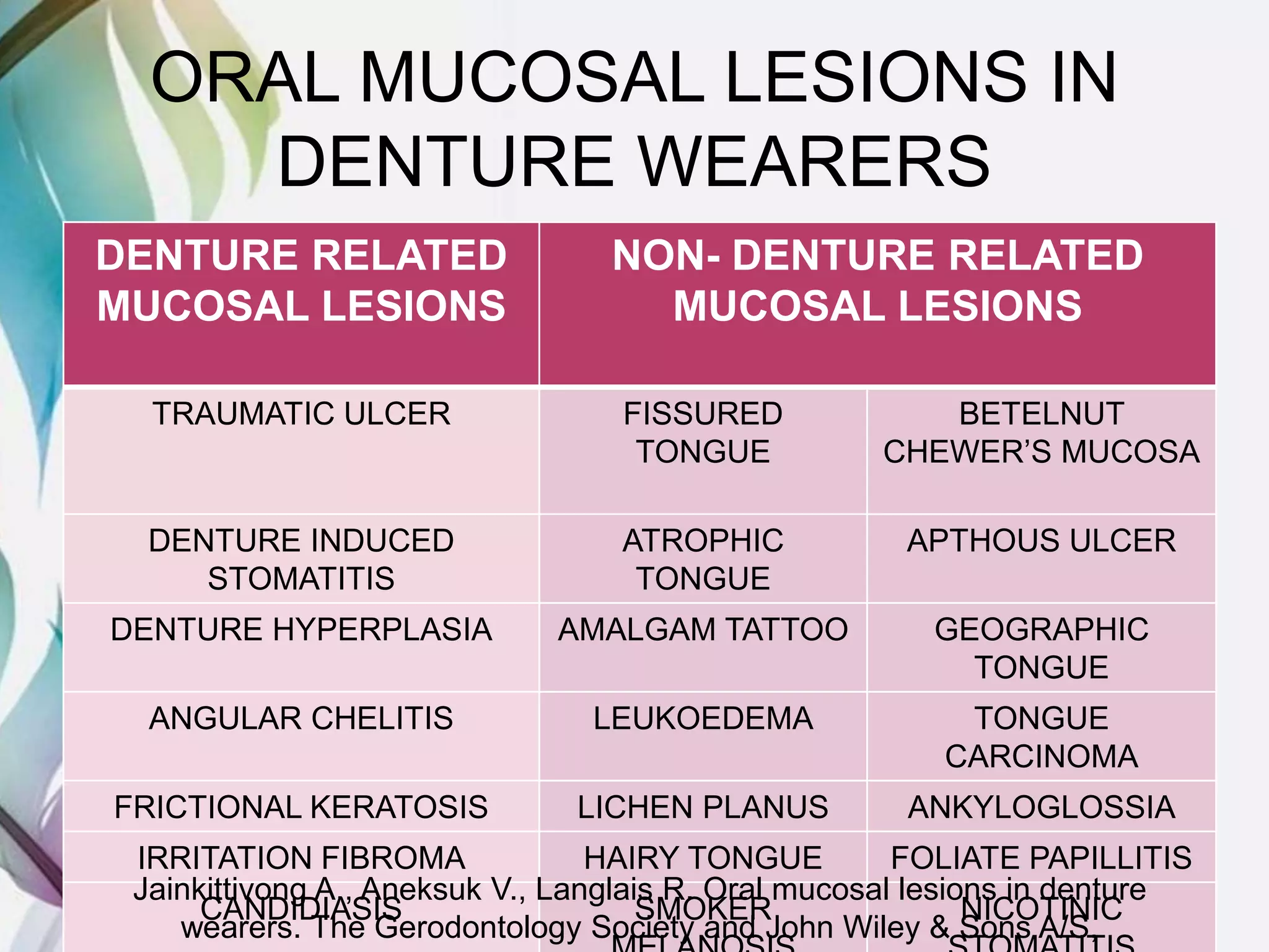
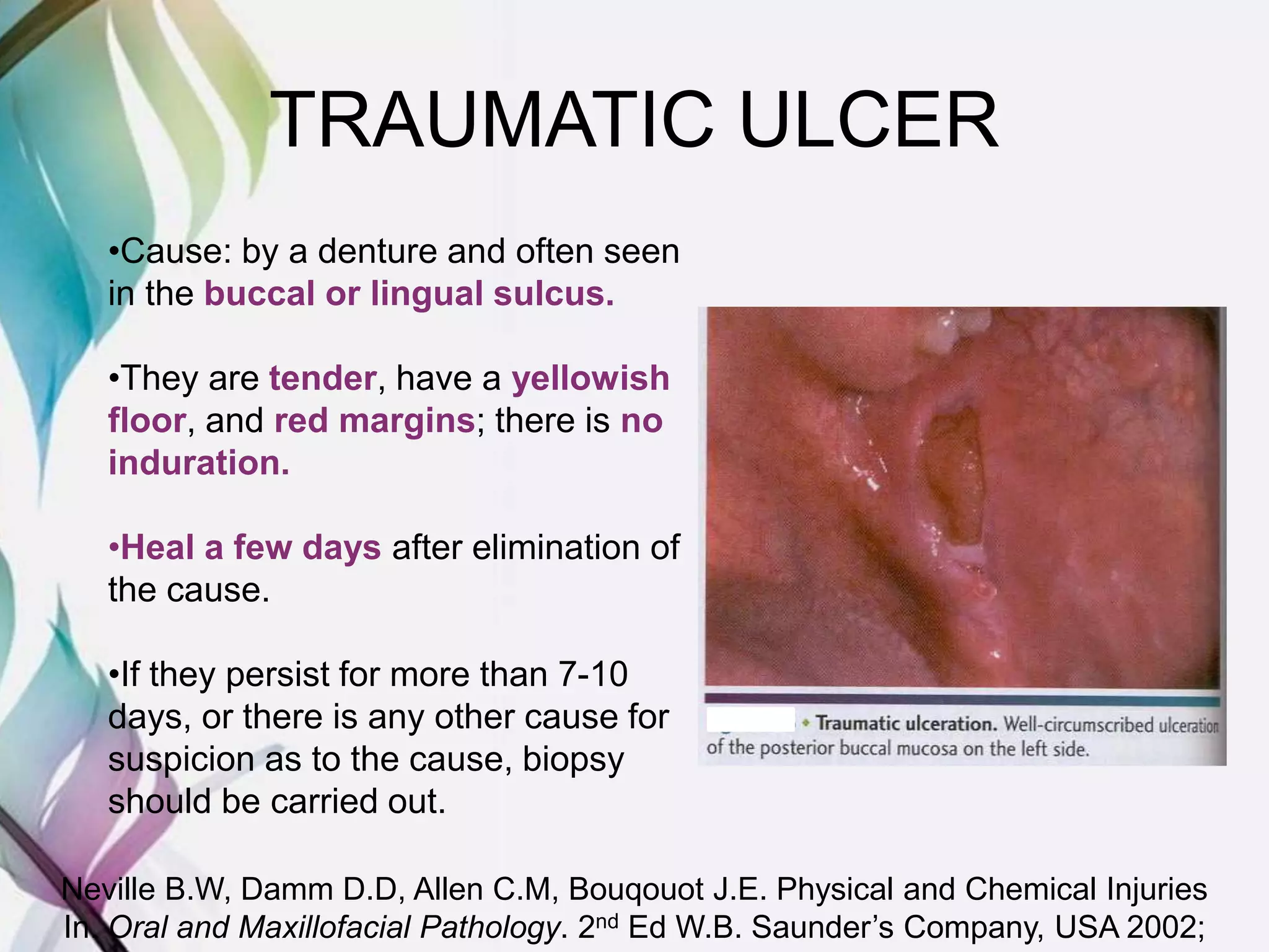
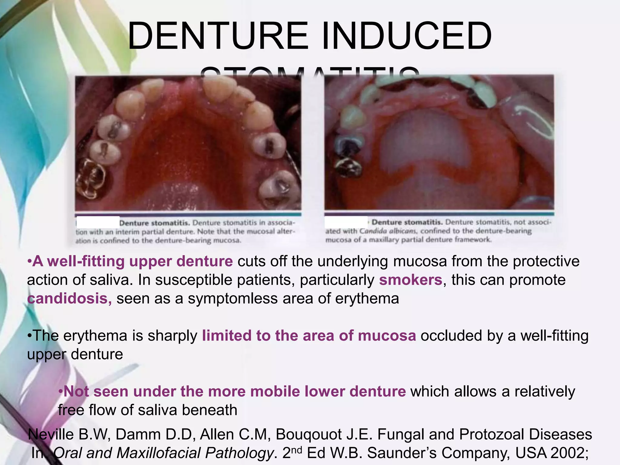
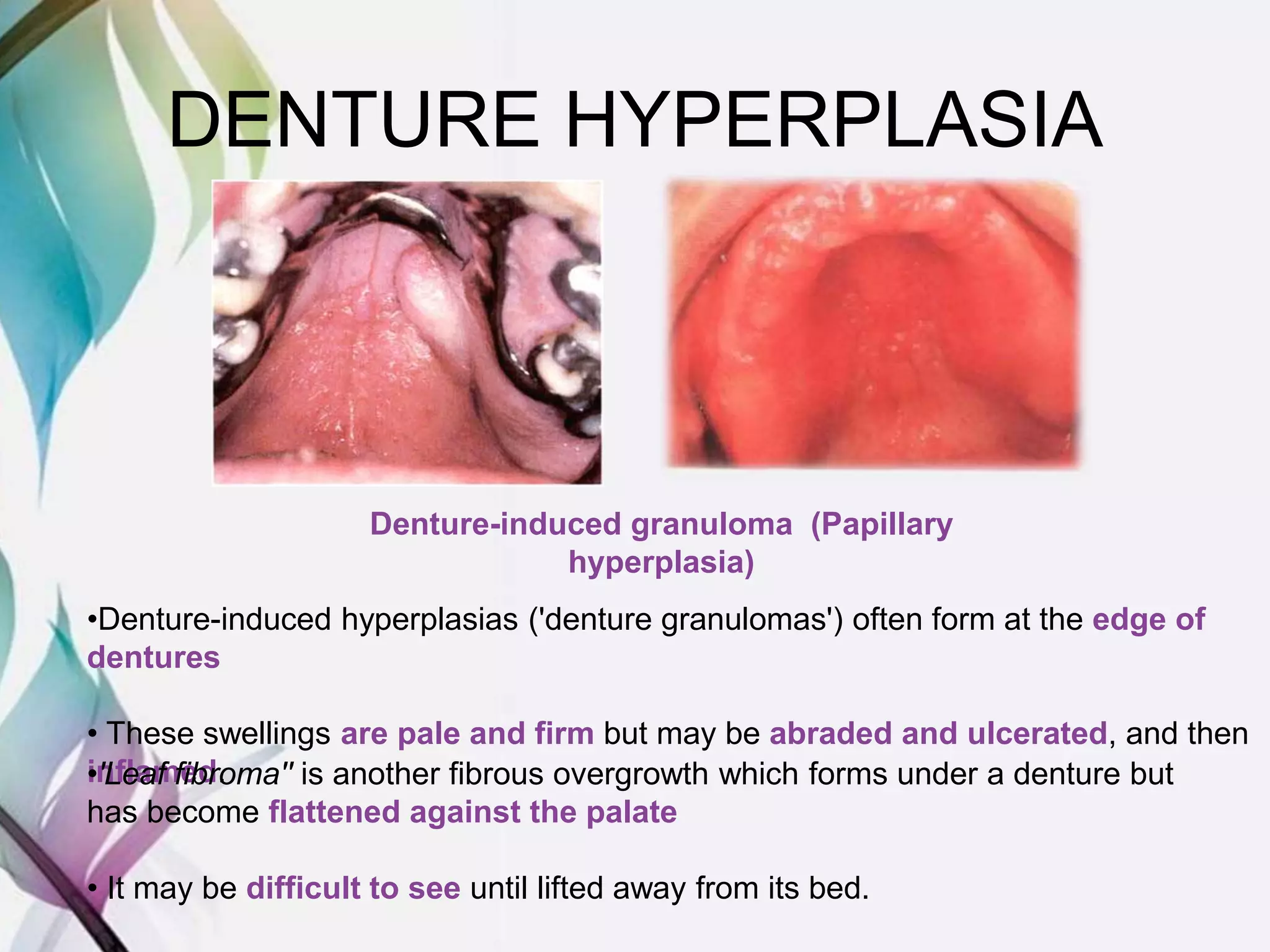
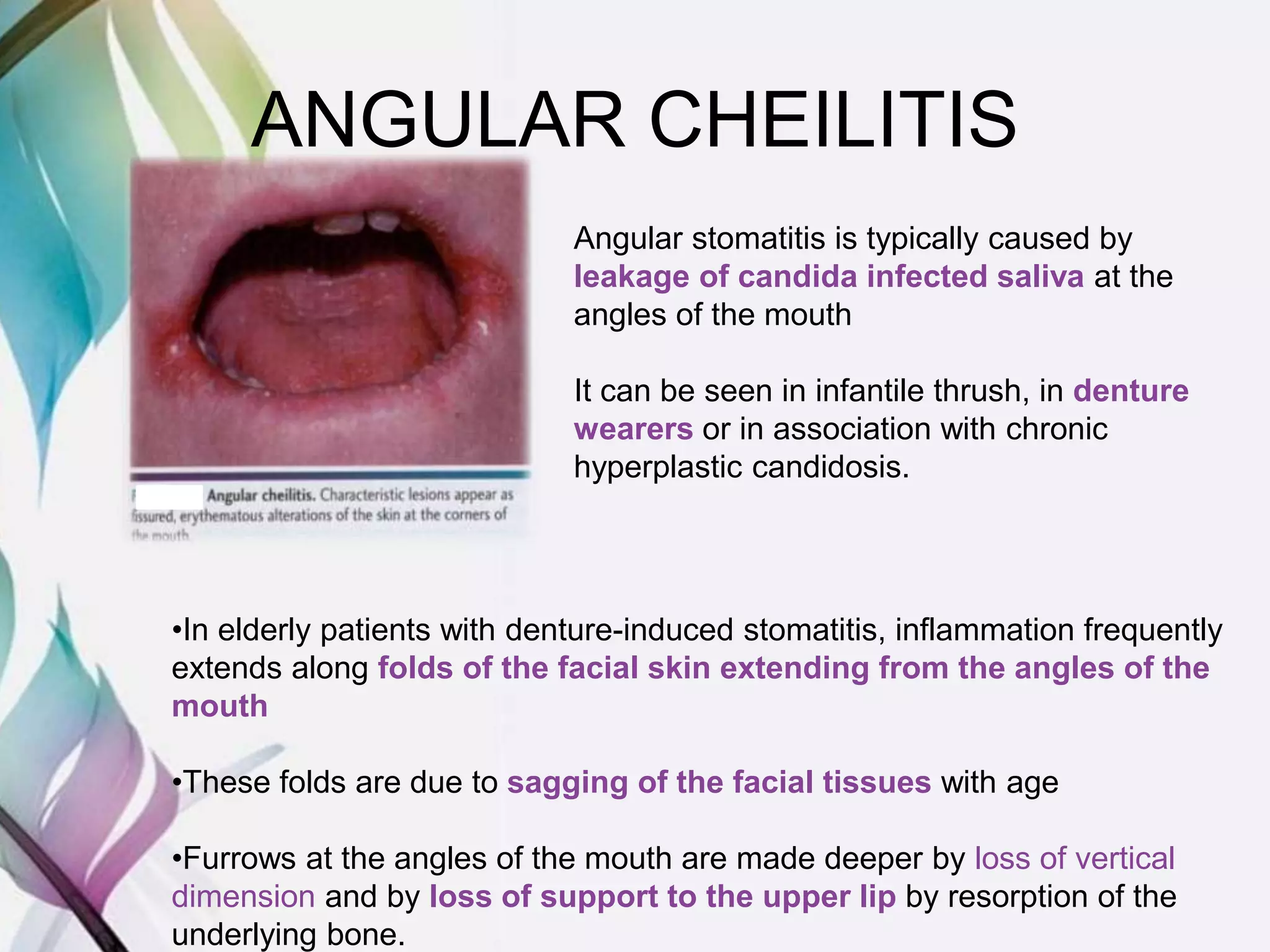
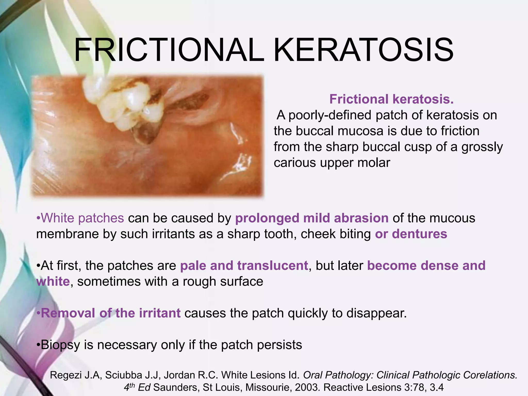
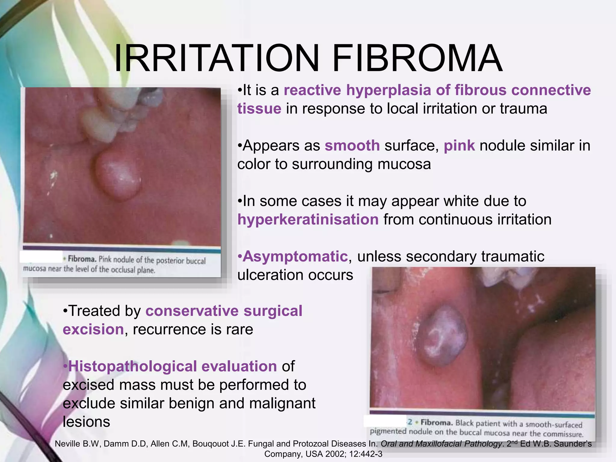
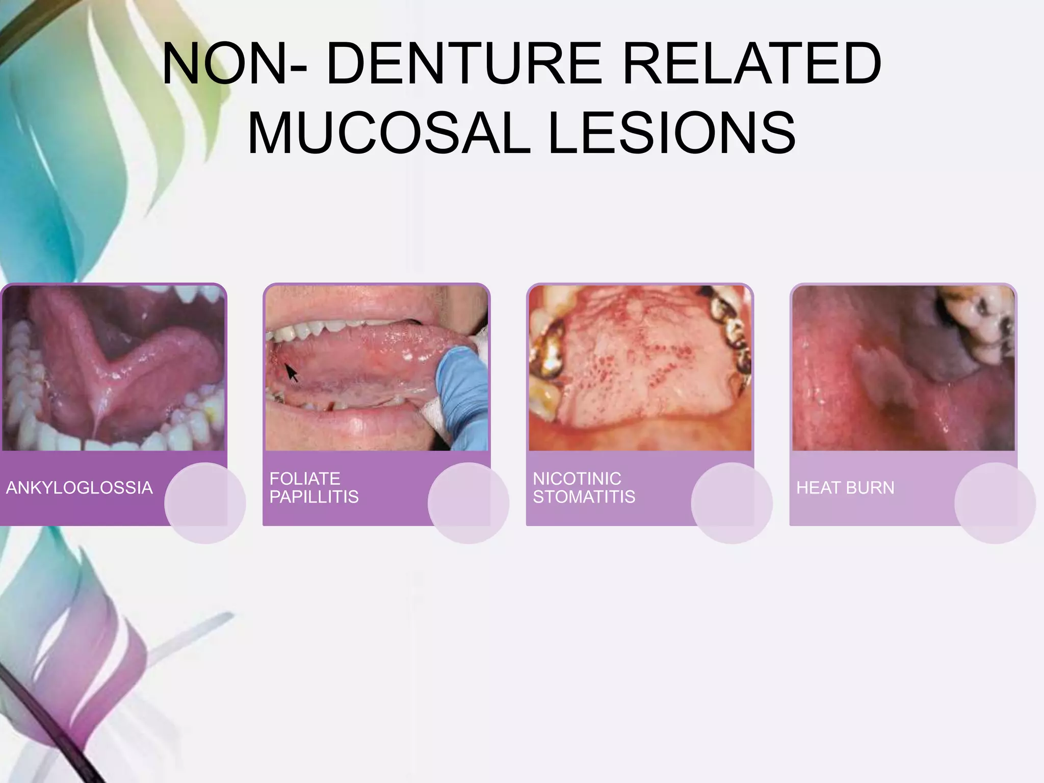
ORAL MUCOSAL LESIONS
IN DENTURE WEARERS
Aree Jainkittivong, Vilaiwan Aneksuk and Robert
P. Langlais
The Gerodontology Society and John Wiley & Sons A/S, Gerodontology 2010; 27: 26–
32
AAMIR ZAHID GODIL
FIRST YEAR P.G.
DEPARTMENT OF PROSTHODONTICS
M.A.R.D.C.
INTRODUCTION
• Communication is often the key problem in
the management of the elderly
• There is a great variety of oral complaints
that they may suffer. In extreme cases for
example, an elderly patient may be so
intellectually impaired, deaf and depressed
as to make communication excessively
difficult but may also have oral cancer
Cawson.R.A, Odell E.W. Intellectual and Physical Disability In: Essentials of
Oral Pathology and Oral Medicine, Edition 7. Spain 2002. 33: 367
• The prevalence of oral mucosal lesions
(OMLs) is usually reported to be higher in
denture wearers than in non-wearers
• The area of the oral mucosa covered by a
complete denture is greater than that covered
by a partial denture and may therefore
increase the risk of Denture Related Mucosal
Lesions (DMLs)
Coelho CM, Sousa YT, Dare AM. Denture-related oral mucosal lesions in a
Brazilian school of dentistry. J Oral Rehabil 2004; 31: 135–139.
ORAL MUCOSAL LESIONS IN
DENTURE WEARERS
DENTURE RELATED
MUCOSAL LESIONS
NON- DENTURE RELATED
MUCOSAL LESIONS
TRAUMATIC ULCER FISSURED
TONGUE
BETELNUT
CHEWER’S MUCOSA
DENTURE INDUCED
STOMATITIS
ATROPHIC
TONGUE
APTHOUS ULCER
DENTURE HYPERPLASIA AMALGAM TATTOO GEOGRAPHIC
TONGUE
ANGULAR CHELITIS LEUKOEDEMA TONGUE
CARCINOMA
FRICTIONAL KERATOSIS LICHEN PLANUS ANKYLOGLOSSIA
IRRITATION FIBROMA HAIRY TONGUE FOLIATE PAPILLITIS
CANDIDIASIS SMOKER NICOTINIC
Jainkittivong A., Aneksuk V., Langlais R. Oral mucosal lesions in denture
wearers. The Gerodontology Society and John Wiley & Sons A/S,
DENTURE RELATED
MUCOSAL LESIONS
TRAUMATIC ULCER
•Cause: by a denture and often seen
in the buccal or lingual sulcus.
•They are tender, have a yellowish
floor, and red margins; there is no
induration.
•Heal a few days after elimination of
the cause.
•If they persist for more than 7-10
days, or there is any other cause for
suspicion as to the cause, biopsy
should be carried out.
Neville B.W, Damm D.D, Allen C.M, Bouqouot J.E. Physical and Chemical Injuries
In. Oral and Maxillofacial Pathology. 2nd Ed W.B. Saunder’s Company, USA 2002;
DENTURE INDUCED
STOMATITIS
•A well-fitting upper denture cuts off the underlying mucosa from the protective
action of saliva. In susceptible patients, particularly smokers, this can promote
candidosis, seen as a symptomless area of erythema
•The erythema is sharply limited to the area of mucosa occluded by a well-fitting
upper denture
•Not seen under the more mobile lower denture which allows a relatively
free flow of saliva beneath
Neville B.W, Damm D.D, Allen C.M, Bouqouot J.E. Fungal and Protozoal Diseases
In. Oral and Maxillofacial Pathology. 2nd Ed W.B. Saunder’s Company, USA 2002;
Management
• The clinical picture is distinctive but the
diagnosis can be confirmed by finding
candidal hyphae in a Gram-stained smear
taken from the inflamed mucosa or the fitting
surface of the denture.
• The infection responds to antifungal drugs,
but topical agents such as nystatin or
amphotericin can only gain access to the
palate if the patient leaves out the denture
while the tablets are allowed to dissolve in
the mouth.
Cawson.R.A, Odell E.W. Diseases of Oral Mucosa: Introduction and Mucosal
Infections In: Essentials of Oral Pathology and Oral Medicine, Edition 7. Spain
2002. 12:187-8
DENTURE HYPERPLASIA
Denture-induced granuloma (Papillary
hyperplasia)
•Denture-induced hyperplasias ('denture granulomas') often form at the edge of
dentures
• These swellings are pale and firm but may be abraded and ulcerated, and then
inflamed.•'Leaf fibroma'' is another fibrous overgrowth which forms under a denture but
has become flattened against the palate
• It may be difficult to see until lifted away from its bed.
'Leaf fibroma‘
Flat lesions formed between the denture and mucosa are often termed leaf fibromas
because of their shape. Raising this example with a probe reveals its pedunculated
shape.
Management
•Fibrous nodules should be excised together with the small base of
normal tissue from which they arise
•There should be no recurrence if this is done thoroughly and the
source of irritation is removed
Cawson.R.A, Odell E.W. Diseases of Oral Mucosa: Introduction and Mucosal
Infections In: Essentials of Oral Pathology and Oral Medicine, Edition 7. Spain
ANGULAR CHEILITIS
Angular stomatitis is typically caused by
leakage of candida infected saliva at the
angles of the mouth
It can be seen in infantile thrush, in denture
wearers or in association with chronic
hyperplastic candidosis.
•In elderly patients with denture-induced stomatitis, inflammation frequently
extends along folds of the facial skin extending from the angles of the
mouth
•These folds are due to sagging of the facial tissues with age
•Furrows at the angles of the mouth are made deeper by loss of vertical
dimension and by loss of support to the upper lip by resorption of the
underlying bone.
Management
• Though establishment of correct vertical dimension and increasing
the thickness of the labial flange of the upper denture can slightly
lessen these furrows, they can rarely be eliminated in this way.
• Plastic surgery is required when patients are anxious to have these
signs of age removed
• Treatment of intraoral candidal infection alone causes angular
stomatitis to resolve
– Apply miconazole gel24 mg/ml QDS to the angles of the mouth 10-14
days or fusidic acid cream
Angular stomatitis
Cracking and erythema at the commissure
is due to leakage of saliva containing C.
albicans, constantly reinfecting the lesion.
Cawson.R.A, Odell E.W. Diseases of Oral Mucosa: Introduction and Mucosal
Infections In: Essentials of Oral Pathology and Oral Medicine, Edition 7. Spain
2002. 12:186
FRICTIONAL KERATOSIS
•White patches can be caused by prolonged mild abrasion of the mucous
membrane by such irritants as a sharp tooth, cheek biting or dentures
•At first, the patches are pale and translucent, but later become dense and
white, sometimes with a rough surface
•Removal of the irritant causes the patch quickly to disappear.
•Biopsy is necessary only if the patch persists
Frictional keratosis.
A poorly-defined patch of keratosis on
the buccal mucosa is due to friction
from the sharp buccal cusp of a grossly
carious upper molar
Regezi J.A, Sciubba J.J, Jordan R.C. White Lesions Id. Oral Pathology: Clinical Pathologic Corelations.
4th Ed Saunders, St Louis, Missourie, 2003. Reactive Lesions 3:78, 3.4
IRRITATION FIBROMA
•It is a reactive hyperplasia of fibrous connective
tissue in response to local irritation or trauma
•Appears as smooth surface, pink nodule similar in
color to surrounding mucosa
•In some cases it may appear white due to
hyperkeratinisation from continuous irritation
•Asymptomatic, unless secondary traumatic
ulceration occurs
•Treated by conservative surgical
excision, recurrence is rare
•Histopathological evaluation of
excised mass must be performed to
exclude similar benign and malignant
lesions
Neville B.W, Damm D.D, Allen C.M, Bouqouot J.E. Fungal and Protozoal Diseases In. Oral and Maxillofacial Pathology. 2nd Ed W.B. Saunder’s
Company, USA 2002; 12:442-3
CANDIDIASIS
•In nonimmunocompromised, denture wearing patients, it appears
as generalised area of red atrophic tissue commonly found under
maxillary dentures on the palate
Management
• If a denture is worn:
– Cease night-time wear
– Check denture hygiene and advise
– Soak denture overnight in antifungal (dilute hypochlorite,
chlorhexidine mouthwash) or, less effective, apply
miconazole gel to denture fit surface while worn
• Drug of choice and regime:
– Nystatin 100 000 units QDS for 7-10 days as suspension
or pastilles or
– Amphotericin 10 mg QDS as lozenges or suspension10-
14 days.
Cawson.R.A, Odell E.W. Diseases of Oral Mucosa: Introduction and Mucosal
Infections In: Essentials of Oral Pathology and Oral Medicine, Edition 7. Spain
2002. 12:189, 12.1
NON- DENTURE RELATED
MUCOSAL LESIONS
NON- DENTURE RELATED
MUCOSAL LESIONS
FISSURED
TONGUE
ATROPHIC
TONGUE
AMALGAM
TATTOO
LEUKOEDEMA
NON- DENTURE RELATED
MUCOSAL LESIONS
LICHEN
PLANUS
HAIRY
TONGUE
SMOKER’S
MELANOSIS
LEUKOPLAKIA
NON- DENTURE RELATED
MUCOSAL LESIONS
BETELNUT
CHEWER’S
MUCOSA
APTHOUS
ULCER
GEOGRAPHIC
TONGUE
TONGUE
CARCINOMA
NON- DENTURE RELATED
MUCOSAL LESIONS
ANKYLOGLOSSIA
FOLIATE
PAPILLITIS
NICOTINIC
STOMATITIS
HEAT BURN
STATISTICAL ANALYSIS
• The most common DMLs were traumatic ulcer (19.5%) and
denture-induced stomatitis (18.1%)
• When analysed by type, traumatic ulcer, denture hyperplasia,
frictional keratosis and candidiasis were more common in
complete denture wearers, whereas denture-induced stomatitis
was more common in partial denture wearers
• Frictional keratosis was more common in men than in women
• The prevalence of OMLs not related to denture wearing was
higher in complete denture wearers than in partial denture
wearers, and the most common OML was fissured
tongue(27.6%)
• No association between DMLs and systemic conditions or
xerostomic drugs was noted.
Jainkittivong A., Aneksuk V., Langlais R. Oral mucosal lesions in denture
wearers. The Gerodontology Society and John Wiley & Sons A/S,
Table: Number and percentage of oral biopsied lesions in elderly and non-elderly patie
CorreaL.etal.Orallesionsinelderlypopulation:a
biopsysurveyusing2250histopathologicalrecords.The
GerodontologyAssociationandBlackwellMunksgaard
Ltd,Gerodontology2006;23:48–54
CONCLUSION
• Importance of long term follow up
• Patient counseling and compliance
• Minimize the risk of deleterious and
debilitating effects due to dentures
CRITIQUE
• Repetitive with random and unorganized style of
writing
• Replica of an earlier study done by the same authors,
without a specified purpose
• Devoid of clinical differentiating factors (e.g.
candidiasis and denture sore mouth)
• Lack of association between the lesions and systemic
conditions or xerostomic drugs is questionable
• Association with allergies, nutritional deficiencies and
atrophy of masticatory muscles not evaluated
REFERENCES
• Correa L. et al. Oral lesions in elderly population: a biopsy survey using 2250
histopathological records. The Gerodontology Association and Blackwell Munksgaard
Ltd, Gerodontology 2006; 23: 48–54
• Coelho CM, Sousa YT, Dare AM. Denture-related oral mucosal lesions in a Brazilian
school of dentistry. J Oral Rehabil 2004; 31: 135–139.
• Cawson.R.A, Odell E.W. Diseases of Oral Mucosa: Introduction and Mucosal
Infections In: Essentials of Oral Pathology and Oral Medicine, Edition 7. Spain 2002.
• Regezi J.A, Sciubba J.J, Jordan R.C. White Lesions Id. Oral Pathology: Clinical
Pathologic Corelations. 4th Ed Saunders, St Louis, Missourie, 2003.
• Neville B.W, Damm D.D, Allen C.M, Bouqouot J.E. Physical and Chemical Injuries In.
Oral and Maxillofacial Pathology. 2nd Ed W.B. Saunder’s Company, USA 2002
• Shafer, Hine, Levy. Physical and Chemical Injuries of Oral Cavity In. Rajendran R.,
Sivapathasundharam B. ed. Shafer’s Textbook of Oral Pathology. 7th Ed; Elsiever
New Delhi, India 2012
Thank You

Oral mucosal lesions in denture wearers

  • 1.
    ORAL MUCOSAL LESIONS INDENTURE WEARERS Aree Jainkittivong, Vilaiwan Aneksuk and Robert P. Langlais The Gerodontology Society and John Wiley & Sons A/S, Gerodontology 2010; 27: 26– 32 AAMIR ZAHID GODIL FIRST YEAR P.G. DEPARTMENT OF PROSTHODONTICS M.A.R.D.C.
  • 2.
    INTRODUCTION • Communication isoften the key problem in the management of the elderly • There is a great variety of oral complaints that they may suffer. In extreme cases for example, an elderly patient may be so intellectually impaired, deaf and depressed as to make communication excessively difficult but may also have oral cancer Cawson.R.A, Odell E.W. Intellectual and Physical Disability In: Essentials of Oral Pathology and Oral Medicine, Edition 7. Spain 2002. 33: 367
  • 3.
    • The prevalenceof oral mucosal lesions (OMLs) is usually reported to be higher in denture wearers than in non-wearers • The area of the oral mucosa covered by a complete denture is greater than that covered by a partial denture and may therefore increase the risk of Denture Related Mucosal Lesions (DMLs) Coelho CM, Sousa YT, Dare AM. Denture-related oral mucosal lesions in a Brazilian school of dentistry. J Oral Rehabil 2004; 31: 135–139.
  • 4.
    ORAL MUCOSAL LESIONSIN DENTURE WEARERS DENTURE RELATED MUCOSAL LESIONS NON- DENTURE RELATED MUCOSAL LESIONS TRAUMATIC ULCER FISSURED TONGUE BETELNUT CHEWER’S MUCOSA DENTURE INDUCED STOMATITIS ATROPHIC TONGUE APTHOUS ULCER DENTURE HYPERPLASIA AMALGAM TATTOO GEOGRAPHIC TONGUE ANGULAR CHELITIS LEUKOEDEMA TONGUE CARCINOMA FRICTIONAL KERATOSIS LICHEN PLANUS ANKYLOGLOSSIA IRRITATION FIBROMA HAIRY TONGUE FOLIATE PAPILLITIS CANDIDIASIS SMOKER NICOTINIC Jainkittivong A., Aneksuk V., Langlais R. Oral mucosal lesions in denture wearers. The Gerodontology Society and John Wiley & Sons A/S,
  • 5.
  • 6.
    TRAUMATIC ULCER •Cause: bya denture and often seen in the buccal or lingual sulcus. •They are tender, have a yellowish floor, and red margins; there is no induration. •Heal a few days after elimination of the cause. •If they persist for more than 7-10 days, or there is any other cause for suspicion as to the cause, biopsy should be carried out. Neville B.W, Damm D.D, Allen C.M, Bouqouot J.E. Physical and Chemical Injuries In. Oral and Maxillofacial Pathology. 2nd Ed W.B. Saunder’s Company, USA 2002;
  • 7.
    DENTURE INDUCED STOMATITIS •A well-fittingupper denture cuts off the underlying mucosa from the protective action of saliva. In susceptible patients, particularly smokers, this can promote candidosis, seen as a symptomless area of erythema •The erythema is sharply limited to the area of mucosa occluded by a well-fitting upper denture •Not seen under the more mobile lower denture which allows a relatively free flow of saliva beneath Neville B.W, Damm D.D, Allen C.M, Bouqouot J.E. Fungal and Protozoal Diseases In. Oral and Maxillofacial Pathology. 2nd Ed W.B. Saunder’s Company, USA 2002;
  • 8.
    Management • The clinicalpicture is distinctive but the diagnosis can be confirmed by finding candidal hyphae in a Gram-stained smear taken from the inflamed mucosa or the fitting surface of the denture. • The infection responds to antifungal drugs, but topical agents such as nystatin or amphotericin can only gain access to the palate if the patient leaves out the denture while the tablets are allowed to dissolve in the mouth. Cawson.R.A, Odell E.W. Diseases of Oral Mucosa: Introduction and Mucosal Infections In: Essentials of Oral Pathology and Oral Medicine, Edition 7. Spain 2002. 12:187-8
  • 9.
    DENTURE HYPERPLASIA Denture-induced granuloma(Papillary hyperplasia) •Denture-induced hyperplasias ('denture granulomas') often form at the edge of dentures • These swellings are pale and firm but may be abraded and ulcerated, and then inflamed.•'Leaf fibroma'' is another fibrous overgrowth which forms under a denture but has become flattened against the palate • It may be difficult to see until lifted away from its bed.
  • 10.
    'Leaf fibroma‘ Flat lesionsformed between the denture and mucosa are often termed leaf fibromas because of their shape. Raising this example with a probe reveals its pedunculated shape. Management •Fibrous nodules should be excised together with the small base of normal tissue from which they arise •There should be no recurrence if this is done thoroughly and the source of irritation is removed Cawson.R.A, Odell E.W. Diseases of Oral Mucosa: Introduction and Mucosal Infections In: Essentials of Oral Pathology and Oral Medicine, Edition 7. Spain
  • 11.
    ANGULAR CHEILITIS Angular stomatitisis typically caused by leakage of candida infected saliva at the angles of the mouth It can be seen in infantile thrush, in denture wearers or in association with chronic hyperplastic candidosis. •In elderly patients with denture-induced stomatitis, inflammation frequently extends along folds of the facial skin extending from the angles of the mouth •These folds are due to sagging of the facial tissues with age •Furrows at the angles of the mouth are made deeper by loss of vertical dimension and by loss of support to the upper lip by resorption of the underlying bone.
  • 12.
    Management • Though establishmentof correct vertical dimension and increasing the thickness of the labial flange of the upper denture can slightly lessen these furrows, they can rarely be eliminated in this way. • Plastic surgery is required when patients are anxious to have these signs of age removed • Treatment of intraoral candidal infection alone causes angular stomatitis to resolve – Apply miconazole gel24 mg/ml QDS to the angles of the mouth 10-14 days or fusidic acid cream Angular stomatitis Cracking and erythema at the commissure is due to leakage of saliva containing C. albicans, constantly reinfecting the lesion. Cawson.R.A, Odell E.W. Diseases of Oral Mucosa: Introduction and Mucosal Infections In: Essentials of Oral Pathology and Oral Medicine, Edition 7. Spain 2002. 12:186
  • 13.
    FRICTIONAL KERATOSIS •White patchescan be caused by prolonged mild abrasion of the mucous membrane by such irritants as a sharp tooth, cheek biting or dentures •At first, the patches are pale and translucent, but later become dense and white, sometimes with a rough surface •Removal of the irritant causes the patch quickly to disappear. •Biopsy is necessary only if the patch persists Frictional keratosis. A poorly-defined patch of keratosis on the buccal mucosa is due to friction from the sharp buccal cusp of a grossly carious upper molar Regezi J.A, Sciubba J.J, Jordan R.C. White Lesions Id. Oral Pathology: Clinical Pathologic Corelations. 4th Ed Saunders, St Louis, Missourie, 2003. Reactive Lesions 3:78, 3.4
  • 14.
    IRRITATION FIBROMA •It isa reactive hyperplasia of fibrous connective tissue in response to local irritation or trauma •Appears as smooth surface, pink nodule similar in color to surrounding mucosa •In some cases it may appear white due to hyperkeratinisation from continuous irritation •Asymptomatic, unless secondary traumatic ulceration occurs •Treated by conservative surgical excision, recurrence is rare •Histopathological evaluation of excised mass must be performed to exclude similar benign and malignant lesions Neville B.W, Damm D.D, Allen C.M, Bouqouot J.E. Fungal and Protozoal Diseases In. Oral and Maxillofacial Pathology. 2nd Ed W.B. Saunder’s Company, USA 2002; 12:442-3
  • 15.
    CANDIDIASIS •In nonimmunocompromised, denturewearing patients, it appears as generalised area of red atrophic tissue commonly found under maxillary dentures on the palate
  • 16.
    Management • If adenture is worn: – Cease night-time wear – Check denture hygiene and advise – Soak denture overnight in antifungal (dilute hypochlorite, chlorhexidine mouthwash) or, less effective, apply miconazole gel to denture fit surface while worn • Drug of choice and regime: – Nystatin 100 000 units QDS for 7-10 days as suspension or pastilles or – Amphotericin 10 mg QDS as lozenges or suspension10- 14 days. Cawson.R.A, Odell E.W. Diseases of Oral Mucosa: Introduction and Mucosal Infections In: Essentials of Oral Pathology and Oral Medicine, Edition 7. Spain 2002. 12:189, 12.1
  • 17.
  • 18.
    NON- DENTURE RELATED MUCOSALLESIONS FISSURED TONGUE ATROPHIC TONGUE AMALGAM TATTOO LEUKOEDEMA
  • 19.
    NON- DENTURE RELATED MUCOSALLESIONS LICHEN PLANUS HAIRY TONGUE SMOKER’S MELANOSIS LEUKOPLAKIA
  • 20.
    NON- DENTURE RELATED MUCOSALLESIONS BETELNUT CHEWER’S MUCOSA APTHOUS ULCER GEOGRAPHIC TONGUE TONGUE CARCINOMA
  • 21.
    NON- DENTURE RELATED MUCOSALLESIONS ANKYLOGLOSSIA FOLIATE PAPILLITIS NICOTINIC STOMATITIS HEAT BURN
  • 22.
    STATISTICAL ANALYSIS • Themost common DMLs were traumatic ulcer (19.5%) and denture-induced stomatitis (18.1%) • When analysed by type, traumatic ulcer, denture hyperplasia, frictional keratosis and candidiasis were more common in complete denture wearers, whereas denture-induced stomatitis was more common in partial denture wearers • Frictional keratosis was more common in men than in women • The prevalence of OMLs not related to denture wearing was higher in complete denture wearers than in partial denture wearers, and the most common OML was fissured tongue(27.6%) • No association between DMLs and systemic conditions or xerostomic drugs was noted. Jainkittivong A., Aneksuk V., Langlais R. Oral mucosal lesions in denture wearers. The Gerodontology Society and John Wiley & Sons A/S,
  • 23.
    Table: Number andpercentage of oral biopsied lesions in elderly and non-elderly patie CorreaL.etal.Orallesionsinelderlypopulation:a biopsysurveyusing2250histopathologicalrecords.The GerodontologyAssociationandBlackwellMunksgaard Ltd,Gerodontology2006;23:48–54
  • 24.
    CONCLUSION • Importance oflong term follow up • Patient counseling and compliance • Minimize the risk of deleterious and debilitating effects due to dentures
  • 25.
    CRITIQUE • Repetitive withrandom and unorganized style of writing • Replica of an earlier study done by the same authors, without a specified purpose • Devoid of clinical differentiating factors (e.g. candidiasis and denture sore mouth) • Lack of association between the lesions and systemic conditions or xerostomic drugs is questionable • Association with allergies, nutritional deficiencies and atrophy of masticatory muscles not evaluated
  • 26.
    REFERENCES • Correa L.et al. Oral lesions in elderly population: a biopsy survey using 2250 histopathological records. The Gerodontology Association and Blackwell Munksgaard Ltd, Gerodontology 2006; 23: 48–54 • Coelho CM, Sousa YT, Dare AM. Denture-related oral mucosal lesions in a Brazilian school of dentistry. J Oral Rehabil 2004; 31: 135–139. • Cawson.R.A, Odell E.W. Diseases of Oral Mucosa: Introduction and Mucosal Infections In: Essentials of Oral Pathology and Oral Medicine, Edition 7. Spain 2002. • Regezi J.A, Sciubba J.J, Jordan R.C. White Lesions Id. Oral Pathology: Clinical Pathologic Corelations. 4th Ed Saunders, St Louis, Missourie, 2003. • Neville B.W, Damm D.D, Allen C.M, Bouqouot J.E. Physical and Chemical Injuries In. Oral and Maxillofacial Pathology. 2nd Ed W.B. Saunder’s Company, USA 2002 • Shafer, Hine, Levy. Physical and Chemical Injuries of Oral Cavity In. Rajendran R., Sivapathasundharam B. ed. Shafer’s Textbook of Oral Pathology. 7th Ed; Elsiever New Delhi, India 2012
  • 27.