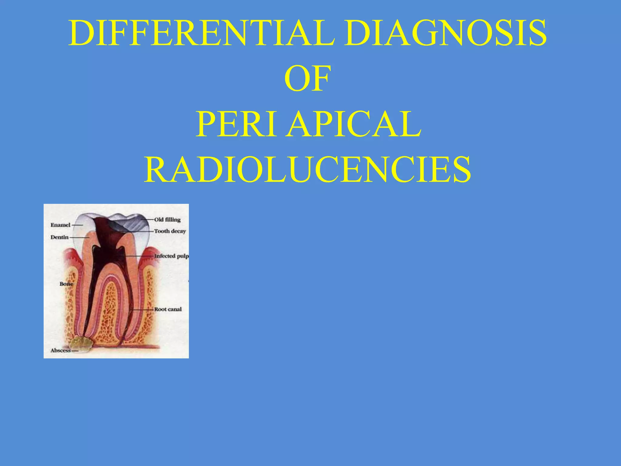
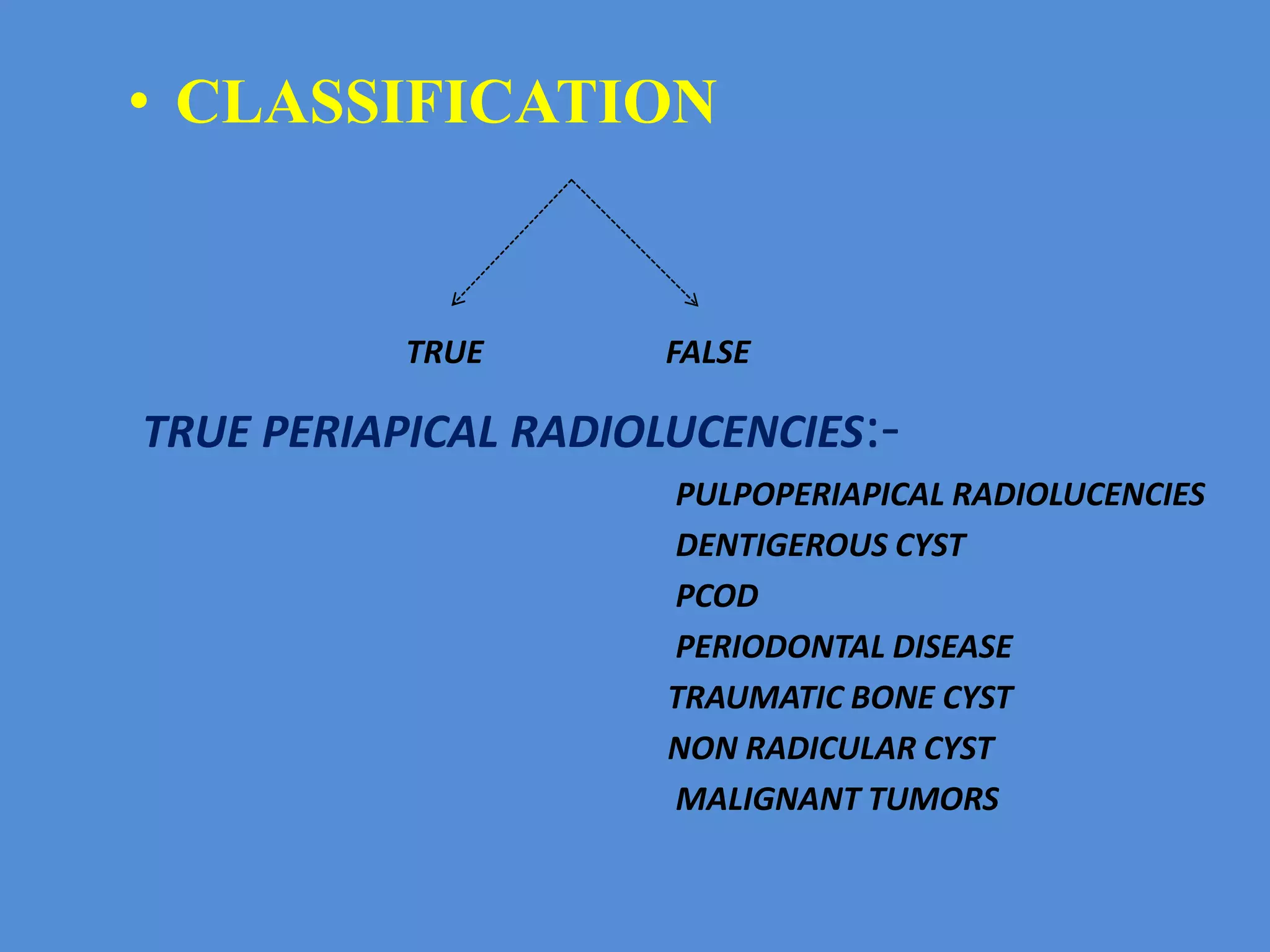
4. ANATOMIC
RADIOLUCENCIES
• False periapical radiolucencies are produced by
anatomic varients that do not contact the apex
of the tooth.
• These radiolucencies may be shifted from the
periapex by taking additional periapical
radiographs at different angle.
• If radiolucencies are anatomic in origin,a
comparision with the radiographs of the
opposite side frequently reveals an identical
situation.
10. APICAL PERIODONTITIS
• Apical periodontitis is the inflammation of
the periodontal ligament around the root
apex.
• It is of 2 types :-
Acute
Chronic
11. ACUTE APICAL
PERIODONTITIS
• Definition:-Acute apical periodontitis is a painful inflammation of
periodontium as a result of trauma, irritation [or] infection through root
canal regardless of whether pulp is vital or non-vital.
• ETIOLOGY:-
In a vital tooth:
Occlusal trauma
Wedging of foreign object b/w the teeth
Blow to the teeth
12. • In a non-vital tooth:
As a sequlae to pulpitis
Iotrogenic
Forcing of medicaments
Extension of obturating material
Over instrumentation during cleaning &
shaping
13. Clinical features
pain
Tooth is slightly elevated from the socket
Tenderness on percussion
Tooth may be slightly sore or may become
sore on percussion
Thermal changes does not induce pain.
16. PERI APICAL GRANULOMA
• Most common type of pathologic radiolucency
C/F :-
• Tooth is non vital
• It sounds dull on percussion due to granulation tissue at the
apex.
• Pt complains of mild pain on chewing.
• R/F
• Well circumscribed rl surrounding apex
• Involved tooth may reveal deep rest`ns extensive caries.
• Swelling or expansion of cortical plates is unusual.
17.
18. Differential diagnosis
Radicular cyst:- Cyst is larger than
granuloma but it is may not always right.
If radiolucency is1.6cm or more it is more
likely to be cyst.
Surgical defects:-previous history should
taken
PCOD:-pulp is vital & frequently involves
lower anteriors
TRAUMATIC BONE CYST:-pulp vital,
mostly seen in lower posteriors,LD intact
20. ABSCESS
• Abscess is an localised collection of pus
surrounded by an area inflammed tissue in
which hyperemia & infiltration of leucocytes
is mark
• ETIOLOGY:- trauma
• chemical or mechanical irritation
• pulpal infection
24. RADIOGRAPHIC FEATURES
• LOCATION:- present at the apex of
involved tooth
• PHERIPHERY:-ill defined
• INTERNAL STRUCTURE:-radiolucent
• SURROUNDING STRUCTURES:-loss of
LD in the peri apical region
28. PERI APICAL CYST
CLINICAL FEATURES:-
usually asymptomatic
mostly seen in maxillary incisors
if large produces swelling
29.
30. RADIOGRAPHIC FEATURES
• LOCATION:-Presents at apex of tooth
• PHERIPHERY & SHAPE:-well defined
pheriphery with cortical border, outline is
curved or circular
• INTERNAL STRUCTURE:-Radiolucent
• EFFECT ON SURROUNDING
STRUTURES:-If cyst is large,displacement &
resorption of adjacent tooth may occur
33. MANAGEMENT
• ROOT CANAL THERAPY
• EXTRACTION
• FOR LARGE CYST WHERE BONE DESTROYED
1.surical ennucleation
2. surgical ennucleation & restoration of defect
with graft
3. marsupilization
4. Decompression
5.decompression with delayed ennucleation
6.creation of a common chamber with maxillary
sinus or nasal cavity
34.
35. PERI APICAL SCAR
• Peri apical scar is a dense fibrous tissue
situated at the periapex of non vital tooth.
Features :-
well circumscribed radiolucency i.e.,
more or less round resembles
granuloma/cyst & it is usually small.
mostly in anterior of maxilla.
rl remains constant in size/ shrink
slightly.
36.
37.
38. 5.SURGICAL DEFECT
It is an area that fails to fill in with osseous tissue after surgery.
Seen periapically after root resection procedures when both
labial & lingual plates have been destroyed.
Mucosal scar due to previous surgery.
R/F
Usually round in app, smoothly contoured,well defined
borders.
Rl not more than 1cm in diameter.
D/D
SCAR
39.
40. OSTEOMYELITIS
• Defined as inflammation of bone & marrow
components.
• Streptococci, staphy.aureus, staphy.albus &
anaerobes like bacteroides, prevotella.
Predisposing factors:-
Fractures due to trauma.
Road traffic accidents.
Gun shot wounds &Radiation damage
Pagets disease & osteopetrosis
Sys cond. Leukemia,malnutn,diabetes
41. • Clinical features:-
30 to 80 yrs.
Mostly seen in mandible.
Tooth is non-vital may be associated
with acute/chronic periapical abscess.
Sinus is seen mucosa & skin.
42. RADIOGRAPHIC FEATURES
• LOCATION:-Post.body of mandible.
• Periphery & shape ;-Irregularly shaped with
poor or ragged borders.
• Internal struc:-Radiolucent.
• Effect on surrounding struc:-LD lost.
Can stimulate either resorption /
formation of bone.
46. HYPERPLASIA OF MAXILLARY
SINUS LINING
• It appear as grey shadows that may be dome
shaped in maxillary sinus floor
• Radicular cyst can pouch into the sinus &
may show a thin curved radioopaque rim of
bone seperating the cyst from the sinus
cavity
47. DENTIGEROUS CYST
• It is an odontogenic cyst assosiated with
crown of unerupted tooth
CLINICAL FEATURES:-
Clinical examination reveals a
missing tooth & a hard swelling results in
facial asymmetry
48. RADIOGRAPHIC FEATURES
• LOCATION:-Present above the crown of
involved tooth
• PHERIPHERY:-Well defined
• INTERNAL STRUCTURE:-Radiolucent
except for crown of unerupted
• SURROUNDING STRUCTURES:-Can
displace & resorb the adjacent teeth
52. MANAGEMENT
• Smaller lesions can surgically removed
• Larger lesions –insertion of surgical drain or
marsupilization
53. PERIAPICAL CEMENTO OSSEOUS
DYSPLASIA
• SYNONYMS:- Sclerosing cementum
Periapical osteo fibrosis
Fibrocementoma
Periapical fibrosarcoma
ETIOLOGY:- Trauma or Local irritation
54. • CLINICAL FEATURES:-
. Mostly present in mandibular anterior
region
. No history of pain/sensitivity
. Occasionally lesion near the mental foramen
and impinge on mental nerve & produces
pain /parasthesia /even anaesthsia
.Tooth have vital pulp
55. RADIOGRAPHIC FEATURES
• LOCATION:-Apex of the tooth
• PHERIPHERY:-Well defined
• INTERNAL STRUTURE:-Radiolucent
surrounded by hyperostotic border
• Loss of lamina dura
58. TRAUMATIC BONE CYST
• SYNONYMS:- Solitory cone cyst
Hemorrhagic cyst
Extravasation cyst
Unicameral bone cyst
Simple bone cyst
Idiopathic bone cyst
ETIOLOGY:- Trauma
59. • CLINICAL FEAATURES:-
Mostly seen in young persons
More male predilection
Present mostly in posterior mandible
Occasional tender on percussion
60. RADIOGRAPHIC FEATURES
• LOCATION:-mandible posterior part
• PHERIPHERY:-well defined delicate cortex
to ill defined border that blends into
surrrouding structure
• INTERNAL STRUCTURE:-total radiolucent
• SURROUNDING STRUTURE:-sometimes
root resorption & displacement may present
67. • FEATURES:-
More common in middle & old age
May be pain
Involve may retain their vitaliity
Advance cases :-tooth migration, loosening ,
tipping, spreading
Gingival bleeding may also present
paresthesia/anesthesia of the soft tissues
Expansion of jaw in advanced cases
68. RADIOGRAPHIC FEATURES
• Well defined or poorly defined radiolucency
or a large ragged well defined radiolucent
tumor
• Root resorption & band like widening of
periodontal ligament space
69.
70.
71. MANAGEMENT:-
Proper diagnosis has to be done to
treat the affected tooth .
Extensive management is
recommended if microscopic study of
periapical tissue after root resection is
diagnosed as malignancy.