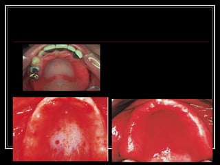
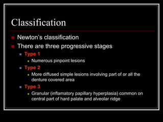
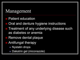
Denture Induced
Stomatitis
Denture Induced Stomatitis
 Also known as
 Denture stomatits
 Denture sore mouth
 Chronic atrophic candidiasis
Introduction
 Mild erythema or redness of the mucosa
under the denture.
 Usually occurs in the maxilla
 Common in complete denture wearers
 Incidence
 Age: middle to old age
 More common in females
 Has been found in up to 70% of denture wearers
Predisposing Factors
 Wearing dentures at night
 Dry mouth
 Diabetes
 Increased carbohydrate diet
 HIV
Etiology
 Changes in the oral environment related to
presence of dentures or removable
orthodontic appliance
 Poor oral and denture hygiene
 Candida is the main cause (70%)
 Can also occur because of mechanical
irritation and bacterial infections
Pathogenesis
Accumulation of plaque colonization
by candida albicans increased
candidal enzymatic activity due to
salivary flow and Ph
inflammation
Clinical features
 Marked redness or erythema and odema of
the mucosa in contact with dentures
 Restricted to the denture bearing area often
with a sharply defined edge
 Patient has no complaints of pain
 Uncommon complications
 Angular stomatits
 Papillary hyperplasia in palatal vault
Classification
 Newton’s classification
 There are three progressive stages
 Type 1
 Numerous pinpoint lesions
 Type 2
 More diffused simple lesions involving part of or all the
denture covered area
 Type 3
 Granular (inflamatory papillary hyperplasia) common on
central part of hard palate and alveolar ridge
Diagnosis
 Depends on clinical findings
 In presence of angular stomatitis or other
systemic lesions further investigations are
required
 Blood picture
 Smears and culture
 Biopsy in persistent or atypical lesions and
 HIV serology
Management
 Patient education
 Oral and denture hygiene instructions
 Treatment of any underlying disease such
as diabetes or anemia
 Remove dental plaque
 Antifungal therapy
 Nystatin drops
 Dakatrin gel (miconazole)

Denture Stomatitis

  • 1.
  • 2.
    Denture Induced Stomatitis Also known as  Denture stomatits  Denture sore mouth  Chronic atrophic candidiasis
  • 3.
    Introduction  Mild erythemaor redness of the mucosa under the denture.  Usually occurs in the maxilla  Common in complete denture wearers  Incidence  Age: middle to old age  More common in females  Has been found in up to 70% of denture wearers
  • 4.
    Predisposing Factors  Wearingdentures at night  Dry mouth  Diabetes  Increased carbohydrate diet  HIV
  • 5.
    Etiology  Changes inthe oral environment related to presence of dentures or removable orthodontic appliance  Poor oral and denture hygiene  Candida is the main cause (70%)  Can also occur because of mechanical irritation and bacterial infections
  • 6.
    Pathogenesis Accumulation of plaquecolonization by candida albicans increased candidal enzymatic activity due to salivary flow and Ph inflammation
  • 7.
    Clinical features  Markedredness or erythema and odema of the mucosa in contact with dentures  Restricted to the denture bearing area often with a sharply defined edge  Patient has no complaints of pain  Uncommon complications  Angular stomatits  Papillary hyperplasia in palatal vault
  • 9.
    Classification  Newton’s classification There are three progressive stages  Type 1  Numerous pinpoint lesions  Type 2  More diffused simple lesions involving part of or all the denture covered area  Type 3  Granular (inflamatory papillary hyperplasia) common on central part of hard palate and alveolar ridge
  • 10.
    Diagnosis  Depends onclinical findings  In presence of angular stomatitis or other systemic lesions further investigations are required  Blood picture  Smears and culture  Biopsy in persistent or atypical lesions and  HIV serology
  • 11.
    Management  Patient education Oral and denture hygiene instructions  Treatment of any underlying disease such as diabetes or anemia  Remove dental plaque  Antifungal therapy  Nystatin drops  Dakatrin gel (miconazole)