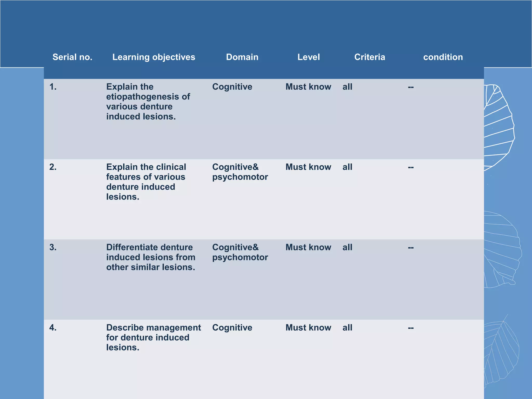
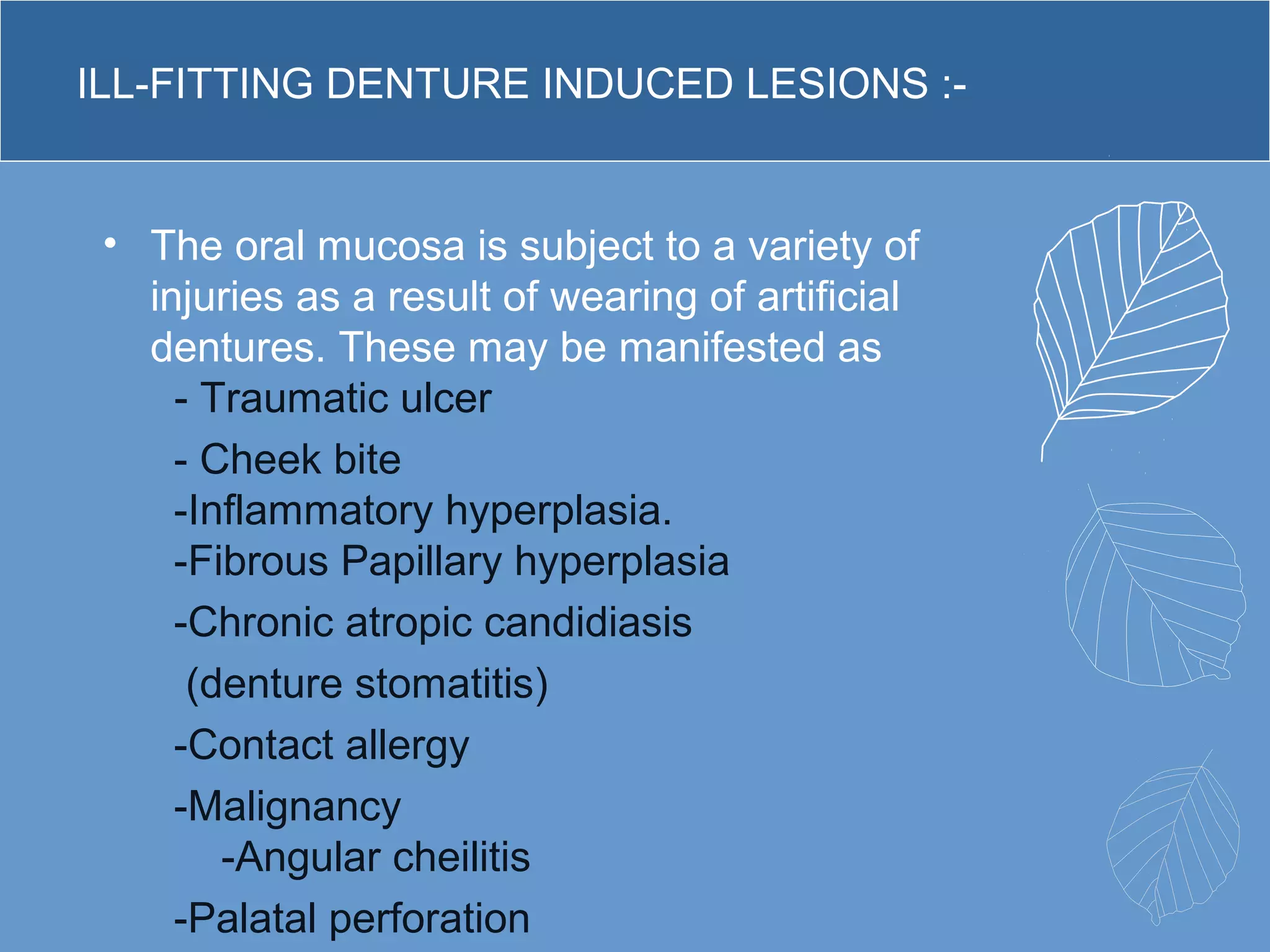
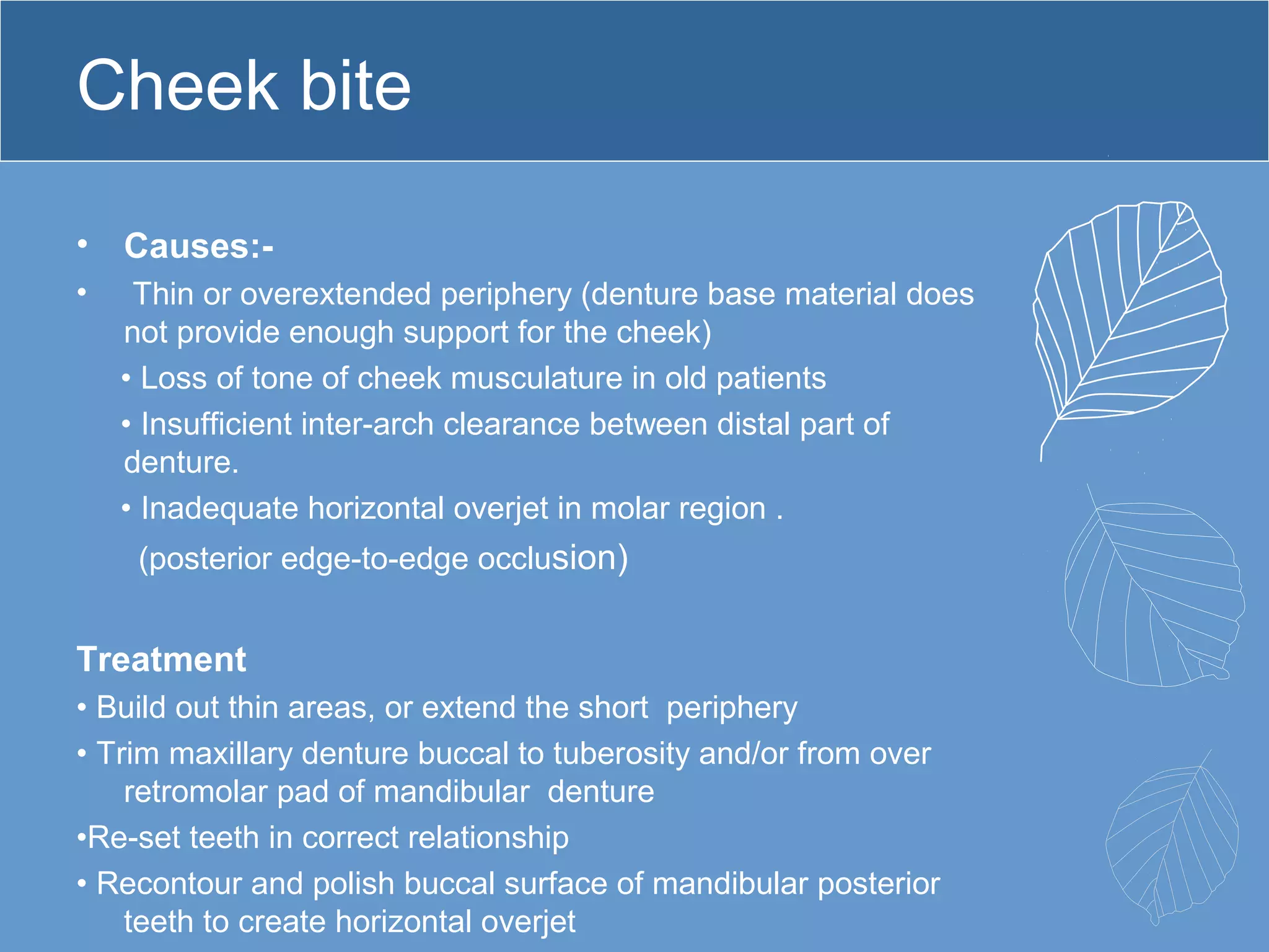
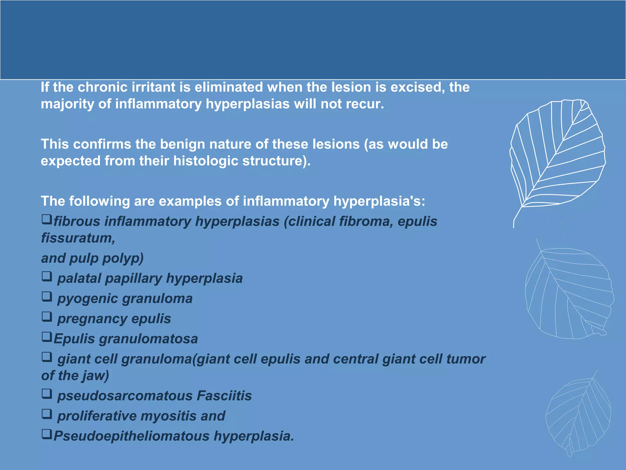
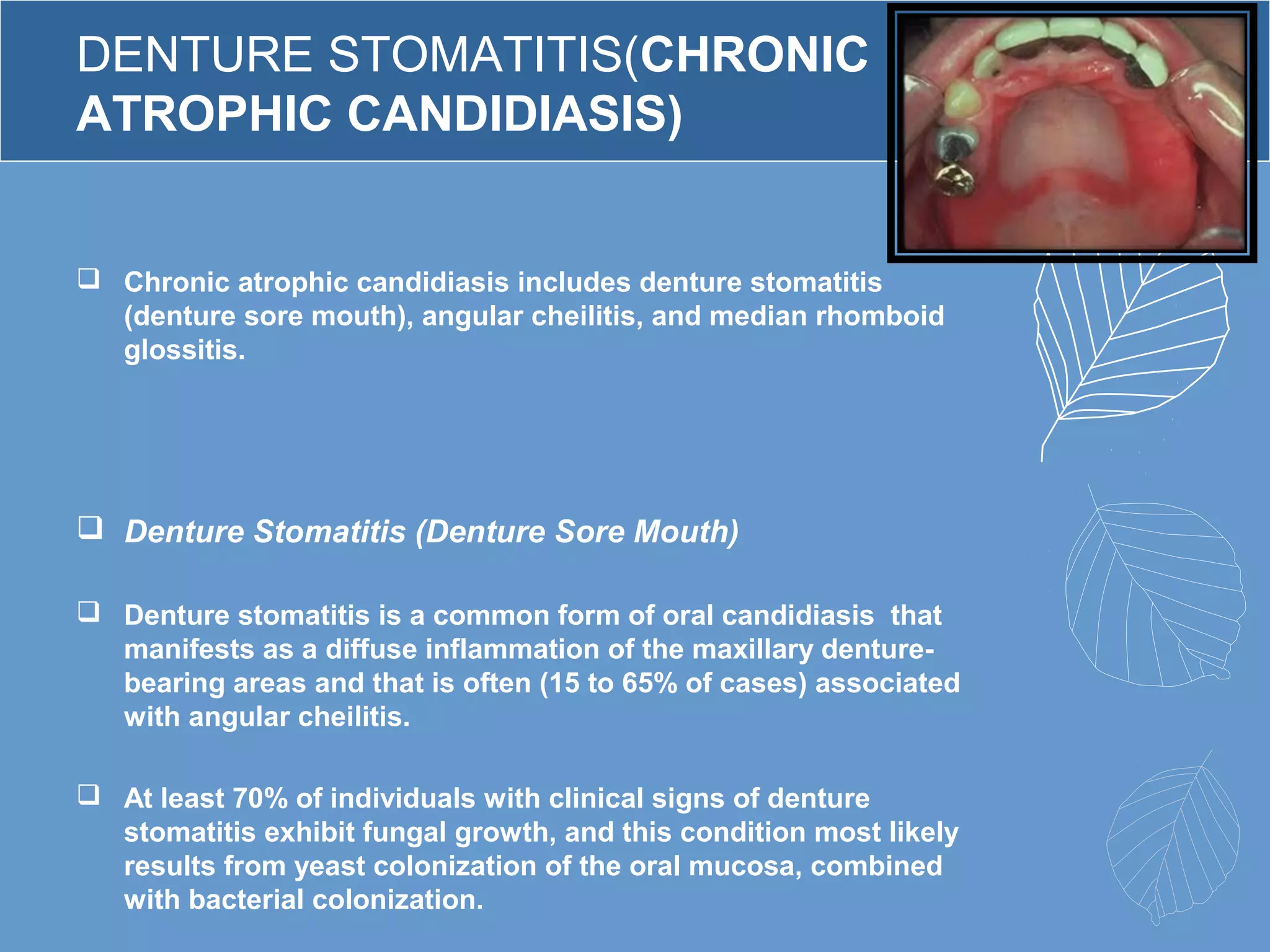
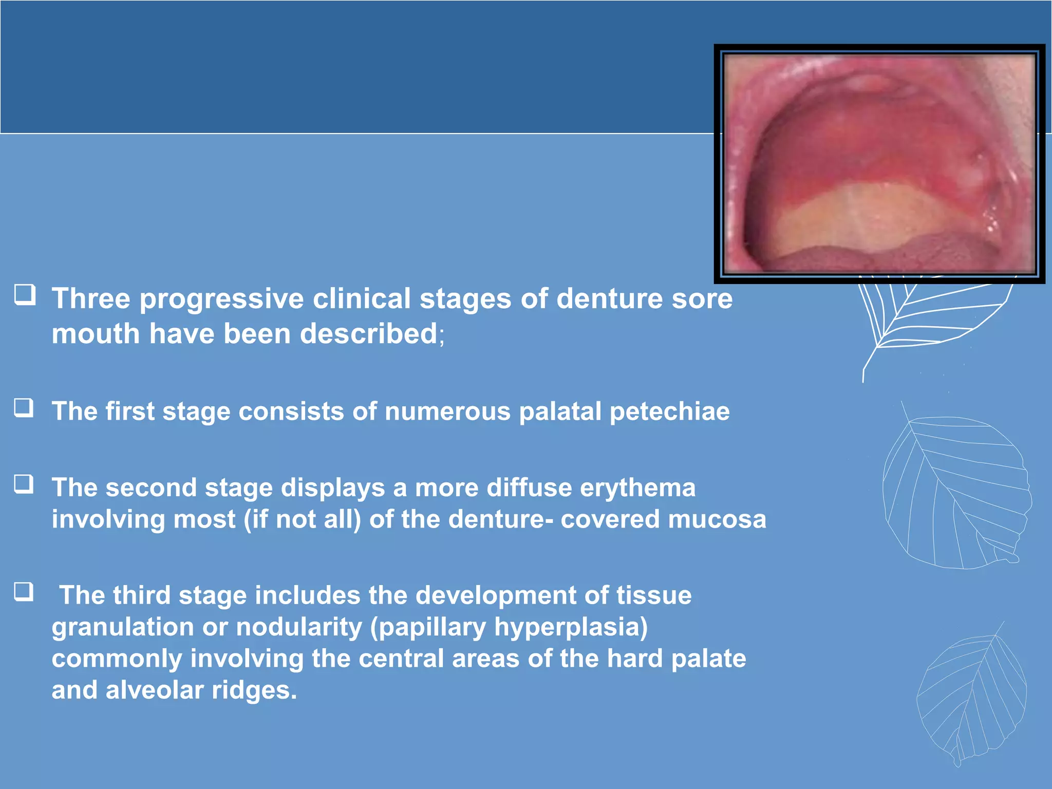
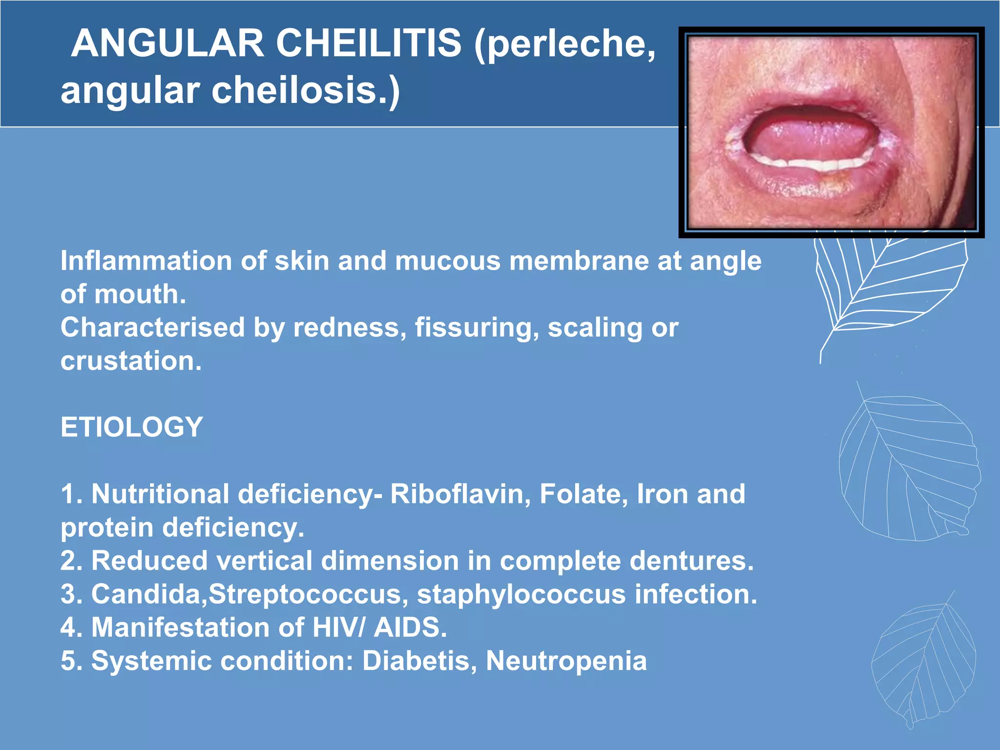
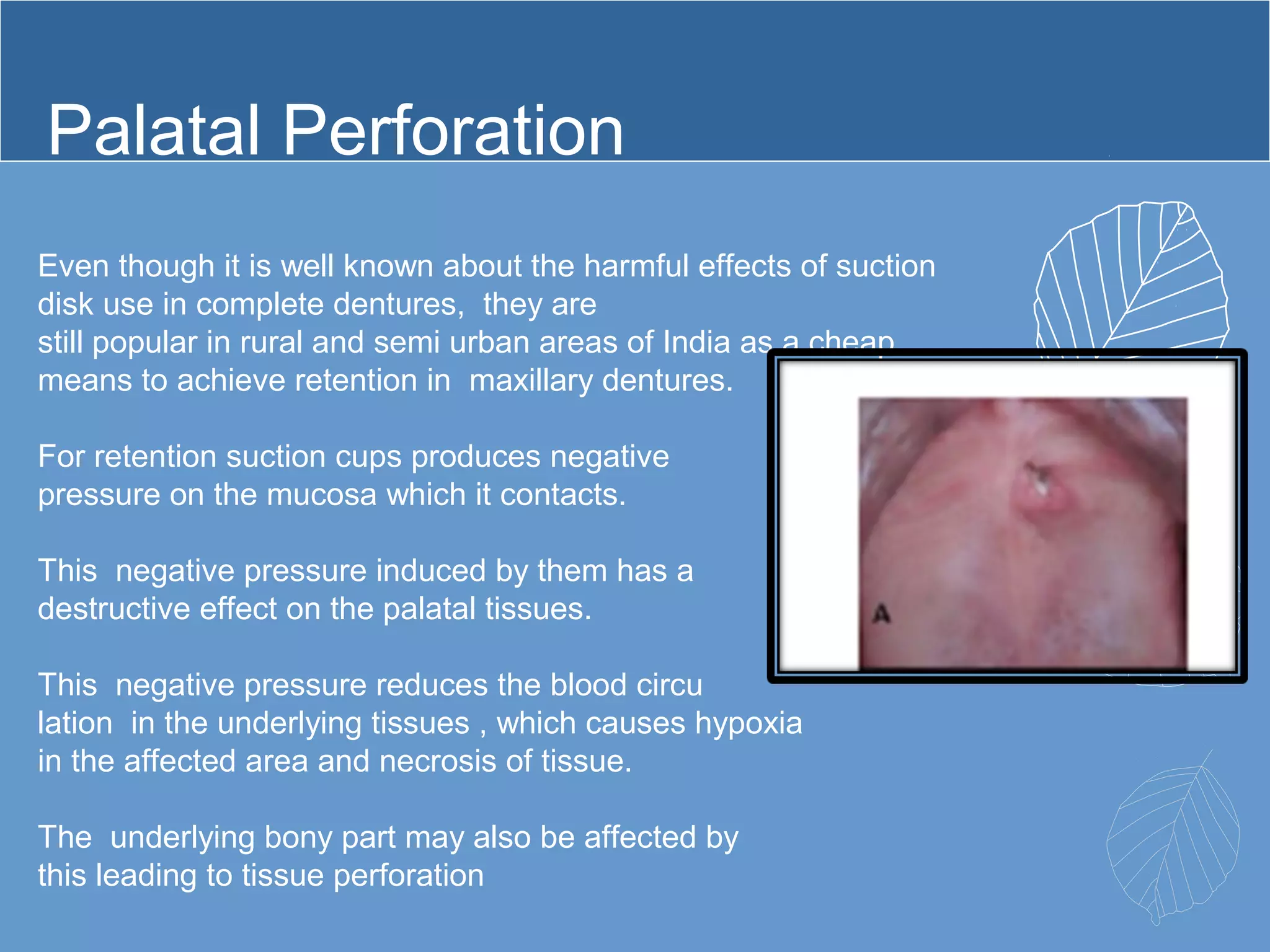
SlideShare a Scribd company logo
SEMINAR
ILL- FITTING
DENTURE
INDUCED
LESIONS
PRESENTED BY :-
AARTI DUBEY
GUIDED BY :-
DR S.B. DANGORE
DR S.S. DEGWEKAR
DR R.R. BHOWATE
PURPOSE STATEMENT
• At the end of the presentation the
learners should be able to
describe etiopathogenesis, clinical
features, management &
differential diagnosis of various
denture induced lesions.
Serial no. Learning objectives Domain Level Criteria condition
1. Explain the
etiopathogenesis of
various denture
induced lesions.
Cognitive Must know all --
2. Explain the clinical
features of various
denture induced
lesions.
Cognitive&
psychomotor
Must know all --
3. Differentiate denture
induced lesions from
other similar lesions.
Cognitive&
psychomotor
Must know all --
4. Describe management
for denture induced
lesions.
Cognitive Must know all --
ILL-FITTING DENTURE INDUCED LESIONS :-
• The oral mucosa is subject to a variety of
injuries as a result of wearing of artificial
dentures. These may be manifested as
- Traumatic ulcer
- Cheek bite
-Inflammatory hyperplasia.
-Fibrous Papillary hyperplasia
-Chronic atropic candidiasis
(denture stomatitis)
-Contact allergy
-Malignancy
-Angular cheilitis
-Palatal perforation
1.TRAUMATIC ULCER ( sore spots)
Clinical Features
1. Develops within a day or two after the insertion of a new denture.
2. Due to over-extension of the flanges, sequestration or spicules of
bone under the denture or roughened or “high” spot on the inner
surface of the denture.
3. These are small, painful, irregularly shaped lesions covered by a
delicate grey necrotic membrane and surrounded by an inflammatory
halo.
Management
1. Correction of underlying cause.
2. Relief of the flanges.
4. Relief of high spots
Cheek bite
• Causes:-
• Thin or overextended periphery (denture base material does
not provide enough support for the cheek)
• Loss of tone of cheek musculature in old patients
• Insufficient inter-arch clearance between distal part of
denture.
• Inadequate horizontal overjet in molar region .
(posterior edge-to-edge occlusion)
Treatment
• Build out thin areas, or extend the short periphery
• Trim maxillary denture buccal to tuberosity and/or from over
retromolar pad of mandibular denture
•Re-set teeth in correct relationship
• Recontour and polish buccal surface of mandibular posterior
teeth to create horizontal overjet
INFLAMMATORY HYPERPLASIA
 The term “inflammatory hyperplasia” is used to describe a large
range of commonly occurring nodular growths of the oral
mucosa that histologically represent inflamed fibrous and
granulation tissues.
 The major etiologic factor for these lesions is generally assumed
to be chronic trauma (such as that produced by ill-fitting
dentures, calculus, overhanging dental restorations, acute or
chronic tissue injury from biting, and fractured teeth), and
chronic irritants can be convincingly demonstrated in many
cases (eg, palatal papillary hyperplasia associated with aged
maxillary dentures).
 With some of these lesions, (eg, pregnancy epulis and the central
giant cell tumor associated with hyperparathyroidism), the levels
of circulating hormones also undoubtedly play a role.
 The majority of lesions occur on the surface of the oral mucous
membrane, where irritants are quite common.
If the chronic irritant is eliminated when the lesion is excised, the
majority of inflammatory hyperplasias will not recur.
This confirms the benign nature of these lesions (as would be
expected from their histologic structure).
The following are examples of inflammatory hyperplasia's:
fibrous inflammatory hyperplasias (clinical fibroma, epulis
fissuratum,
and pulp polyp)
 palatal papillary hyperplasia
 pyogenic granuloma
 pregnancy epulis
Epulis granulomatosa
 giant cell granuloma(giant cell epulis and central giant cell tumor
of the jaw)
 pseudosarcomatous Fasciitis
 proliferative myositis and
Pseudoepitheliomatous hyperplasia.
Fibrous Inflammatory Hyperplasias
and Traumatic Fibromas
 Fibrous inflammatory hyperplasias may occur as either
pedunculated or sessile (broad-based) growths on any
surface of the oral mucous membrane.
 They are called fibromas if they are sessile, firm, and
covered by thin squamous epithelium.
 On the gingiva, a similar lesion is often referred to as
an epulis.
 The majority remain small, and lesions that are > 1 cm
in diameter are rare.
 An exception to this rule occurs with a lesion that is
associated with the periphery of illfitting dentures, the so-
called epulis fissuratum, in which the growth is often split
by the edge of the denture, one part of the lesion lying
under the denture and the other part lying between the lip
or cheek and the outer denture surface.
 This lesion may extend the full length of one side of the
denture. Many such hyperplastic growths will become
less edematous and inflamed following the removal of the
associated chronic irritant, but they rarely resolve
entirely.
 In the preparation of the mouth to receive dentures, these
lesions are excised to prevent further irritation and to
ensure a soft-tissue seal for the denture periphery.
 The differential diagnosis of fibrous inflammatory
hyperplasia should include consideration of the
possibility that the lesion is a true papilloma (a
cauliflower-like mass made up of multiple fingerlike
projections of stratified squamous epithelium with a
central core of vascular connective tissue) or a small
verrucous carcinoma.
 Fibrous inflammatory hyperplasias have no malignant
potential, and recurrences following excision are almost
always a result of the failure to eliminate the particular
form of chronic irritation involved.
 The occasional report of squamous cell carcinoma
arising in an area of chronic denture irritation, however,
underlines the fact that no oral growth, even those
associated with an obvious chronic irritant, can be
assumed to be benign until proven so by histologic
study.
 Thus,whenever possible, all fibrous inflammatory
hyperplasias of the oral cavity should be treated by local
excision, with microscopic examination of the excised
tissue.
EPULIS FISSURATUM ( inflammatory
Fibrous Hyperplassia, Denture Injury tumour,
Denture epulis.)
-It is a tumour like
hyperplasia of fibrous
connective tissue.
-Develops in association
with the flange of ill fitting
complete or partial
dentures.
CLINICAL FEATURES
-Appears as single or multiple folds of hyperplastic tissue
in the alveolar vestibule.
-Flange of denture fits in to the fissure between these
folds.
-Tissue is firm and fibrous.
-Some lesions appear erythematous and ulcerated.
-Size of lesion vary from localized hyperplasia's less than
1 cm in size to massive lesions involve the entire length
of vestibule.
-Anterior portion of jaws is affected more.
-Female predilection.
-Another lesion called fibro epithelial polyp or
leaf like denture fibroma occurs on hard
palate beneath maxillary denture.
-It is a flattened pink mass attached to palate
by narrow stalk.
-It can easily lifted up with a probe
demonstrate its pedunculated nature.
-Edge of lesion is serrated and resembles a
leaf.
-If minor salivary glands are included they
show chronic sialadenitis.
-Unusual appearing products known as
osseous and chondromatous metaplasia is a
reactive phenomenon caused by chronic
irritation by the ill fitting denture.
MANAGEMENT
1. Surgical removal.
2. Ill fitting denture should be remade or
relined.
• The frequency of occurrence of epulis
fissuratum far exceeds that of any other
exophytic lesion at the periphery of dentures.
• However the possibility of malignancy must be
considered in each case: squamous and
verrucous carcinomas, minor salivary gland
tumours, metastatic tumours, osteosarcoma,
and down reaching maxillary sinus
malignancies.
PALATAL PAPILLARY HYPERPLASIA
 Palatal papillary hyperplasia (denture papillomatosis) is a
common lesion with a characteristic clinical appearance that
develops on the hard palate in response to chronic denture
irritation in approximately 3 to 4% of denture wearers.
 Full dentures in which relief areas or “suction chambers” are cut
in the palatal seating surface appear to be the strongest stimuli,
but the lesion is also seen under partial dentures, and occasional
case reports have described the lesion in patients who have
never worn dentures.
 The palatal lesion is usually associated with some degree of
denture sore mouth (stomatitis) due to chronic candidal
infection, which influences the appearance of the papillary
hyperplasia.
 When complicated by candidal infection, the lesion may
be red to scarlet, and the swollen and tightly packed
projections resemble the surface of an overripe berry.
Such lesions are friable, often bleed with minimal trauma,
and may be covered with a thin whitish exudate.
 When the candidal infection is eliminated, either by
removing the denture or by topical administration of an
antifungal agent, the papillary lesion becomes little
different in color from the rest of the palate and consists
of more or less tightly packed nodular projections.
 If tiny, the nodular projections simply give a feltlike
texture to that portion of the palate, and the lesion may
even pass unnoticed unless it is stroked with an
instrument or disturbed by a jet of air.
 It was first classified by Newton (1962) according to its
clinical appearance as:
Type 1: A localized simple inflammation or pinpoint
hyperaemia
Type 2: An erythematous or generalized simple type seen as
more diffuse erythema involving a part or the entire denture
covered mucosa
Type 3: A granular type (inflammatory papillary hyperplasia)
commonly involving the central part of the hard palate and
the alveolar ridges
• Type III often is seen in association with type I or type II.
• Type III denture stomatitis involves the epithelial response
to chronic inflammatory stimulation secondary to yeast
 Despite their sometimes bizarre clinical
appearance, these lesions have almost no
neoplastic potential, a finding that is borne
out by the absence of atypia and cellular
dysplasia in biopsy specimens.
 If the alveolar ridges are surgically
prepared for new dentures, papillary
hyperplasia lesions are usually excised or
removed (by electrocautery, cryosurgery,
or laser surgery), and the old denture or a
palatal splint is used to maintain a
postoperative surgical dressing over the
denuded area.
 If florid papillomatosis of the palate occurs
or persists in the absence of dentures, the
differential diagnosis should also consider
several granulomatous diseases that may
manifest intraorally in this fashion (eg,
infectious granulomas,Cowden disease,
and verrucous carcinoma), particularly
when the papillary lesions are white and
extend beyond the palatal vault and onto
the alveolar mucosa.
• Nicotine stomatitis may also feature multiple small
nodules on palate, which are reddish before
hyperkeratosis develop.
• The following observations help in the differentiation of
this condition from IPH;
1. Nicotine stomatitis on the hard palate occurs almost
exclusively in pipe smokers who do not wear full
maxillary dentures.
2. The pattern in nicotine stomatitis is linear and
angular, and the segments are flatter and broader but
less elevated.
3. The segments in nicotine stomatitis have a
characteristic red dot in their approximate center, which
is not seen in PHP.
DENTURE STOMATITIS(CHRONIC
ATROPHIC CANDIDIASIS)
 Chronic atrophic candidiasis includes denture stomatitis
(denture sore mouth), angular cheilitis, and median rhomboid
glossitis.
 Denture Stomatitis (Denture Sore Mouth)
 Denture stomatitis is a common form of oral candidiasis that
manifests as a diffuse inflammation of the maxillary denture-
bearing areas and that is often (15 to 65% of cases) associated
with angular cheilitis.
 At least 70% of individuals with clinical signs of denture
stomatitis exhibit fungal growth, and this condition most likely
results from yeast colonization of the oral mucosa, combined
with bacterial colonization.
 Three progressive clinical stages of denture sore
mouth have been described;
 The first stage consists of numerous palatal petechiae
 The second stage displays a more diffuse erythema
involving most (if not all) of the denture- covered mucosa
 The third stage includes the development of tissue
granulation or nodularity (papillary hyperplasia)
commonly involving the central areas of the hard palate
and alveolar ridges.
 Antifungal treatment will modify the bright red
appearance of denture sore mouth and papillary
hyperplasia specifically but will not resolve the basic
papillomatous lesion, especially if the lesions have been
present for more than 1 year.
 Antifungal therapy and cessation of denture wearing
usually is advisable before surgical excision since
elimination of the mucosal inflammation often reduces
the amount of tissue that needs to be excised.
 Yeast attached to the denture plays an important etiologic
role in chronic atrophic candidiasis
 The attachment of yeast to the patient’s appliances is
increased by mucus and serum and decreased by the
presence of salivary pellicle, suggesting an explanation
for the severity of candidiasis in xerostomic patients.
 Rinsing the appliance with a dilute (10%) solution of
household bleach, soaking it in boric acid, or applying
nystatin cream before inserting the denture will eliminate
the yeast.
 Disinfection of the appliance is an important part of the
treatment of denture sore mouth.
 Soft liners in dentures provide a porous
surface and an opportunity for additional
mechanical locking of plaque and yeast to
the appliance.
 In general, soft liners are considered to be
an additional hazard for patients who are
susceptible to oral candidiasis.
 Denture sore mouth is rarely found under a
mandibular denture.
 One possible explanation for this is that
the negative pressure that forms under the
maxillary denture excludes salivary
antibody from this region, and yeast may
reproduce, undisturbed, in the space
between the denture and mucosa.
 The closer adaptation of the maxillary
denture and palate may also bring the large
number of yeasts adhering to the denture
surface into contact with the mucosa.
• The clinical picture of denture
stomatits is rather specific; few if
any other diseases appear the same.
• Infections by other organisms,
however could be responsible for a
similar diffuse redness either alone
or I association with candida.
• Contact allergy to denture base
occurs occasionally.
• In such cases redness will not be
restricted to tissue under the
denture, but all mucosal surfaces in
contact with the acrylic will be red.
• Epicutaneous tests of the material
will be diagnostic .
ANGULAR CHEILITIS (perleche,
angular cheilosis.)
Inflammation of skin and mucous membrane at angle
of mouth.
Characterised by redness, fissuring, scaling or
crustation.
ETIOLOGY
1. Nutritional deficiency- Riboflavin, Folate, Iron and
protein deficiency.
2. Reduced vertical dimension in complete dentures.
3. Candida,Streptococcus, staphylococcus infection.
4. Manifestation of HIV/ AIDS.
5. Systemic condition: Diabetis, Neutropenia
 CLINICAL FEATURES
-Occurs as an area of redness, linear deep
fissure, peeling of skin, dry crust at the
angle of mouth.
- Symptomatic.
-Pain and bleeding at site.
-Difficulty in mouth opening.
-Occurs bilaterally.
-RHAGADES :- linear furrow or fissures
radiating from the angle of mouth are seen
in severe forms, especially in denture
wearers.
 MANAGEMENT
1. Removal of the cause.
2. Nutritional supplement:- Vitamin B and
Iron supplements.
3. Ketoconazole: 200mg tab once daily for
1-2 weeks or
Fluconazole: 100mg tab once daily for 1-2
weeks.
4. For severe candidiasis - Fluconazole is
drug of choice.
Palatal Perforation
Even though it is well known about the harmful effects of suction
disk use in complete dentures, they are
still popular in rural and semi urban areas of India as a cheap
means to achieve retention in maxillary dentures.
For retention suction cups produces negative
pressure on the mucosa which it contacts.
This negative pressure induced by them has a
destructive effect on the palatal tissues.
This negative pressure reduces the blood circu
lation in the underlying tissues , which causes hypoxia
in the affected area and necrosis of tissue.
The underlying bony part may also be affected by
this leading to tissue perforation
• These pathological changes are more severe if the
patient is in the habit of continuously wearing the
dentures 24 hrs a day.
TREATMENT:-
• surgically close the opening and allow complete
healing using a healing plate and followed by fabrication
of new complete denture.
Precautions to be taken by the
Dentist to avoid these problems
• There should be adequate relief .
• There should be no overextension of the
lingual flange into the lateral throat form.
• The vertical dimension must be accurate.
• There shouldn’t be any deflective occlusal
contacts.
• The denture borders should not be
overextended.
• There should be sufficient horizontal overlap of
posterior teeth.
• Note that the borders are rounded and
smooth, and the palate is highly polished and
the proper thickness.
CONCLUSION
Proper denture care is important for both the
health of dentures and patients mouth.
Handle dentures with great care.
To avoid accidentally dropping them,
stand over a folded towel or a full
sink of water when handling
dentures.
Brush and rinse dentures daily:-
•Like natural teeth, dentures must be brushed daily to remove
food and plaque.
• Brushing also helps prevent the development of permanent
stains on the dentures.
•Use a brush with soft bristles that is specifically designed for
cleaning dentures.
•Avoid using a hard-bristled brush as it can damage or wear down
dentures.
•Gently brush all surfaces of the denture and be careful not to
damage the plastic or bend attachments.
• In between brushings, rinse dentures after every meal.
Denture care when not being worn:-
•Dentures need to be kept moist when not being worn so
they do not dry out or lose their shape.
•When not worn, dentures should be placed in a denture
cleanser soaking solution or in water.
•However, if the denture has metal attachments, the
attachments could tarnish if placed in a soaking solution.
•Dentures should never be placed in hot water, as it can
cause them to warp.
Clean with a denture cleaner:-
• Hand soap or mild dishwashing liquid can be used for
cleaning dentures.
• Household cleansers and many toothpastes may be too
abrasive for dentures and should not be used.
•Also, avoid using bleach, as this may whiten the pink
portion of the denture.
•Ultrasonic cleaners can be used to care for dentures.
•These cleaners are small bathtub-like devices that contain
a cleaning solution.
• The denture is immersed in the tub and then
sound waves create a wave motion that
dislodges the undesirable deposits.
• Use of an ultrasonic cleaner, however, does
not replace a thorough daily brushing.
• Products with the American Dental Association
(ADA) Seal of Acceptance are recommended
since they have been evaluated for safety and
effectiveness.
REFERENCES
• MARTIN S. GREENBERG,MICHAEL GLICK; Burket’s oral medicine
Diagnosis & Treatment Tenth Edition
• Norman.k Wood, Paul.W.Goaz; DIFFERENTIAL DIAGNOSIS of
ORAL and MAXILLOFACIAL LESIONS;5th
edition
• B. C. Webb,C. J. Thomas et al; Candida-associated denture
stomatitis . Aetiology and management : A review. Part 2 Oral
diseases caused by candida species Australian Dental Journal
1998;43:(3):160-6
• Tatiana PEREIRA-CENCI, Altair Antoninha DEL BEL CURY, Wim
CRIELAAR, Jacob Martien TEN CATE DEVELOPMENT OF
CANDIDA-ASSOCIATED DENTURE STOMATITIS: NEW INSIGHTS;
J Appl Oral Sci. 2008;16(2):86-94
• Hrizdana Hadjieva, Mariana Dimova, S. Todorov; STOMATITIS
PROSTHETICA-A POLYETIOLOGIC DISORDER; Journal of IMAB -
Annual Proceeding (Scientific Papers) 2006, book 2
• GAUTAM BAGCHI, DEVENDRA MANDHYAN AND DILIP KUMAR
NATH; REHABILITATION OF A PATIENT WITH PALATAL PERFORATION DUE TO
PROLONGED USE OF DENTURE WITH SUCTION DISK: A CASE REPORT; (JRAAS)
27:62-64, 2012
THANKYOU!

More Related Content

What's hot

Abutment selection in FPD
Abutment selection in FPDAbutment selection in FPD
Abutment selection in FPD
Dr. Anshul Sahu
 
benign and malignant tumors of connective tissue origin
benign and malignant tumors of connective tissue originbenign and malignant tumors of connective tissue origin
benign and malignant tumors of connective tissue origin
madhusudhan reddy
 
Oral lesions associated with the use of tobacco
Oral lesions associated with the use of tobaccoOral lesions associated with the use of tobacco
Oral lesions associated with the use of tobacco
Mangai Yarkkarasi
 
White Lesions
White LesionsWhite Lesions
Non odontogenic cysts or fissural cysts
Non odontogenic cysts or fissural cystsNon odontogenic cysts or fissural cysts
Non odontogenic cysts or fissural cysts
madhusudhan reddy
 
Peridontal pocket
Peridontal pocketPeridontal pocket
Peridontal pocket
Parth Thakkar
 
Caries diagnosis
Caries diagnosisCaries diagnosis
Caries diagnosis
D Venkatesh Kumar
 
Management of Ellis Class IV Fracture
Management of Ellis Class IV FractureManagement of Ellis Class IV Fracture
Management of Ellis Class IV Fracture
Muskan Agarwal
 
Dentinogenesis Imperfecta
Dentinogenesis ImperfectaDentinogenesis Imperfecta
Dentinogenesis Imperfecta
shabeel pn
 
Major Connectors
Major ConnectorsMajor Connectors
Major Connectors
Apurva Thampi
 
Denture Stomatitis
Denture StomatitisDenture Stomatitis
Denture Stomatitis
Arsalan Wahid Malik
 
periapical pathologies- pulpal origin
periapical pathologies- pulpal originperiapical pathologies- pulpal origin
periapical pathologies- pulpal origin
Gurmeen Kaur
 
Measurement of Periodontal Attachment Loss
Measurement of Periodontal Attachment LossMeasurement of Periodontal Attachment Loss
Measurement of Periodontal Attachment Loss
Iraqi Dental Academy
 
Oral Lichen Planus (OLP)
Oral Lichen Planus (OLP)Oral Lichen Planus (OLP)
Oral Lichen Planus (OLP)
Ashish Dr.Angural
 
Odontogeniccysts OKC
Odontogeniccysts OKCOdontogeniccysts OKC
Odontogeniccysts OKC
Maryam Arbab
 
 Traumatic bone cyst
 Traumatic bone cyst Traumatic bone cyst
 Traumatic bone cyst
Amin Abusallamah
 
Sequelae of dental caries
Sequelae of dental cariesSequelae of dental caries
Sequelae of dental caries
Sushant Pandey
 
Oral mucosal lesions in denture wearers
Oral mucosal lesions in denture wearersOral mucosal lesions in denture wearers
Oral mucosal lesions in denture wearers
Aamir Godil
 
Pindborgs Tumour
Pindborgs TumourPindborgs Tumour
Pindborgs Tumour
Shravya Kishore
 
Endodontic Diagnosis: Pulp Vitality Tests
Endodontic Diagnosis: Pulp Vitality TestsEndodontic Diagnosis: Pulp Vitality Tests
Endodontic Diagnosis: Pulp Vitality Tests
Iraqi Dental Academy
 

What's hot (20)

Abutment selection in FPD
Abutment selection in FPDAbutment selection in FPD
Abutment selection in FPD
 
benign and malignant tumors of connective tissue origin
benign and malignant tumors of connective tissue originbenign and malignant tumors of connective tissue origin
benign and malignant tumors of connective tissue origin
 
Oral lesions associated with the use of tobacco
Oral lesions associated with the use of tobaccoOral lesions associated with the use of tobacco
Oral lesions associated with the use of tobacco
 
White Lesions
White LesionsWhite Lesions
White Lesions
 
Non odontogenic cysts or fissural cysts
Non odontogenic cysts or fissural cystsNon odontogenic cysts or fissural cysts
Non odontogenic cysts or fissural cysts
 
Peridontal pocket
Peridontal pocketPeridontal pocket
Peridontal pocket
 
Caries diagnosis
Caries diagnosisCaries diagnosis
Caries diagnosis
 
Management of Ellis Class IV Fracture
Management of Ellis Class IV FractureManagement of Ellis Class IV Fracture
Management of Ellis Class IV Fracture
 
Dentinogenesis Imperfecta
Dentinogenesis ImperfectaDentinogenesis Imperfecta
Dentinogenesis Imperfecta
 
Major Connectors
Major ConnectorsMajor Connectors
Major Connectors
 
Denture Stomatitis
Denture StomatitisDenture Stomatitis
Denture Stomatitis
 
periapical pathologies- pulpal origin
periapical pathologies- pulpal originperiapical pathologies- pulpal origin
periapical pathologies- pulpal origin
 
Measurement of Periodontal Attachment Loss
Measurement of Periodontal Attachment LossMeasurement of Periodontal Attachment Loss
Measurement of Periodontal Attachment Loss
 
Oral Lichen Planus (OLP)
Oral Lichen Planus (OLP)Oral Lichen Planus (OLP)
Oral Lichen Planus (OLP)
 
Odontogeniccysts OKC
Odontogeniccysts OKCOdontogeniccysts OKC
Odontogeniccysts OKC
 
 Traumatic bone cyst
 Traumatic bone cyst Traumatic bone cyst
 Traumatic bone cyst
 
Sequelae of dental caries
Sequelae of dental cariesSequelae of dental caries
Sequelae of dental caries
 
Oral mucosal lesions in denture wearers
Oral mucosal lesions in denture wearersOral mucosal lesions in denture wearers
Oral mucosal lesions in denture wearers
 
Pindborgs Tumour
Pindborgs TumourPindborgs Tumour
Pindborgs Tumour
 
Endodontic Diagnosis: Pulp Vitality Tests
Endodontic Diagnosis: Pulp Vitality TestsEndodontic Diagnosis: Pulp Vitality Tests
Endodontic Diagnosis: Pulp Vitality Tests
 

Viewers also liked

Mucosal Response To Oral Prostheses
Mucosal Response To Oral ProsthesesMucosal Response To Oral Prostheses
Mucosal Response To Oral Prostheses
Dr Aaron Sarwal
 
Hiperplasia
Hiperplasia Hiperplasia
Hiperplasia
Dvno Mkz
 
Lesiones hiperplasicas de tejido fibroso. rm m ayhuasca
Lesiones hiperplasicas de tejido fibroso. rm m ayhuascaLesiones hiperplasicas de tejido fibroso. rm m ayhuasca
Lesiones hiperplasicas de tejido fibroso. rm m ayhuasca
Univ Peruana Los Andes
 
Lesiones ulcerativas e hiperplásicas
Lesiones ulcerativas e hiperplásicasLesiones ulcerativas e hiperplásicas
Lesiones ulcerativas e hiperplásicas
Cat Lunac
 
Complete denture instructions
Complete denture instructionsComplete denture instructions
Complete denture instructions
Muaiyed Mahmoud Buzayan
 
Sequelae of wearing complete dentures/ orthodontics training courses
Sequelae of wearing complete dentures/ orthodontics training coursesSequelae of wearing complete dentures/ orthodontics training courses
Sequelae of wearing complete dentures/ orthodontics training courses
Indian dental academy
 
Hiperplasia papilar inflamatoria (2)
Hiperplasia papilar inflamatoria (2)Hiperplasia papilar inflamatoria (2)
Hiperplasia papilar inflamatoria (2)
MIGUEL CHAVEZ
 
Post insertion problems in complete denture 2 tissue response
Post insertion problems in complete denture 2  tissue response Post insertion problems in complete denture 2  tissue response
Post insertion problems in complete denture 2 tissue response
Muaiyed Mahmoud Buzayan
 
HIPERPLASIA FIBROSA INFLAMATORIA TRATADA CON VESTIBULOPLASTIA MODIFICADA: REP...
HIPERPLASIA FIBROSA INFLAMATORIA TRATADA CON VESTIBULOPLASTIA MODIFICADA: REP...HIPERPLASIA FIBROSA INFLAMATORIA TRATADA CON VESTIBULOPLASTIA MODIFICADA: REP...
HIPERPLASIA FIBROSA INFLAMATORIA TRATADA CON VESTIBULOPLASTIA MODIFICADA: REP...
Edwin José Calderón Flores
 
Common complaints of complete denture wearers
Common complaints of complete denture wearersCommon complaints of complete denture wearers
Common complaints of complete denture wearers
Arubuola Olawale
 
Estomatitis nicotinica
Estomatitis nicotinicaEstomatitis nicotinica
Estomatitis nicotinica
CDCLAUDIA
 
Hiperqueratosis focal (friccional)
Hiperqueratosis focal (friccional)Hiperqueratosis focal (friccional)
Hiperqueratosis focal (friccional)
Maria Alvarado
 
pateint istruction, prob, solution-complete denture insertion
pateint istruction, prob, solution-complete denture insertionpateint istruction, prob, solution-complete denture insertion
pateint istruction, prob, solution-complete denture insertion
nikunj999
 
Lesiones ulcerativas hiperplasicas de la cavidad bucal
Lesiones ulcerativas hiperplasicas  de la cavidad bucalLesiones ulcerativas hiperplasicas  de la cavidad bucal
Lesiones ulcerativas hiperplasicas de la cavidad bucal
Cat Lunac
 
Patologías en prótesis completa
Patologías en prótesis completa Patologías en prótesis completa
Patologías en prótesis completa
Isabel Neyra Neira
 
30.insertion and followup
30.insertion and followup30.insertion and followup
Management of Epulis fissuratum
Management of  Epulis fissuratumManagement of  Epulis fissuratum
Management of Epulis fissuratum
Amin Abusallamah
 
epulis fissuratum
 epulis fissuratum epulis fissuratum
epulis fissuratum
Aya Guzman
 
Lesiones blancas
Lesiones blancasLesiones blancas
Lesiones blancas
Yoy Rangel
 
Post Denture insertion complaints
Post Denture insertion complaintsPost Denture insertion complaints
Post Denture insertion complaints
IAU Dent
 

Viewers also liked (20)

Mucosal Response To Oral Prostheses
Mucosal Response To Oral ProsthesesMucosal Response To Oral Prostheses
Mucosal Response To Oral Prostheses
 
Hiperplasia
Hiperplasia Hiperplasia
Hiperplasia
 
Lesiones hiperplasicas de tejido fibroso. rm m ayhuasca
Lesiones hiperplasicas de tejido fibroso. rm m ayhuascaLesiones hiperplasicas de tejido fibroso. rm m ayhuasca
Lesiones hiperplasicas de tejido fibroso. rm m ayhuasca
 
Lesiones ulcerativas e hiperplásicas
Lesiones ulcerativas e hiperplásicasLesiones ulcerativas e hiperplásicas
Lesiones ulcerativas e hiperplásicas
 
Complete denture instructions
Complete denture instructionsComplete denture instructions
Complete denture instructions
 
Sequelae of wearing complete dentures/ orthodontics training courses
Sequelae of wearing complete dentures/ orthodontics training coursesSequelae of wearing complete dentures/ orthodontics training courses
Sequelae of wearing complete dentures/ orthodontics training courses
 
Hiperplasia papilar inflamatoria (2)
Hiperplasia papilar inflamatoria (2)Hiperplasia papilar inflamatoria (2)
Hiperplasia papilar inflamatoria (2)
 
Post insertion problems in complete denture 2 tissue response
Post insertion problems in complete denture 2  tissue response Post insertion problems in complete denture 2  tissue response
Post insertion problems in complete denture 2 tissue response
 
HIPERPLASIA FIBROSA INFLAMATORIA TRATADA CON VESTIBULOPLASTIA MODIFICADA: REP...
HIPERPLASIA FIBROSA INFLAMATORIA TRATADA CON VESTIBULOPLASTIA MODIFICADA: REP...HIPERPLASIA FIBROSA INFLAMATORIA TRATADA CON VESTIBULOPLASTIA MODIFICADA: REP...
HIPERPLASIA FIBROSA INFLAMATORIA TRATADA CON VESTIBULOPLASTIA MODIFICADA: REP...
 
Common complaints of complete denture wearers
Common complaints of complete denture wearersCommon complaints of complete denture wearers
Common complaints of complete denture wearers
 
Estomatitis nicotinica
Estomatitis nicotinicaEstomatitis nicotinica
Estomatitis nicotinica
 
Hiperqueratosis focal (friccional)
Hiperqueratosis focal (friccional)Hiperqueratosis focal (friccional)
Hiperqueratosis focal (friccional)
 
pateint istruction, prob, solution-complete denture insertion
pateint istruction, prob, solution-complete denture insertionpateint istruction, prob, solution-complete denture insertion
pateint istruction, prob, solution-complete denture insertion
 
Lesiones ulcerativas hiperplasicas de la cavidad bucal
Lesiones ulcerativas hiperplasicas  de la cavidad bucalLesiones ulcerativas hiperplasicas  de la cavidad bucal
Lesiones ulcerativas hiperplasicas de la cavidad bucal
 
Patologías en prótesis completa
Patologías en prótesis completa Patologías en prótesis completa
Patologías en prótesis completa
 
30.insertion and followup
30.insertion and followup30.insertion and followup
30.insertion and followup
 
Management of Epulis fissuratum
Management of  Epulis fissuratumManagement of  Epulis fissuratum
Management of Epulis fissuratum
 
epulis fissuratum
 epulis fissuratum epulis fissuratum
epulis fissuratum
 
Lesiones blancas
Lesiones blancasLesiones blancas
Lesiones blancas
 
Post Denture insertion complaints
Post Denture insertion complaintsPost Denture insertion complaints
Post Denture insertion complaints
 

Similar to Denture induced lesions- Aarti Dubey

Denture induced lesions /orthodontic courses by Indian dental academy 
Denture induced lesions /orthodontic courses by Indian dental academy Denture induced lesions /orthodontic courses by Indian dental academy 
Denture induced lesions /orthodontic courses by Indian dental academy 
Indian dental academy
 
gingival enlargements / dental implant courses
gingival enlargements / dental implant coursesgingival enlargements / dental implant courses
gingival enlargements / dental implant courses
Indian dental academy
 
BENIGN SOFT TISSUE TUMOURS.pptx
BENIGN SOFT TISSUE TUMOURS.pptxBENIGN SOFT TISSUE TUMOURS.pptx
BENIGN SOFT TISSUE TUMOURS.pptx
RebekahChibborah1
 
Management of apical lesions
Management of apical lesionsManagement of apical lesions
Management of apical lesions
Vikram Perakath
 
3.gingival enlargement.ppt
3.gingival enlargement.ppt3.gingival enlargement.ppt
3.gingival enlargement.ppt
DrNavyadidla
 
Aberrant frenum and its treatment
Aberrant frenum and its treatmentAberrant frenum and its treatment
Aberrant frenum and its treatment
Sneha Viswanathan
 
Gingivalenlargment HOD SIR.pptx
Gingivalenlargment HOD SIR.pptxGingivalenlargment HOD SIR.pptx
Gingivalenlargment HOD SIR.pptx
DentalYoutube
 
Gingival enlrgment
Gingival enlrgmentGingival enlrgment
Gingival enlrgment
Imran Bhatt
 
Tuberculosis of the oral cavity and facial bones (orofacial tuberculosis)
Tuberculosis of the oral cavity and facial bones (orofacial tuberculosis)Tuberculosis of the oral cavity and facial bones (orofacial tuberculosis)
Tuberculosis of the oral cavity and facial bones (orofacial tuberculosis)
Oleksandr Ivashchenko
 
Gingival enlargement
Gingival  enlargementGingival  enlargement
Gingival enlargement
ManishaSinha17
 
CYSTS OF HEAD AND NECK
CYSTS OF HEAD AND NECKCYSTS OF HEAD AND NECK
CYSTS OF HEAD AND NECK
Karishma Sirimulla
 
Dentigerous cyst
Dentigerous cystDentigerous cyst
Dentigerous cyst
HIMANI THAWALE
 
11- gingival and periodontal diseses.pdf
11- gingival and periodontal diseses.pdf11- gingival and periodontal diseses.pdf
11- gingival and periodontal diseses.pdf
Eslam Elghazouly
 
Odontogenic Infection
Odontogenic InfectionOdontogenic Infection
Odontogenic Infection
Ahmed Adawy
 
Endo-Perio relationship
Endo-Perio relationshipEndo-Perio relationship
Endo-Perio relationship
Aya Guzman
 
Infections of the facial spaces.pptx
Infections of the facial spaces.pptxInfections of the facial spaces.pptx
Infections of the facial spaces.pptx
AlexJames725570
 
Pigmented lesions of oral mucosa
Pigmented lesions of oral mucosaPigmented lesions of oral mucosa
osteomyelitis
osteomyelitisosteomyelitis
Interrelationship between periodontics and endodontics
Interrelationship between periodontics and endodonticsInterrelationship between periodontics and endodontics
Interrelationship between periodontics and endodontics
University
 
Pericoronitis
Pericoronitis  Pericoronitis
Pericoronitis
Parth Thakkar
 

Similar to Denture induced lesions- Aarti Dubey (20)

Denture induced lesions /orthodontic courses by Indian dental academy 
Denture induced lesions /orthodontic courses by Indian dental academy Denture induced lesions /orthodontic courses by Indian dental academy 
Denture induced lesions /orthodontic courses by Indian dental academy 
 
gingival enlargements / dental implant courses
gingival enlargements / dental implant coursesgingival enlargements / dental implant courses
gingival enlargements / dental implant courses
 
BENIGN SOFT TISSUE TUMOURS.pptx
BENIGN SOFT TISSUE TUMOURS.pptxBENIGN SOFT TISSUE TUMOURS.pptx
BENIGN SOFT TISSUE TUMOURS.pptx
 
Management of apical lesions
Management of apical lesionsManagement of apical lesions
Management of apical lesions
 
3.gingival enlargement.ppt
3.gingival enlargement.ppt3.gingival enlargement.ppt
3.gingival enlargement.ppt
 
Aberrant frenum and its treatment
Aberrant frenum and its treatmentAberrant frenum and its treatment
Aberrant frenum and its treatment
 
Gingivalenlargment HOD SIR.pptx
Gingivalenlargment HOD SIR.pptxGingivalenlargment HOD SIR.pptx
Gingivalenlargment HOD SIR.pptx
 
Gingival enlrgment
Gingival enlrgmentGingival enlrgment
Gingival enlrgment
 
Tuberculosis of the oral cavity and facial bones (orofacial tuberculosis)
Tuberculosis of the oral cavity and facial bones (orofacial tuberculosis)Tuberculosis of the oral cavity and facial bones (orofacial tuberculosis)
Tuberculosis of the oral cavity and facial bones (orofacial tuberculosis)
 
Gingival enlargement
Gingival  enlargementGingival  enlargement
Gingival enlargement
 
CYSTS OF HEAD AND NECK
CYSTS OF HEAD AND NECKCYSTS OF HEAD AND NECK
CYSTS OF HEAD AND NECK
 
Dentigerous cyst
Dentigerous cystDentigerous cyst
Dentigerous cyst
 
11- gingival and periodontal diseses.pdf
11- gingival and periodontal diseses.pdf11- gingival and periodontal diseses.pdf
11- gingival and periodontal diseses.pdf
 
Odontogenic Infection
Odontogenic InfectionOdontogenic Infection
Odontogenic Infection
 
Endo-Perio relationship
Endo-Perio relationshipEndo-Perio relationship
Endo-Perio relationship
 
Infections of the facial spaces.pptx
Infections of the facial spaces.pptxInfections of the facial spaces.pptx
Infections of the facial spaces.pptx
 
Pigmented lesions of oral mucosa
Pigmented lesions of oral mucosaPigmented lesions of oral mucosa
Pigmented lesions of oral mucosa
 
osteomyelitis
osteomyelitisosteomyelitis
osteomyelitis
 
Interrelationship between periodontics and endodontics
Interrelationship between periodontics and endodonticsInterrelationship between periodontics and endodontics
Interrelationship between periodontics and endodontics
 
Pericoronitis
Pericoronitis  Pericoronitis
Pericoronitis
 

Recently uploaded

Bringing AI into a Mid-Sized Company: A structured Approach
Bringing AI into a Mid-Sized Company: A structured ApproachBringing AI into a Mid-Sized Company: A structured Approach
Bringing AI into a Mid-Sized Company: A structured Approach
Brian Frerichs
 
INFECTION OF THE BRAIN -ENCEPHALITIS ( PPT)
INFECTION OF THE BRAIN -ENCEPHALITIS ( PPT)INFECTION OF THE BRAIN -ENCEPHALITIS ( PPT)
INFECTION OF THE BRAIN -ENCEPHALITIS ( PPT)
blessyjannu21
 
Pneumothorax and role of Physiotherapy in it.
Pneumothorax and role of Physiotherapy in it.Pneumothorax and role of Physiotherapy in it.
Pneumothorax and role of Physiotherapy in it.
Vishal kr Thakur
 
Dr. David Greene R3 stem cell Breakthroughs: Stem Cell Therapy in Cardiology
Dr. David Greene R3 stem cell Breakthroughs: Stem Cell Therapy in CardiologyDr. David Greene R3 stem cell Breakthroughs: Stem Cell Therapy in Cardiology
Dr. David Greene R3 stem cell Breakthroughs: Stem Cell Therapy in Cardiology
R3 Stem Cell
 
定制(wsu毕业证书)美国华盛顿州立大学毕业证学位证书实拍图原版一模一样
定制(wsu毕业证书)美国华盛顿州立大学毕业证学位证书实拍图原版一模一样定制(wsu毕业证书)美国华盛顿州立大学毕业证学位证书实拍图原版一模一样
定制(wsu毕业证书)美国华盛顿州立大学毕业证学位证书实拍图原版一模一样
khvdq584
 
Tips for Pet Care in winters How to take care of pets.
Tips for Pet Care in winters How to take care of pets.Tips for Pet Care in winters How to take care of pets.
Tips for Pet Care in winters How to take care of pets.
Dinesh Chauhan
 
The Power of Superfoods and Exercise.pdf
The Power of Superfoods and Exercise.pdfThe Power of Superfoods and Exercise.pdf
The Power of Superfoods and Exercise.pdf
Dr Rachana Gujar
 
NEEDLE STICK INJURY - JOURNAL CLUB PRESENTATION - DR SHAMIN EABENSON
NEEDLE STICK INJURY - JOURNAL CLUB PRESENTATION - DR SHAMIN EABENSONNEEDLE STICK INJURY - JOURNAL CLUB PRESENTATION - DR SHAMIN EABENSON
NEEDLE STICK INJURY - JOURNAL CLUB PRESENTATION - DR SHAMIN EABENSON
SHAMIN EABENSON
 
PrudentRx's Function in the Management of Chronic Illnesses
PrudentRx's Function in the Management of Chronic IllnessesPrudentRx's Function in the Management of Chronic Illnesses
PrudentRx's Function in the Management of Chronic Illnesses
PrudentRx Program
 
Rate Controlled Drug Delivery Systems.pdf
Rate Controlled Drug Delivery Systems.pdfRate Controlled Drug Delivery Systems.pdf
Rate Controlled Drug Delivery Systems.pdf
Rajarambapu College of Pharmacy Kasegaon Dist Sangli
 
LGBTQ+ Adults: Unique Opportunities and Inclusive Approaches to Care
LGBTQ+ Adults: Unique Opportunities and Inclusive Approaches to CareLGBTQ+ Adults: Unique Opportunities and Inclusive Approaches to Care
LGBTQ+ Adults: Unique Opportunities and Inclusive Approaches to Care
VITASAuthor
 
Michigan HealthTech Market Map 2024 with Policy Makers, Academic Innovation C...
Michigan HealthTech Market Map 2024 with Policy Makers, Academic Innovation C...Michigan HealthTech Market Map 2024 with Policy Makers, Academic Innovation C...
Michigan HealthTech Market Map 2024 with Policy Makers, Academic Innovation C...
Levi Shapiro
 
Gemma Wean- Nutritional solution for Artemia
Gemma Wean- Nutritional solution for ArtemiaGemma Wean- Nutritional solution for Artemia
Gemma Wean- Nutritional solution for Artemia
smuskaan0008
 
Professional Secrecy: Forensic Medicine Lecture
Professional Secrecy: Forensic Medicine LectureProfessional Secrecy: Forensic Medicine Lecture
Professional Secrecy: Forensic Medicine Lecture
DIVYANSHU740006
 
MBC Support Group for Black Women – Insights in Genetic Testing.pdf
MBC Support Group for Black Women – Insights in Genetic Testing.pdfMBC Support Group for Black Women – Insights in Genetic Testing.pdf
MBC Support Group for Black Women – Insights in Genetic Testing.pdf
bkling
 
DRAFT Ventilator Rapid Reference version 2.4.pdf
DRAFT Ventilator Rapid Reference  version  2.4.pdfDRAFT Ventilator Rapid Reference  version  2.4.pdf
DRAFT Ventilator Rapid Reference version 2.4.pdf
Robert Cole
 
Time line.ppQAWSDRFTGYUIOPÑLKIUYTREWASDFTGY
Time line.ppQAWSDRFTGYUIOPÑLKIUYTREWASDFTGYTime line.ppQAWSDRFTGYUIOPÑLKIUYTREWASDFTGY
Time line.ppQAWSDRFTGYUIOPÑLKIUYTREWASDFTGY
DianaRodriguez639773
 
DELIRIUM BY DR JAGMOHAN PRAJAPATI.......
DELIRIUM BY DR JAGMOHAN PRAJAPATI.......DELIRIUM BY DR JAGMOHAN PRAJAPATI.......
DELIRIUM BY DR JAGMOHAN PRAJAPATI.......
DR Jag Mohan Prajapati
 
Under Pressure : Kenneth Kruk's Strategy
Under Pressure : Kenneth Kruk's StrategyUnder Pressure : Kenneth Kruk's Strategy
Under Pressure : Kenneth Kruk's Strategy
Kenneth Kruk
 
PET CT beginners Guide covers some of the underrepresented topics in PET CT
PET CT  beginners Guide  covers some of the underrepresented topics  in PET CTPET CT  beginners Guide  covers some of the underrepresented topics  in PET CT
PET CT beginners Guide covers some of the underrepresented topics in PET CT
MiadAlsulami
 

Recently uploaded (20)

Bringing AI into a Mid-Sized Company: A structured Approach
Bringing AI into a Mid-Sized Company: A structured ApproachBringing AI into a Mid-Sized Company: A structured Approach
Bringing AI into a Mid-Sized Company: A structured Approach
 
INFECTION OF THE BRAIN -ENCEPHALITIS ( PPT)
INFECTION OF THE BRAIN -ENCEPHALITIS ( PPT)INFECTION OF THE BRAIN -ENCEPHALITIS ( PPT)
INFECTION OF THE BRAIN -ENCEPHALITIS ( PPT)
 
Pneumothorax and role of Physiotherapy in it.
Pneumothorax and role of Physiotherapy in it.Pneumothorax and role of Physiotherapy in it.
Pneumothorax and role of Physiotherapy in it.
 
Dr. David Greene R3 stem cell Breakthroughs: Stem Cell Therapy in Cardiology
Dr. David Greene R3 stem cell Breakthroughs: Stem Cell Therapy in CardiologyDr. David Greene R3 stem cell Breakthroughs: Stem Cell Therapy in Cardiology
Dr. David Greene R3 stem cell Breakthroughs: Stem Cell Therapy in Cardiology
 
定制(wsu毕业证书)美国华盛顿州立大学毕业证学位证书实拍图原版一模一样
定制(wsu毕业证书)美国华盛顿州立大学毕业证学位证书实拍图原版一模一样定制(wsu毕业证书)美国华盛顿州立大学毕业证学位证书实拍图原版一模一样
定制(wsu毕业证书)美国华盛顿州立大学毕业证学位证书实拍图原版一模一样
 
Tips for Pet Care in winters How to take care of pets.
Tips for Pet Care in winters How to take care of pets.Tips for Pet Care in winters How to take care of pets.
Tips for Pet Care in winters How to take care of pets.
 
The Power of Superfoods and Exercise.pdf
The Power of Superfoods and Exercise.pdfThe Power of Superfoods and Exercise.pdf
The Power of Superfoods and Exercise.pdf
 
NEEDLE STICK INJURY - JOURNAL CLUB PRESENTATION - DR SHAMIN EABENSON
NEEDLE STICK INJURY - JOURNAL CLUB PRESENTATION - DR SHAMIN EABENSONNEEDLE STICK INJURY - JOURNAL CLUB PRESENTATION - DR SHAMIN EABENSON
NEEDLE STICK INJURY - JOURNAL CLUB PRESENTATION - DR SHAMIN EABENSON
 
PrudentRx's Function in the Management of Chronic Illnesses
PrudentRx's Function in the Management of Chronic IllnessesPrudentRx's Function in the Management of Chronic Illnesses
PrudentRx's Function in the Management of Chronic Illnesses
 
Rate Controlled Drug Delivery Systems.pdf
Rate Controlled Drug Delivery Systems.pdfRate Controlled Drug Delivery Systems.pdf
Rate Controlled Drug Delivery Systems.pdf
 
LGBTQ+ Adults: Unique Opportunities and Inclusive Approaches to Care
LGBTQ+ Adults: Unique Opportunities and Inclusive Approaches to CareLGBTQ+ Adults: Unique Opportunities and Inclusive Approaches to Care
LGBTQ+ Adults: Unique Opportunities and Inclusive Approaches to Care
 
Michigan HealthTech Market Map 2024 with Policy Makers, Academic Innovation C...
Michigan HealthTech Market Map 2024 with Policy Makers, Academic Innovation C...Michigan HealthTech Market Map 2024 with Policy Makers, Academic Innovation C...
Michigan HealthTech Market Map 2024 with Policy Makers, Academic Innovation C...
 
Gemma Wean- Nutritional solution for Artemia
Gemma Wean- Nutritional solution for ArtemiaGemma Wean- Nutritional solution for Artemia
Gemma Wean- Nutritional solution for Artemia
 
Professional Secrecy: Forensic Medicine Lecture
Professional Secrecy: Forensic Medicine LectureProfessional Secrecy: Forensic Medicine Lecture
Professional Secrecy: Forensic Medicine Lecture
 
MBC Support Group for Black Women – Insights in Genetic Testing.pdf
MBC Support Group for Black Women – Insights in Genetic Testing.pdfMBC Support Group for Black Women – Insights in Genetic Testing.pdf
MBC Support Group for Black Women – Insights in Genetic Testing.pdf
 
DRAFT Ventilator Rapid Reference version 2.4.pdf
DRAFT Ventilator Rapid Reference  version  2.4.pdfDRAFT Ventilator Rapid Reference  version  2.4.pdf
DRAFT Ventilator Rapid Reference version 2.4.pdf
 
Time line.ppQAWSDRFTGYUIOPÑLKIUYTREWASDFTGY
Time line.ppQAWSDRFTGYUIOPÑLKIUYTREWASDFTGYTime line.ppQAWSDRFTGYUIOPÑLKIUYTREWASDFTGY
Time line.ppQAWSDRFTGYUIOPÑLKIUYTREWASDFTGY
 
DELIRIUM BY DR JAGMOHAN PRAJAPATI.......
DELIRIUM BY DR JAGMOHAN PRAJAPATI.......DELIRIUM BY DR JAGMOHAN PRAJAPATI.......
DELIRIUM BY DR JAGMOHAN PRAJAPATI.......
 
Under Pressure : Kenneth Kruk's Strategy
Under Pressure : Kenneth Kruk's StrategyUnder Pressure : Kenneth Kruk's Strategy
Under Pressure : Kenneth Kruk's Strategy
 
PET CT beginners Guide covers some of the underrepresented topics in PET CT
PET CT  beginners Guide  covers some of the underrepresented topics  in PET CTPET CT  beginners Guide  covers some of the underrepresented topics  in PET CT
PET CT beginners Guide covers some of the underrepresented topics in PET CT
 

Denture induced lesions- Aarti Dubey

  • 1. SEMINAR ILL- FITTING DENTURE INDUCED LESIONS PRESENTED BY :- AARTI DUBEY GUIDED BY :- DR S.B. DANGORE DR S.S. DEGWEKAR DR R.R. BHOWATE
  • 2. PURPOSE STATEMENT • At the end of the presentation the learners should be able to describe etiopathogenesis, clinical features, management & differential diagnosis of various denture induced lesions.
  • 3. Serial no. Learning objectives Domain Level Criteria condition 1. Explain the etiopathogenesis of various denture induced lesions. Cognitive Must know all -- 2. Explain the clinical features of various denture induced lesions. Cognitive& psychomotor Must know all -- 3. Differentiate denture induced lesions from other similar lesions. Cognitive& psychomotor Must know all -- 4. Describe management for denture induced lesions. Cognitive Must know all --
  • 4. ILL-FITTING DENTURE INDUCED LESIONS :- • The oral mucosa is subject to a variety of injuries as a result of wearing of artificial dentures. These may be manifested as - Traumatic ulcer - Cheek bite -Inflammatory hyperplasia. -Fibrous Papillary hyperplasia -Chronic atropic candidiasis (denture stomatitis) -Contact allergy -Malignancy -Angular cheilitis -Palatal perforation
  • 5. 1.TRAUMATIC ULCER ( sore spots) Clinical Features 1. Develops within a day or two after the insertion of a new denture. 2. Due to over-extension of the flanges, sequestration or spicules of bone under the denture or roughened or “high” spot on the inner surface of the denture. 3. These are small, painful, irregularly shaped lesions covered by a delicate grey necrotic membrane and surrounded by an inflammatory halo. Management 1. Correction of underlying cause. 2. Relief of the flanges. 4. Relief of high spots
  • 6. Cheek bite • Causes:- • Thin or overextended periphery (denture base material does not provide enough support for the cheek) • Loss of tone of cheek musculature in old patients • Insufficient inter-arch clearance between distal part of denture. • Inadequate horizontal overjet in molar region . (posterior edge-to-edge occlusion) Treatment • Build out thin areas, or extend the short periphery • Trim maxillary denture buccal to tuberosity and/or from over retromolar pad of mandibular denture •Re-set teeth in correct relationship • Recontour and polish buccal surface of mandibular posterior teeth to create horizontal overjet
  • 7. INFLAMMATORY HYPERPLASIA  The term “inflammatory hyperplasia” is used to describe a large range of commonly occurring nodular growths of the oral mucosa that histologically represent inflamed fibrous and granulation tissues.  The major etiologic factor for these lesions is generally assumed to be chronic trauma (such as that produced by ill-fitting dentures, calculus, overhanging dental restorations, acute or chronic tissue injury from biting, and fractured teeth), and chronic irritants can be convincingly demonstrated in many cases (eg, palatal papillary hyperplasia associated with aged maxillary dentures).  With some of these lesions, (eg, pregnancy epulis and the central giant cell tumor associated with hyperparathyroidism), the levels of circulating hormones also undoubtedly play a role.  The majority of lesions occur on the surface of the oral mucous membrane, where irritants are quite common.
  • 8. If the chronic irritant is eliminated when the lesion is excised, the majority of inflammatory hyperplasias will not recur. This confirms the benign nature of these lesions (as would be expected from their histologic structure). The following are examples of inflammatory hyperplasia's: fibrous inflammatory hyperplasias (clinical fibroma, epulis fissuratum, and pulp polyp)  palatal papillary hyperplasia  pyogenic granuloma  pregnancy epulis Epulis granulomatosa  giant cell granuloma(giant cell epulis and central giant cell tumor of the jaw)  pseudosarcomatous Fasciitis  proliferative myositis and Pseudoepitheliomatous hyperplasia.
  • 9. Fibrous Inflammatory Hyperplasias and Traumatic Fibromas  Fibrous inflammatory hyperplasias may occur as either pedunculated or sessile (broad-based) growths on any surface of the oral mucous membrane.  They are called fibromas if they are sessile, firm, and covered by thin squamous epithelium.  On the gingiva, a similar lesion is often referred to as an epulis.  The majority remain small, and lesions that are > 1 cm in diameter are rare.
  • 10.  An exception to this rule occurs with a lesion that is associated with the periphery of illfitting dentures, the so- called epulis fissuratum, in which the growth is often split by the edge of the denture, one part of the lesion lying under the denture and the other part lying between the lip or cheek and the outer denture surface.  This lesion may extend the full length of one side of the denture. Many such hyperplastic growths will become less edematous and inflamed following the removal of the associated chronic irritant, but they rarely resolve entirely.  In the preparation of the mouth to receive dentures, these lesions are excised to prevent further irritation and to ensure a soft-tissue seal for the denture periphery.
  • 11.  The differential diagnosis of fibrous inflammatory hyperplasia should include consideration of the possibility that the lesion is a true papilloma (a cauliflower-like mass made up of multiple fingerlike projections of stratified squamous epithelium with a central core of vascular connective tissue) or a small verrucous carcinoma.  Fibrous inflammatory hyperplasias have no malignant potential, and recurrences following excision are almost always a result of the failure to eliminate the particular form of chronic irritation involved.
  • 12.  The occasional report of squamous cell carcinoma arising in an area of chronic denture irritation, however, underlines the fact that no oral growth, even those associated with an obvious chronic irritant, can be assumed to be benign until proven so by histologic study.  Thus,whenever possible, all fibrous inflammatory hyperplasias of the oral cavity should be treated by local excision, with microscopic examination of the excised tissue.
  • 13. EPULIS FISSURATUM ( inflammatory Fibrous Hyperplassia, Denture Injury tumour, Denture epulis.) -It is a tumour like hyperplasia of fibrous connective tissue. -Develops in association with the flange of ill fitting complete or partial dentures.
  • 14. CLINICAL FEATURES -Appears as single or multiple folds of hyperplastic tissue in the alveolar vestibule. -Flange of denture fits in to the fissure between these folds. -Tissue is firm and fibrous. -Some lesions appear erythematous and ulcerated. -Size of lesion vary from localized hyperplasia's less than 1 cm in size to massive lesions involve the entire length of vestibule. -Anterior portion of jaws is affected more. -Female predilection.
  • 15. -Another lesion called fibro epithelial polyp or leaf like denture fibroma occurs on hard palate beneath maxillary denture. -It is a flattened pink mass attached to palate by narrow stalk. -It can easily lifted up with a probe demonstrate its pedunculated nature. -Edge of lesion is serrated and resembles a leaf. -If minor salivary glands are included they show chronic sialadenitis. -Unusual appearing products known as osseous and chondromatous metaplasia is a reactive phenomenon caused by chronic irritation by the ill fitting denture.
  • 16. MANAGEMENT 1. Surgical removal. 2. Ill fitting denture should be remade or relined.
  • 17. • The frequency of occurrence of epulis fissuratum far exceeds that of any other exophytic lesion at the periphery of dentures. • However the possibility of malignancy must be considered in each case: squamous and verrucous carcinomas, minor salivary gland tumours, metastatic tumours, osteosarcoma, and down reaching maxillary sinus malignancies.
  • 18. PALATAL PAPILLARY HYPERPLASIA  Palatal papillary hyperplasia (denture papillomatosis) is a common lesion with a characteristic clinical appearance that develops on the hard palate in response to chronic denture irritation in approximately 3 to 4% of denture wearers.  Full dentures in which relief areas or “suction chambers” are cut in the palatal seating surface appear to be the strongest stimuli, but the lesion is also seen under partial dentures, and occasional case reports have described the lesion in patients who have never worn dentures.  The palatal lesion is usually associated with some degree of denture sore mouth (stomatitis) due to chronic candidal infection, which influences the appearance of the papillary hyperplasia.
  • 19.  When complicated by candidal infection, the lesion may be red to scarlet, and the swollen and tightly packed projections resemble the surface of an overripe berry. Such lesions are friable, often bleed with minimal trauma, and may be covered with a thin whitish exudate.  When the candidal infection is eliminated, either by removing the denture or by topical administration of an antifungal agent, the papillary lesion becomes little different in color from the rest of the palate and consists of more or less tightly packed nodular projections.  If tiny, the nodular projections simply give a feltlike texture to that portion of the palate, and the lesion may even pass unnoticed unless it is stroked with an instrument or disturbed by a jet of air.
  • 20.  It was first classified by Newton (1962) according to its clinical appearance as: Type 1: A localized simple inflammation or pinpoint hyperaemia Type 2: An erythematous or generalized simple type seen as more diffuse erythema involving a part or the entire denture covered mucosa Type 3: A granular type (inflammatory papillary hyperplasia) commonly involving the central part of the hard palate and the alveolar ridges • Type III often is seen in association with type I or type II. • Type III denture stomatitis involves the epithelial response to chronic inflammatory stimulation secondary to yeast
  • 21.  Despite their sometimes bizarre clinical appearance, these lesions have almost no neoplastic potential, a finding that is borne out by the absence of atypia and cellular dysplasia in biopsy specimens.  If the alveolar ridges are surgically prepared for new dentures, papillary hyperplasia lesions are usually excised or removed (by electrocautery, cryosurgery, or laser surgery), and the old denture or a palatal splint is used to maintain a postoperative surgical dressing over the denuded area.
  • 22.  If florid papillomatosis of the palate occurs or persists in the absence of dentures, the differential diagnosis should also consider several granulomatous diseases that may manifest intraorally in this fashion (eg, infectious granulomas,Cowden disease, and verrucous carcinoma), particularly when the papillary lesions are white and extend beyond the palatal vault and onto the alveolar mucosa.
  • 23. • Nicotine stomatitis may also feature multiple small nodules on palate, which are reddish before hyperkeratosis develop. • The following observations help in the differentiation of this condition from IPH; 1. Nicotine stomatitis on the hard palate occurs almost exclusively in pipe smokers who do not wear full maxillary dentures. 2. The pattern in nicotine stomatitis is linear and angular, and the segments are flatter and broader but less elevated. 3. The segments in nicotine stomatitis have a characteristic red dot in their approximate center, which is not seen in PHP.
  • 24. DENTURE STOMATITIS(CHRONIC ATROPHIC CANDIDIASIS)  Chronic atrophic candidiasis includes denture stomatitis (denture sore mouth), angular cheilitis, and median rhomboid glossitis.  Denture Stomatitis (Denture Sore Mouth)  Denture stomatitis is a common form of oral candidiasis that manifests as a diffuse inflammation of the maxillary denture- bearing areas and that is often (15 to 65% of cases) associated with angular cheilitis.  At least 70% of individuals with clinical signs of denture stomatitis exhibit fungal growth, and this condition most likely results from yeast colonization of the oral mucosa, combined with bacterial colonization.
  • 25.  Three progressive clinical stages of denture sore mouth have been described;  The first stage consists of numerous palatal petechiae  The second stage displays a more diffuse erythema involving most (if not all) of the denture- covered mucosa  The third stage includes the development of tissue granulation or nodularity (papillary hyperplasia) commonly involving the central areas of the hard palate and alveolar ridges.
  • 26.  Antifungal treatment will modify the bright red appearance of denture sore mouth and papillary hyperplasia specifically but will not resolve the basic papillomatous lesion, especially if the lesions have been present for more than 1 year.  Antifungal therapy and cessation of denture wearing usually is advisable before surgical excision since elimination of the mucosal inflammation often reduces the amount of tissue that needs to be excised.  Yeast attached to the denture plays an important etiologic role in chronic atrophic candidiasis
  • 27.  The attachment of yeast to the patient’s appliances is increased by mucus and serum and decreased by the presence of salivary pellicle, suggesting an explanation for the severity of candidiasis in xerostomic patients.  Rinsing the appliance with a dilute (10%) solution of household bleach, soaking it in boric acid, or applying nystatin cream before inserting the denture will eliminate the yeast.  Disinfection of the appliance is an important part of the treatment of denture sore mouth.
  • 28.  Soft liners in dentures provide a porous surface and an opportunity for additional mechanical locking of plaque and yeast to the appliance.  In general, soft liners are considered to be an additional hazard for patients who are susceptible to oral candidiasis.  Denture sore mouth is rarely found under a mandibular denture.
  • 29.  One possible explanation for this is that the negative pressure that forms under the maxillary denture excludes salivary antibody from this region, and yeast may reproduce, undisturbed, in the space between the denture and mucosa.  The closer adaptation of the maxillary denture and palate may also bring the large number of yeasts adhering to the denture surface into contact with the mucosa.
  • 30. • The clinical picture of denture stomatits is rather specific; few if any other diseases appear the same. • Infections by other organisms, however could be responsible for a similar diffuse redness either alone or I association with candida.
  • 31. • Contact allergy to denture base occurs occasionally. • In such cases redness will not be restricted to tissue under the denture, but all mucosal surfaces in contact with the acrylic will be red. • Epicutaneous tests of the material will be diagnostic .
  • 32.
  • 33. ANGULAR CHEILITIS (perleche, angular cheilosis.) Inflammation of skin and mucous membrane at angle of mouth. Characterised by redness, fissuring, scaling or crustation. ETIOLOGY 1. Nutritional deficiency- Riboflavin, Folate, Iron and protein deficiency. 2. Reduced vertical dimension in complete dentures. 3. Candida,Streptococcus, staphylococcus infection. 4. Manifestation of HIV/ AIDS. 5. Systemic condition: Diabetis, Neutropenia
  • 34.  CLINICAL FEATURES -Occurs as an area of redness, linear deep fissure, peeling of skin, dry crust at the angle of mouth. - Symptomatic. -Pain and bleeding at site. -Difficulty in mouth opening. -Occurs bilaterally. -RHAGADES :- linear furrow or fissures radiating from the angle of mouth are seen in severe forms, especially in denture wearers.
  • 35.  MANAGEMENT 1. Removal of the cause. 2. Nutritional supplement:- Vitamin B and Iron supplements. 3. Ketoconazole: 200mg tab once daily for 1-2 weeks or Fluconazole: 100mg tab once daily for 1-2 weeks. 4. For severe candidiasis - Fluconazole is drug of choice.
  • 36. Palatal Perforation Even though it is well known about the harmful effects of suction disk use in complete dentures, they are still popular in rural and semi urban areas of India as a cheap means to achieve retention in maxillary dentures. For retention suction cups produces negative pressure on the mucosa which it contacts. This negative pressure induced by them has a destructive effect on the palatal tissues. This negative pressure reduces the blood circu lation in the underlying tissues , which causes hypoxia in the affected area and necrosis of tissue. The underlying bony part may also be affected by this leading to tissue perforation
  • 37. • These pathological changes are more severe if the patient is in the habit of continuously wearing the dentures 24 hrs a day. TREATMENT:- • surgically close the opening and allow complete healing using a healing plate and followed by fabrication of new complete denture.
  • 38.
  • 39. Precautions to be taken by the Dentist to avoid these problems • There should be adequate relief . • There should be no overextension of the lingual flange into the lateral throat form. • The vertical dimension must be accurate. • There shouldn’t be any deflective occlusal contacts. • The denture borders should not be overextended. • There should be sufficient horizontal overlap of posterior teeth. • Note that the borders are rounded and smooth, and the palate is highly polished and the proper thickness.
  • 40. CONCLUSION Proper denture care is important for both the health of dentures and patients mouth. Handle dentures with great care. To avoid accidentally dropping them, stand over a folded towel or a full sink of water when handling dentures.
  • 41. Brush and rinse dentures daily:- •Like natural teeth, dentures must be brushed daily to remove food and plaque. • Brushing also helps prevent the development of permanent stains on the dentures. •Use a brush with soft bristles that is specifically designed for cleaning dentures. •Avoid using a hard-bristled brush as it can damage or wear down dentures. •Gently brush all surfaces of the denture and be careful not to damage the plastic or bend attachments. • In between brushings, rinse dentures after every meal.
  • 42. Denture care when not being worn:- •Dentures need to be kept moist when not being worn so they do not dry out or lose their shape. •When not worn, dentures should be placed in a denture cleanser soaking solution or in water. •However, if the denture has metal attachments, the attachments could tarnish if placed in a soaking solution. •Dentures should never be placed in hot water, as it can cause them to warp.
  • 43. Clean with a denture cleaner:- • Hand soap or mild dishwashing liquid can be used for cleaning dentures. • Household cleansers and many toothpastes may be too abrasive for dentures and should not be used. •Also, avoid using bleach, as this may whiten the pink portion of the denture. •Ultrasonic cleaners can be used to care for dentures. •These cleaners are small bathtub-like devices that contain a cleaning solution.
  • 44. • The denture is immersed in the tub and then sound waves create a wave motion that dislodges the undesirable deposits. • Use of an ultrasonic cleaner, however, does not replace a thorough daily brushing. • Products with the American Dental Association (ADA) Seal of Acceptance are recommended since they have been evaluated for safety and effectiveness.
  • 45. REFERENCES • MARTIN S. GREENBERG,MICHAEL GLICK; Burket’s oral medicine Diagnosis & Treatment Tenth Edition • Norman.k Wood, Paul.W.Goaz; DIFFERENTIAL DIAGNOSIS of ORAL and MAXILLOFACIAL LESIONS;5th edition • B. C. Webb,C. J. Thomas et al; Candida-associated denture stomatitis . Aetiology and management : A review. Part 2 Oral diseases caused by candida species Australian Dental Journal 1998;43:(3):160-6 • Tatiana PEREIRA-CENCI, Altair Antoninha DEL BEL CURY, Wim CRIELAAR, Jacob Martien TEN CATE DEVELOPMENT OF CANDIDA-ASSOCIATED DENTURE STOMATITIS: NEW INSIGHTS; J Appl Oral Sci. 2008;16(2):86-94 • Hrizdana Hadjieva, Mariana Dimova, S. Todorov; STOMATITIS PROSTHETICA-A POLYETIOLOGIC DISORDER; Journal of IMAB - Annual Proceeding (Scientific Papers) 2006, book 2 • GAUTAM BAGCHI, DEVENDRA MANDHYAN AND DILIP KUMAR NATH; REHABILITATION OF A PATIENT WITH PALATAL PERFORATION DUE TO PROLONGED USE OF DENTURE WITH SUCTION DISK: A CASE REPORT; (JRAAS) 27:62-64, 2012