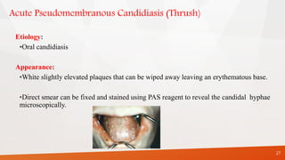
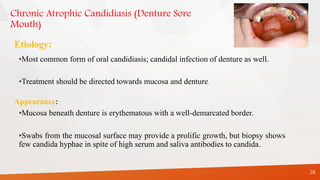
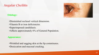
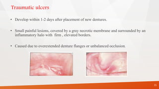
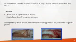
SEQUELAE OF WEARING
COMPLETE DENTURE
Presented by:-
DR. ROHIT PATIL
MDS II
Date :23/11/2021
CONTENTS
1. Introduction
2. Interaction b/w prosthetic materials and oral environment
3. Direct sequelae
4. Indirect sequelae
5. Management
6. Combination syndrome
7. Conclusion
8. References
2
INTRODUCTION
3
• Placement of removable prosthesis in the oral cavity produces profound changes of the
oral environment that may have an adverse effect on the integrity of oral tissues.
• Mucosal reactions could result from a mechanical irritation by the dentures, an
accumulation of microbial plaque on dentures or occasionally a toxic or allergic reaction to
denture base material.
4
• The continuous wearing of denture have a negative effect on residual
ridges form because of bone resorption.
• Furthermore wearing dentures that function poorly and that impair
masticatory function could be a negative factor with regard to
maintenance of adequate muscle function and nutritional status.
Interaction of prosthetic materials and oral environment
• Surface properties of denture base- Irregularities or microporosities - promote
plaque formation and accumulation on the prosthesis.
• Different materials in oral cavity may give rise to electrochemical corrosion .
• Corrosive galvanic currents is seen in oral lichen planus & in altered taste
perception.
5
• Certain microorganisms (yeast) are able to use methyl methacrylate as a carbon
source thereby causing chemical degradation of denture resin.
• Local irritation of mucosa by dentures may increase mucosal permeability to
allergens or microbial antigen thereby capable of eliciting an allergic response.
6
SOFT TISSUE CONSIDERATIONS
Injuries to oral tissues occur principally in 3 areas:
1. Tissues that support and resists stress.
2. Tissues that act to form a seal with denture borders.
3. Tissues that contact the polished surfaces and the teeth.
7
Stress bearing mucosa
Signs and symptoms of traumatic injury to this area is as follows :
1. Lesions occurring in the mucosa that covers the palate and the crest of
residual ridges
- small, well circumscribed &indurated.
- the presence of excessive keratin often causes the area to be white.
8
2.Lesions that appear punched out & the surrounding mucosa hyperemic -
- imperfections of denture base,
- trauma from food particles or
- an injury produced when the dentures were not in mouth
9
3.Lesions that are hyperemic & painful to the pressure of closure-pressure directed
toward an area of exostosis - Spur of bone or a foreign body.
4.Hyperemic , painful and detached areas of epithelium that develop on the slope of
residual ridges - Occlusal disharmony
10
5.Severe irritation and detaching of overlying mucosa occurs occasionally over
mylohyoid ridge, cuspid eminences, alveolar tubercles & areas of exostosis
- Denture flanges during insertion or removal of denture or from excessive
friction during function.
11
Basal seat mucosa
2 problems associated are
- hypertrophy and inflammation.
1. Continuous wear of dentures - Generalized hyperemia of crest and slopes of the ridges
accompanied by pain in the muscles attached to mandible, the production of hyperkeratin.
2. Complete denture opposite natural dentition or partial denture causes localized hyperemia
and edema.
12
3. Presence of loosely attached submucosa results in friction of underlying bone against the
undersurface of the mucosa when the dentures glide on mucosa thereby producing
inflammation..
4. Poor oral hygiene.
5. Unbalanced diet and avitaminosis.
6. Endocrine gland disturbances, systemic diseases.
7. Allergic reactions to denture base materials.
13
Transitional submucosa
• Hypertrophy occur in area of transitional submucosa such as border extensions results
due to unpolished or sharp borders.
• Lesions appear as slit like fissures, varying length and depth, painful and often become
ulcerated.
14
Lining mucosa
Abrasions appear on mucosa of cheeks and lips are frequently the result of :
1. cheek biting.
2. Rough margins on the teeth.
3. Unpolished denture bases.
15
Specialized mucosa
Ulcerations and other lesions appearing on the margins and apex of tongue are
results of:
1. Tongue biting often caused by improper placement of teeth either in horizontal
or vertical position.
2. An unpolished denture base or a too pronounced rugae area.
3. Rough margins on teeth.
16
DIRECT SEQUELAE:
1.Mucosal reactions-
DENTURE
STOMATITIS
CANDIDIASIS
ANGULAR
CHELITIS
EPULIS
FISSURATUM
TRAUMATIC
ULCERS
FLABBY
RIDGE
DENTURE
IRRITATION
HYPERPLASIA
17
DIRECT SEQUEALE
2. Oral galvanic currents
3. Altered taste perception
4. Burning mouth syndrome
5. Gagging
6. Residual ridge reduction
7. Periodontal disease & caries of abutments.
18
INDIRECT SEQUALAE
1. Atrophy of masticatory muscles.
2. Nutritional deficiences.
19
Denture stomatitis
Newton’s classification :
Type I: Localized simple inflammation or pinpoint hyperemia.
Type II: An erythematous or generalized simple type seen as more diffuse erythema
involving a part or entire denture covered mucosa.
Type III: Granular type involving central part of hard palate and alveolar ridges.
20
Joa ˜o Milton Rocha Gusma˜o et al;Treatment protocol for denture stomatitis,The journal of Gerodontology2013
Etiology
• Type I - trauma induced
• Types II & III -presence of microbial plaque accumulation(bacteria or yeast) on the fitting
denture surface & the underlying mucosa
The direct predisposing factor for candida associated denture stomatitis is presence of
dentures in oral cavity.
Kulak Y,Arikan A(1993) found that there was a statistically significant relationship between
denture stomatitis and denture hygiene, smoking habits, candidal formation and colonization.
21
• It is seen the major part of microorganisms of denture plaque are also involved.
• In addition trauma could stimulate turnover of palatal epithelial cells thereby reducing the
degree of keratinization and barrier function of epithelium thus the penetration of fungal
and bacterial antigens takes place more easily.
22
CANDIDIASIS
23
Four fungal organisms: Candida albicans, Candida stellatoidea, Candida tropicalis, and
Candida pseudotropicalis.
Candida albicans is most common.
Morphologically, presents in 3 forms: yeast cell, hypha and mycelium (last form is
pathogenic phase).
Carriers of oral candida do not show the mycelial phase.
Etiology
Colonization of fitting denture surface by candida species depends on :
1. Adherence of yeast cells
2. Interaction with oral commensal bacteria
3. Surface properties of acrylic resins
4. Poor oral hygeine
5. High carbohydrate intake
6. Reduced salivary flow
7. Continous denture wear
24
25
The important factors that can modulate the host-parasite relationship and increases
susceptibility to candida-assosciated denture stomatitis:
1. Ageing
2. Malnutrition
3. Immunosuppression
4. Radiation therapy
5. Diabetes mellitus
6. Antibiotics
Classification of Oral Candidiasis
• Acute pseudomembranous candidiasis (moniliasis, thrush).
• Acute atrophic candidiasis (antibiotic sore tongue).
• Chronic atrophic candidiasis (denture stomatitis).
• Chronic hyperplastic candidiasis (candidal leukoplakia, median rhomboid
glossitis).
• Angular cheilitis
• Chronic mucocutaneous candidiasis
26
Acute Pseudomembranous Candidiasis (Thrush)
Etiology:
•Oral candidiasis
Appearance:
•White slightly elevated plaques that can be wiped away leaving an erythematous base.
•Direct smear can be fixed and stained using PAS reagent to reveal the candidal hyphae
microscopically.
27
Chronic Atrophic Candidiasis (Denture Sore
Mouth)
Etiology:
•Most common form of oral candidiasis; candidal infection of denture as well.
•Treatment should be directed towards mucosa and denture.
Appearance:
•Mucosa beneath denture is erythematous with a well-demarcated border.
•Swabs from the mucosal surface may provide a prolific growth, but biopsy shows
few candida hyphae in spite of high serum and saliva antibodies to candida.
28
Angular Cheilitis
Etiology:
•Diminished occlusal vertical dimension.
•Vitamin B or iron deficiencies.
•Superimposed candidiasis.
•Affects approximately 6% of General Population.
Appearance:
•Wrinkled and sagging skin at the lip commisures.
•Desiccation and mucosal cracking.
29
Rx for Dentures
• Improve oral hygiene of appliance.
• Keep denture out of mouth for extended periods and while sleeping.
• Soak for 30 min in solutions containing benzoic acid, 0.12% chlorhexidine, or
1% sodium hypochlorite and thoroughly rinse.
• Apply a few drops of Nystatin oral suspension or a thin film of Nystatin
ointment to inner surface of denture after each meal.
30
Rx for Refractory Candidiasis
1. Fluconazole 100 mg (20 tabs; 2 tabs stat, then 1 tab daily).
2. Itraconazole 100 mg (20 tabs; 1 tab bid).
3. 2-4 weeks of Ketoconazole 200 mg (20 tabs, 1 tab daily).
31
Flabby ridge
• Flabby ridge(mobile or extremely resilient alveolar ridge) is due replacement of bone by
fibrous tissue.
• Seen in anterior part of maxilla, probably sequelae of excessive load of residual ridge
and unstable occlusal conditions.
32
• Marked fibrosis, inflammation and resorption of
underlying bone is seen.
• They provide poor support for denture.
33
34
• To improve stability of denture and minimize ridge resorption , the tissue
should be surgically removed.
• In situation of extreme atrophy of maxillary alveolar ridge,flabby ridges
should not be totally removed because the vestibular area would be
eliminated.
Traumatic ulcers
• Develop within 1-2 days after placement of new dentures.
• Small painful lesions, covered by a gray necrotic membrane and surrounded by an
inflammatory halo with firm , elevated borders.
• Caused due to overextended denture flanges or unbalanced occlusion.
35
• Conditions that suppress resistance of mucosa to mechanical irritation are predisposing
factors.
e.g: diabetes mellitus, nutritional deficiences ,radiation therapy xerostomia.
• In a non –compromised host ulcers will heal after correction of dentures.
• When left untreated,it subsequently develops into denture irritation hyperplasia.
36
Denture Irritation Hyperplasia
• Common sequelae of wearing ill-fitting dentures is occurrence of tissue hyperplasia
of mucosa in contact with denture border.
• Lesions are a result of chronic injury by unstable dentures or by thin , overextended
denture flanges.
• Lesions may be single or quite numerous and are composed of flaps of hyperplastic
connective tissue.
37
Inflammation is variable; however in bottom of deep fissures, severe inflammation may
occur.
Treatment
• Adjustment or replacement of denture.
• Surgical excision of hyperplastic tissues.
If lymphadenopathy is present, the denture irritation hyperplasia may simulate a neoplastic
process.
38
Clicking
• A clicking noise when teeth contact during functional movements is a result of
insufficient interocclusal distance,
• Vertical displacement of mandibular denture.
39
Treatment :
1. Correct stability and retention by rebasing or remaking the dentures.
2. If dentures are not loose, if sufficient interocclusal distance exists, and if teeth are
porcelain, replace the porcelain teeth with acrylic resin teeth.
3. When interocclusal distance is not sufficient, alter the occlusal surfaces of teeth with
remount procedures to provide adequate space.
40
EPULIS FISSURATUM
The hyperplasia occur in and around the borders of a denture may be fibrous growth referred
to as epulis fissuratum.
Etiology:
Chronic irritation form ill fitting or overextented denture.Since residual ridge resorbed even
best fitting denture gradually develop over extension.
Site :
It occurs in free mucosa lining the sulcus or at junction of attached and free mucosa.
41
Clinical feature:
Tissues are usually hyperemic and swollen
with slight pain.
Treatment:
• Surgical excision is indicated but only after a
period of prescribed tissue rest to reduce edema.
• Rest to tissue and program of regular and
vigorous massage of the site should be instituted.
• The result is good in 6 weeks and eliminates the
need of surgery.
42
Gagging
Stimulation of sensitive areas in posterior pharyngeal wall, soft palate, uvula, fauces or
the posterior surface of tongue results in series of uncoordinated and spasmodic
movements of swallowing muscles.
This is referred to as gagging.
43
Conny DJ,Tedesco LA:The gagging problem in prosthodontic treatment,Part I:description & causes, J Prosthet Dent
Treatment
• Determine the cause
• Eliminate the biological and mechanical factors that contribute to the problem.
• Prescribe a combination of hyoscine, hyoscyamine and atropine with a sedative
during initial period of denture use.
44
Burning mouth syndrome
• Characterized by a burning sensation in one or several oral structures in contact with
dentures.
• Symptoms often appear for first time in association with placement of new dentures.
• Common sites are tongue and upper denture bearing tissues.
• Less common sites are the lips and lower denture bearing tissues.
• Oral mucosa appears normal.
45
Juliana Cassol Spanember get al Aetiology and therapeutics of burning mouth syndrome: an update; The
journal Gerodontology2012;29: 84–89
Causes
• Local factors: mechanical irritation , allergy due to residual monomer infection , oral
habits and parafunction , myofascial pain.
• Errors in denture design which cause a denture to move excessively over the mucosa
which increase the functional stress on the mucosa or which interfere with the freedom
of movement of the surrounding muscles may initiate a complaint of burning rather
than soreness.
• Seen in 50% of BMS patients
46
Systemic factors: vitamin deficiency
iron deficiency anemia
xerostomia ,menopause,
diabetes, parkinson’s diseases,
medication.
Psychogenic factors: depression, anxiety and psychosocial stressors.
47
Management
• Initial assesment (history/examination/special test)
• Provisional diagnosis
• Initial treatment(elimination of local irritants & investigating &treating haematinic
deficiencies)
• Assessment of initial treatment
• Definitive diagnosis
• Definitive treatment (local/systemic/psychological therapy)
• Follow -up
48
Residual ridge reduction
Residual ridge resorption : A term used for the diminishing quantity and quality of the
residual ridge after teeth are removed.
• Continous bone loss after tooth extraction and placement of complete denture is seen.
• Reduction is a sequel of alveolar remodeling due to altered functional stimulus of bone
tissue.
• It is a progressive and irreversible course that results in impairment of prosthesis and
oral function.
49
• The process of resorption is important in areas with thin cortical bone (e.g
buccal and labial plates of maxilla and lingual plate of mandible).
• The annual rate of reduction in height in mandible is about 0.1-0.2 and in
general four times less in edentulous maxilla.
50
CLASSIFICATIONS OF RESIDUAL RIDGE RESORPTION
According to Brånemark et al in 1985, ridges were classified on the basis of bone
quantity and bone quality by radiographic means.
BONE QUANTITY: (Brånemark)
Class A: Most of the alveolar bone is present
Class B: Moderate Residual Ridge Resorption occurs
Class C: Advance residual ridge resorption occurs
Class D: Moderate resorption of the basal bone is present
Class E: Extreme resorption of the basal bone
51
ATWOOD’S CLASSIFICATION:
Order I - Pre-extraction
Order II - Post – extraction
Order III - High, well rounded
Order IV - Knife edge
Order V - Low, well rounded
Order VI - Depressed
52
Etiological factors of residual ridge reduction
Anatomical factors
• Important in mandible versus maxilla.
• Short and square face associated with elevated masticatory forces
• Alveoloplasty.
Prosthodontic factors
• Intensive denture wearing.
• Unstable occlusal conditions.
• Immediate denture treatment.
53
Metabolic and systemic factors
Osteoporosis
Mechanical factors
Transmitted by dentures or tongue to the residual ridges results in remodeling
process.
54
Consequence of residual ridge reduction
1. Apparent loss of sulcus width and depth
2. Displacement of muscle attachment closer to the crest of residual ridge
3. Loss of vertical dimension of occlusion
4. Reduction of lower face height
5. Anterior rotation of mandible
55
6. Increase in relative prognathia.
7. Changes in inter-alveolar ridge relationship after progression of residual ridge
reduction.
8. Morphological changes of alveolar bone such as sharp, spiny, uneven residual
ridges and location of mental foramen to the top of residual ridge.
56
Tylman (1960) found that in the maxillae, the labial and the buccal alveolar plates resorb
much faster than the palatal plates, while in the mandible the amount of bone resorbed in
the lingual and labial are approximately the same.
Treatment
Pre-prosthetic surgery includes the following:
• Ridge preservation procedure as a preventive measure.
• Corrective or recontouring procedures of the defects and abnormalities.
• Ridge extension procedures:
1. Relative methods Eg. sulcus extension (vestibuloplasty)
2. Absolute methods Eg. Ridge augmentation methods.
.
57
Hillerup S:Preprosthetic surgery in the elderly , J Prosthet Dent 72:551-558,1994
• Reconstruction methods like correction of abnormal ridge relationship.
 Provision of accessory undercuts.
Creating favorable undercuts
Dental implants.
Modified denture construction procedure
Eg. Immediate denture where construction of the denture proceeds surgery.
58
The prosthetic factors to be considered include
 Broad area coverage .
Decreased buccolingual width of teeth, and
 Improved tooth form .
Avoidance of inclined planes.
Centralization of occlusal contacts.
Provision of adequate tongue room.
Adequate inter-occlusal distance during rest jaw relation.
59
Overdenture abutments: caries and periodontal disease
• Wearing of overdentures is often associated with a high risk of caries and
progression of periodontal disease of abutment teeth.
• This is due to bacterial colonization, beneath a close fitting denture is enhanced,
and good plaque control of fitting denture surface is difficult to obtain.
• Predominant micro organisms are streptococcus, lactobacilli and actinomyces.
60
• These species initiate gingivitis after 1-3 days of plaque accumulation when oral
hygiene is discontinued.
• Presence of streptococcus mutans and lactobacilli in dental plaque flora in high
proportions results in caries.
61
Management
• Abstain from wearing dentures in the night.
• Application of flouride-chlorhexidine gel and polishing, mechanical & chemical
plaque control.
• Placement of copings that cover the exposed dentin and root surface is indicated
where caries is more deeply penetrating . This is to reduce risk of new or recurrent
caries.
• Periodontal pockets greater than 4-5 mm should be surgically eliminated
62
Oral Cancer in Denture Wearers
An association between oral carcinoma and chronic irritation of the mucosa
by the dentures has often been claimed, but no definite proof seems to exist.
63
Indirect sequelae: Atrophy of masticatory muscles
• Masticatory function depends on the skeletal muscular force and the facility with which
the patient is able to coordinate oral functional movements during mastication.
• In complete denture wearers, particularly in women atrophy of masseter and medial
pterygoid muscle is seen.
• The decrease in bite force and chewing efficiency results in impaired masticatory function.
64
Preventive measures & management
• Retention of a small number of teeth used as overdenture abutments helps in
maintenance of oral functions.
• In completely edentulous patients , placement of implants is usually followed by
an improvement of masticatory function & an increase of maximal occlusal
forces.
65
Nutritional status and masticatory functions
4 factors are related to dietary selection and nutritional status of wearers of complete
dentures:
1. Masticatory function and oral health
2. General health
3. Socio-economic status
4. Dietary habits
66
Management
1.Re-education of elderly denture wearers regarding dietary habits.
2.Replacement of ill-fitting dentures.
3.Mechanical preparation of food before eating will help mastication and reduce its
influence on food selection.
67
COMBINATION SYNDROME
“The characteristic features that occur when an edentulous maxilla is opposed by
natural mandibular anterior teeth, including loss of bone from the anterior portion of
the maxillary ridge, overgrowth of the tuberosities, papillary hyperplasia of the hard
palatal mucosa, extrusion of mandibular anterior teeth and loss of alveolar bone and
ridge height beneath the mandibular removable partial denture bases.
68
First identified by Kelly in 1972,it is found in patients wearing a complete maxillary
denture, opposing a mandibular distal extension prosthesis.
69
Five signs or symptoms commonly occurred in this situation They include:
70
Saunders later described six additional signs associated with the syndrome
They include:
1. Loss of occlusal vertical dimension
2. Occlusal plane discrepancy.
3. Anterior spatial repositioning of the mandible.
4. Poor adaptation of the prostheses.
5. Epulis fissuratum.
6. Periodontal changes
71
PREVENTION OF COMBINATION SYNDROME:
• Avoid combination of complete maxillary dentures opposing class I mandibular
RPD.
• Retaining weak posterior teeth as abutments by means of endodontic and
periodontic techniques.
• An overdenture on the lower teeth.
72
Conclusion
73
• The essential consequences of wearing complete dentures are reduction of residual
ridges and pathological changes of oral mucosae. This results in poor patient
comfort,destabilization of occlusion , insufficient masticatory function and esthetic
problems.
• Effort should be made to retain some teeth in strategically good positions to serve
as overdenture abutments.The maintenance of tooth roots in mandible is
important.
• The patient should follow a regular follow – up schedule at yearly interval so that
an acceptable fit and stable occlusion can be maintained.
• Patients should be aware of implant supported prosthesis.In young
patients,advantage would be reduced residual ridge reduction.In elderly
patients , the main advantage are improved comfort and maintenance of
masticatory function.
• Patient should be motivated to practice proper denture wearing habits and
maintenance of oral hygiene and follow a program of recall and
maintenance for continuous monitoring of dentures and oral tissues.
74
References
• Zarb –Bolender : Prosthodontic treatment for edentulous patients, 12th edition .
• Arthur.Rahn.O,Charles.Heartwell.M,Jr: Textbook of complete dentures, 5th edition.
• Sheldon Winkler:Essentials of complete denture prosthodontics, 2nd edition .
• Basker RM & Davenport JC: Prosthetic treatment of edentulous patient, 4th edition.
• Tallegren A:The continuing reduction of the residual alveolar ridges in complete
denture wearers:mixed longitudinal study covering 25 yrs,J Prosthet Dent 27:120-
132,1972.
• Budtz-Jorgensen E:Oral mucosal lesions assosciated with wearing of removable
dentures,J Oral Path 10:65-80,1981.
• Conny DJ,Tedesco LA:The gagging problem in prosthodontic treatment,Part
I:description & causes, J Prosthet Dent 49:601-606,1983.
• Hillerup S:Preprosthetic surgery in the elderly , J Prosthet Dent 72:551-558,1994.
75
Thank you....
76

SEMINAR 10 SEQUELAE OF CD.pptx

  • 1.
    SEQUELAE OF WEARING COMPLETEDENTURE Presented by:- DR. ROHIT PATIL MDS II Date :23/11/2021
  • 2.
    CONTENTS 1. Introduction 2. Interactionb/w prosthetic materials and oral environment 3. Direct sequelae 4. Indirect sequelae 5. Management 6. Combination syndrome 7. Conclusion 8. References 2
  • 3.
    INTRODUCTION 3 • Placement ofremovable prosthesis in the oral cavity produces profound changes of the oral environment that may have an adverse effect on the integrity of oral tissues. • Mucosal reactions could result from a mechanical irritation by the dentures, an accumulation of microbial plaque on dentures or occasionally a toxic or allergic reaction to denture base material.
  • 4.
    4 • The continuouswearing of denture have a negative effect on residual ridges form because of bone resorption. • Furthermore wearing dentures that function poorly and that impair masticatory function could be a negative factor with regard to maintenance of adequate muscle function and nutritional status.
  • 5.
    Interaction of prostheticmaterials and oral environment • Surface properties of denture base- Irregularities or microporosities - promote plaque formation and accumulation on the prosthesis. • Different materials in oral cavity may give rise to electrochemical corrosion . • Corrosive galvanic currents is seen in oral lichen planus & in altered taste perception. 5
  • 6.
    • Certain microorganisms(yeast) are able to use methyl methacrylate as a carbon source thereby causing chemical degradation of denture resin. • Local irritation of mucosa by dentures may increase mucosal permeability to allergens or microbial antigen thereby capable of eliciting an allergic response. 6
  • 7.
    SOFT TISSUE CONSIDERATIONS Injuriesto oral tissues occur principally in 3 areas: 1. Tissues that support and resists stress. 2. Tissues that act to form a seal with denture borders. 3. Tissues that contact the polished surfaces and the teeth. 7
  • 8.
    Stress bearing mucosa Signsand symptoms of traumatic injury to this area is as follows : 1. Lesions occurring in the mucosa that covers the palate and the crest of residual ridges - small, well circumscribed &indurated. - the presence of excessive keratin often causes the area to be white. 8
  • 9.
    2.Lesions that appearpunched out & the surrounding mucosa hyperemic - - imperfections of denture base, - trauma from food particles or - an injury produced when the dentures were not in mouth 9
  • 10.
    3.Lesions that arehyperemic & painful to the pressure of closure-pressure directed toward an area of exostosis - Spur of bone or a foreign body. 4.Hyperemic , painful and detached areas of epithelium that develop on the slope of residual ridges - Occlusal disharmony 10
  • 11.
    5.Severe irritation anddetaching of overlying mucosa occurs occasionally over mylohyoid ridge, cuspid eminences, alveolar tubercles & areas of exostosis - Denture flanges during insertion or removal of denture or from excessive friction during function. 11
  • 12.
    Basal seat mucosa 2problems associated are - hypertrophy and inflammation. 1. Continuous wear of dentures - Generalized hyperemia of crest and slopes of the ridges accompanied by pain in the muscles attached to mandible, the production of hyperkeratin. 2. Complete denture opposite natural dentition or partial denture causes localized hyperemia and edema. 12
  • 13.
    3. Presence ofloosely attached submucosa results in friction of underlying bone against the undersurface of the mucosa when the dentures glide on mucosa thereby producing inflammation.. 4. Poor oral hygiene. 5. Unbalanced diet and avitaminosis. 6. Endocrine gland disturbances, systemic diseases. 7. Allergic reactions to denture base materials. 13
  • 14.
    Transitional submucosa • Hypertrophyoccur in area of transitional submucosa such as border extensions results due to unpolished or sharp borders. • Lesions appear as slit like fissures, varying length and depth, painful and often become ulcerated. 14
  • 15.
    Lining mucosa Abrasions appearon mucosa of cheeks and lips are frequently the result of : 1. cheek biting. 2. Rough margins on the teeth. 3. Unpolished denture bases. 15
  • 16.
    Specialized mucosa Ulcerations andother lesions appearing on the margins and apex of tongue are results of: 1. Tongue biting often caused by improper placement of teeth either in horizontal or vertical position. 2. An unpolished denture base or a too pronounced rugae area. 3. Rough margins on teeth. 16
  • 17.
  • 18.
    DIRECT SEQUEALE 2. Oralgalvanic currents 3. Altered taste perception 4. Burning mouth syndrome 5. Gagging 6. Residual ridge reduction 7. Periodontal disease & caries of abutments. 18
  • 19.
    INDIRECT SEQUALAE 1. Atrophyof masticatory muscles. 2. Nutritional deficiences. 19
  • 20.
    Denture stomatitis Newton’s classification: Type I: Localized simple inflammation or pinpoint hyperemia. Type II: An erythematous or generalized simple type seen as more diffuse erythema involving a part or entire denture covered mucosa. Type III: Granular type involving central part of hard palate and alveolar ridges. 20 Joa ˜o Milton Rocha Gusma˜o et al;Treatment protocol for denture stomatitis,The journal of Gerodontology2013
  • 21.
    Etiology • Type I- trauma induced • Types II & III -presence of microbial plaque accumulation(bacteria or yeast) on the fitting denture surface & the underlying mucosa The direct predisposing factor for candida associated denture stomatitis is presence of dentures in oral cavity. Kulak Y,Arikan A(1993) found that there was a statistically significant relationship between denture stomatitis and denture hygiene, smoking habits, candidal formation and colonization. 21
  • 22.
    • It isseen the major part of microorganisms of denture plaque are also involved. • In addition trauma could stimulate turnover of palatal epithelial cells thereby reducing the degree of keratinization and barrier function of epithelium thus the penetration of fungal and bacterial antigens takes place more easily. 22
  • 23.
    CANDIDIASIS 23 Four fungal organisms:Candida albicans, Candida stellatoidea, Candida tropicalis, and Candida pseudotropicalis. Candida albicans is most common. Morphologically, presents in 3 forms: yeast cell, hypha and mycelium (last form is pathogenic phase). Carriers of oral candida do not show the mycelial phase.
  • 24.
    Etiology Colonization of fittingdenture surface by candida species depends on : 1. Adherence of yeast cells 2. Interaction with oral commensal bacteria 3. Surface properties of acrylic resins 4. Poor oral hygeine 5. High carbohydrate intake 6. Reduced salivary flow 7. Continous denture wear 24
  • 25.
    25 The important factorsthat can modulate the host-parasite relationship and increases susceptibility to candida-assosciated denture stomatitis: 1. Ageing 2. Malnutrition 3. Immunosuppression 4. Radiation therapy 5. Diabetes mellitus 6. Antibiotics
  • 26.
    Classification of OralCandidiasis • Acute pseudomembranous candidiasis (moniliasis, thrush). • Acute atrophic candidiasis (antibiotic sore tongue). • Chronic atrophic candidiasis (denture stomatitis). • Chronic hyperplastic candidiasis (candidal leukoplakia, median rhomboid glossitis). • Angular cheilitis • Chronic mucocutaneous candidiasis 26
  • 27.
    Acute Pseudomembranous Candidiasis(Thrush) Etiology: •Oral candidiasis Appearance: •White slightly elevated plaques that can be wiped away leaving an erythematous base. •Direct smear can be fixed and stained using PAS reagent to reveal the candidal hyphae microscopically. 27
  • 28.
    Chronic Atrophic Candidiasis(Denture Sore Mouth) Etiology: •Most common form of oral candidiasis; candidal infection of denture as well. •Treatment should be directed towards mucosa and denture. Appearance: •Mucosa beneath denture is erythematous with a well-demarcated border. •Swabs from the mucosal surface may provide a prolific growth, but biopsy shows few candida hyphae in spite of high serum and saliva antibodies to candida. 28
  • 29.
    Angular Cheilitis Etiology: •Diminished occlusalvertical dimension. •Vitamin B or iron deficiencies. •Superimposed candidiasis. •Affects approximately 6% of General Population. Appearance: •Wrinkled and sagging skin at the lip commisures. •Desiccation and mucosal cracking. 29
  • 30.
    Rx for Dentures •Improve oral hygiene of appliance. • Keep denture out of mouth for extended periods and while sleeping. • Soak for 30 min in solutions containing benzoic acid, 0.12% chlorhexidine, or 1% sodium hypochlorite and thoroughly rinse. • Apply a few drops of Nystatin oral suspension or a thin film of Nystatin ointment to inner surface of denture after each meal. 30
  • 31.
    Rx for RefractoryCandidiasis 1. Fluconazole 100 mg (20 tabs; 2 tabs stat, then 1 tab daily). 2. Itraconazole 100 mg (20 tabs; 1 tab bid). 3. 2-4 weeks of Ketoconazole 200 mg (20 tabs, 1 tab daily). 31
  • 32.
    Flabby ridge • Flabbyridge(mobile or extremely resilient alveolar ridge) is due replacement of bone by fibrous tissue. • Seen in anterior part of maxilla, probably sequelae of excessive load of residual ridge and unstable occlusal conditions. 32
  • 33.
    • Marked fibrosis,inflammation and resorption of underlying bone is seen. • They provide poor support for denture. 33
  • 34.
    34 • To improvestability of denture and minimize ridge resorption , the tissue should be surgically removed. • In situation of extreme atrophy of maxillary alveolar ridge,flabby ridges should not be totally removed because the vestibular area would be eliminated.
  • 35.
    Traumatic ulcers • Developwithin 1-2 days after placement of new dentures. • Small painful lesions, covered by a gray necrotic membrane and surrounded by an inflammatory halo with firm , elevated borders. • Caused due to overextended denture flanges or unbalanced occlusion. 35
  • 36.
    • Conditions thatsuppress resistance of mucosa to mechanical irritation are predisposing factors. e.g: diabetes mellitus, nutritional deficiences ,radiation therapy xerostomia. • In a non –compromised host ulcers will heal after correction of dentures. • When left untreated,it subsequently develops into denture irritation hyperplasia. 36
  • 37.
    Denture Irritation Hyperplasia •Common sequelae of wearing ill-fitting dentures is occurrence of tissue hyperplasia of mucosa in contact with denture border. • Lesions are a result of chronic injury by unstable dentures or by thin , overextended denture flanges. • Lesions may be single or quite numerous and are composed of flaps of hyperplastic connective tissue. 37
  • 38.
    Inflammation is variable;however in bottom of deep fissures, severe inflammation may occur. Treatment • Adjustment or replacement of denture. • Surgical excision of hyperplastic tissues. If lymphadenopathy is present, the denture irritation hyperplasia may simulate a neoplastic process. 38
  • 39.
    Clicking • A clickingnoise when teeth contact during functional movements is a result of insufficient interocclusal distance, • Vertical displacement of mandibular denture. 39
  • 40.
    Treatment : 1. Correctstability and retention by rebasing or remaking the dentures. 2. If dentures are not loose, if sufficient interocclusal distance exists, and if teeth are porcelain, replace the porcelain teeth with acrylic resin teeth. 3. When interocclusal distance is not sufficient, alter the occlusal surfaces of teeth with remount procedures to provide adequate space. 40
  • 41.
    EPULIS FISSURATUM The hyperplasiaoccur in and around the borders of a denture may be fibrous growth referred to as epulis fissuratum. Etiology: Chronic irritation form ill fitting or overextented denture.Since residual ridge resorbed even best fitting denture gradually develop over extension. Site : It occurs in free mucosa lining the sulcus or at junction of attached and free mucosa. 41
  • 42.
    Clinical feature: Tissues areusually hyperemic and swollen with slight pain. Treatment: • Surgical excision is indicated but only after a period of prescribed tissue rest to reduce edema. • Rest to tissue and program of regular and vigorous massage of the site should be instituted. • The result is good in 6 weeks and eliminates the need of surgery. 42
  • 43.
    Gagging Stimulation of sensitiveareas in posterior pharyngeal wall, soft palate, uvula, fauces or the posterior surface of tongue results in series of uncoordinated and spasmodic movements of swallowing muscles. This is referred to as gagging. 43 Conny DJ,Tedesco LA:The gagging problem in prosthodontic treatment,Part I:description & causes, J Prosthet Dent
  • 44.
    Treatment • Determine thecause • Eliminate the biological and mechanical factors that contribute to the problem. • Prescribe a combination of hyoscine, hyoscyamine and atropine with a sedative during initial period of denture use. 44
  • 45.
    Burning mouth syndrome •Characterized by a burning sensation in one or several oral structures in contact with dentures. • Symptoms often appear for first time in association with placement of new dentures. • Common sites are tongue and upper denture bearing tissues. • Less common sites are the lips and lower denture bearing tissues. • Oral mucosa appears normal. 45 Juliana Cassol Spanember get al Aetiology and therapeutics of burning mouth syndrome: an update; The journal Gerodontology2012;29: 84–89
  • 46.
    Causes • Local factors:mechanical irritation , allergy due to residual monomer infection , oral habits and parafunction , myofascial pain. • Errors in denture design which cause a denture to move excessively over the mucosa which increase the functional stress on the mucosa or which interfere with the freedom of movement of the surrounding muscles may initiate a complaint of burning rather than soreness. • Seen in 50% of BMS patients 46
  • 47.
    Systemic factors: vitamindeficiency iron deficiency anemia xerostomia ,menopause, diabetes, parkinson’s diseases, medication. Psychogenic factors: depression, anxiety and psychosocial stressors. 47
  • 48.
    Management • Initial assesment(history/examination/special test) • Provisional diagnosis • Initial treatment(elimination of local irritants & investigating &treating haematinic deficiencies) • Assessment of initial treatment • Definitive diagnosis • Definitive treatment (local/systemic/psychological therapy) • Follow -up 48
  • 49.
    Residual ridge reduction Residualridge resorption : A term used for the diminishing quantity and quality of the residual ridge after teeth are removed. • Continous bone loss after tooth extraction and placement of complete denture is seen. • Reduction is a sequel of alveolar remodeling due to altered functional stimulus of bone tissue. • It is a progressive and irreversible course that results in impairment of prosthesis and oral function. 49
  • 50.
    • The processof resorption is important in areas with thin cortical bone (e.g buccal and labial plates of maxilla and lingual plate of mandible). • The annual rate of reduction in height in mandible is about 0.1-0.2 and in general four times less in edentulous maxilla. 50
  • 51.
    CLASSIFICATIONS OF RESIDUALRIDGE RESORPTION According to Brånemark et al in 1985, ridges were classified on the basis of bone quantity and bone quality by radiographic means. BONE QUANTITY: (Brånemark) Class A: Most of the alveolar bone is present Class B: Moderate Residual Ridge Resorption occurs Class C: Advance residual ridge resorption occurs Class D: Moderate resorption of the basal bone is present Class E: Extreme resorption of the basal bone 51
  • 52.
    ATWOOD’S CLASSIFICATION: Order I- Pre-extraction Order II - Post – extraction Order III - High, well rounded Order IV - Knife edge Order V - Low, well rounded Order VI - Depressed 52
  • 53.
    Etiological factors ofresidual ridge reduction Anatomical factors • Important in mandible versus maxilla. • Short and square face associated with elevated masticatory forces • Alveoloplasty. Prosthodontic factors • Intensive denture wearing. • Unstable occlusal conditions. • Immediate denture treatment. 53
  • 54.
    Metabolic and systemicfactors Osteoporosis Mechanical factors Transmitted by dentures or tongue to the residual ridges results in remodeling process. 54
  • 55.
    Consequence of residualridge reduction 1. Apparent loss of sulcus width and depth 2. Displacement of muscle attachment closer to the crest of residual ridge 3. Loss of vertical dimension of occlusion 4. Reduction of lower face height 5. Anterior rotation of mandible 55
  • 56.
    6. Increase inrelative prognathia. 7. Changes in inter-alveolar ridge relationship after progression of residual ridge reduction. 8. Morphological changes of alveolar bone such as sharp, spiny, uneven residual ridges and location of mental foramen to the top of residual ridge. 56 Tylman (1960) found that in the maxillae, the labial and the buccal alveolar plates resorb much faster than the palatal plates, while in the mandible the amount of bone resorbed in the lingual and labial are approximately the same.
  • 57.
    Treatment Pre-prosthetic surgery includesthe following: • Ridge preservation procedure as a preventive measure. • Corrective or recontouring procedures of the defects and abnormalities. • Ridge extension procedures: 1. Relative methods Eg. sulcus extension (vestibuloplasty) 2. Absolute methods Eg. Ridge augmentation methods. . 57 Hillerup S:Preprosthetic surgery in the elderly , J Prosthet Dent 72:551-558,1994
  • 58.
    • Reconstruction methodslike correction of abnormal ridge relationship.  Provision of accessory undercuts. Creating favorable undercuts Dental implants. Modified denture construction procedure Eg. Immediate denture where construction of the denture proceeds surgery. 58
  • 59.
    The prosthetic factorsto be considered include  Broad area coverage . Decreased buccolingual width of teeth, and  Improved tooth form . Avoidance of inclined planes. Centralization of occlusal contacts. Provision of adequate tongue room. Adequate inter-occlusal distance during rest jaw relation. 59
  • 60.
    Overdenture abutments: cariesand periodontal disease • Wearing of overdentures is often associated with a high risk of caries and progression of periodontal disease of abutment teeth. • This is due to bacterial colonization, beneath a close fitting denture is enhanced, and good plaque control of fitting denture surface is difficult to obtain. • Predominant micro organisms are streptococcus, lactobacilli and actinomyces. 60
  • 61.
    • These speciesinitiate gingivitis after 1-3 days of plaque accumulation when oral hygiene is discontinued. • Presence of streptococcus mutans and lactobacilli in dental plaque flora in high proportions results in caries. 61
  • 62.
    Management • Abstain fromwearing dentures in the night. • Application of flouride-chlorhexidine gel and polishing, mechanical & chemical plaque control. • Placement of copings that cover the exposed dentin and root surface is indicated where caries is more deeply penetrating . This is to reduce risk of new or recurrent caries. • Periodontal pockets greater than 4-5 mm should be surgically eliminated 62
  • 63.
    Oral Cancer inDenture Wearers An association between oral carcinoma and chronic irritation of the mucosa by the dentures has often been claimed, but no definite proof seems to exist. 63
  • 64.
    Indirect sequelae: Atrophyof masticatory muscles • Masticatory function depends on the skeletal muscular force and the facility with which the patient is able to coordinate oral functional movements during mastication. • In complete denture wearers, particularly in women atrophy of masseter and medial pterygoid muscle is seen. • The decrease in bite force and chewing efficiency results in impaired masticatory function. 64
  • 65.
    Preventive measures &management • Retention of a small number of teeth used as overdenture abutments helps in maintenance of oral functions. • In completely edentulous patients , placement of implants is usually followed by an improvement of masticatory function & an increase of maximal occlusal forces. 65
  • 66.
    Nutritional status andmasticatory functions 4 factors are related to dietary selection and nutritional status of wearers of complete dentures: 1. Masticatory function and oral health 2. General health 3. Socio-economic status 4. Dietary habits 66
  • 67.
    Management 1.Re-education of elderlydenture wearers regarding dietary habits. 2.Replacement of ill-fitting dentures. 3.Mechanical preparation of food before eating will help mastication and reduce its influence on food selection. 67
  • 68.
    COMBINATION SYNDROME “The characteristicfeatures that occur when an edentulous maxilla is opposed by natural mandibular anterior teeth, including loss of bone from the anterior portion of the maxillary ridge, overgrowth of the tuberosities, papillary hyperplasia of the hard palatal mucosa, extrusion of mandibular anterior teeth and loss of alveolar bone and ridge height beneath the mandibular removable partial denture bases. 68
  • 69.
    First identified byKelly in 1972,it is found in patients wearing a complete maxillary denture, opposing a mandibular distal extension prosthesis. 69
  • 70.
    Five signs orsymptoms commonly occurred in this situation They include: 70
  • 71.
    Saunders later describedsix additional signs associated with the syndrome They include: 1. Loss of occlusal vertical dimension 2. Occlusal plane discrepancy. 3. Anterior spatial repositioning of the mandible. 4. Poor adaptation of the prostheses. 5. Epulis fissuratum. 6. Periodontal changes 71
  • 72.
    PREVENTION OF COMBINATIONSYNDROME: • Avoid combination of complete maxillary dentures opposing class I mandibular RPD. • Retaining weak posterior teeth as abutments by means of endodontic and periodontic techniques. • An overdenture on the lower teeth. 72
  • 73.
    Conclusion 73 • The essentialconsequences of wearing complete dentures are reduction of residual ridges and pathological changes of oral mucosae. This results in poor patient comfort,destabilization of occlusion , insufficient masticatory function and esthetic problems. • Effort should be made to retain some teeth in strategically good positions to serve as overdenture abutments.The maintenance of tooth roots in mandible is important. • The patient should follow a regular follow – up schedule at yearly interval so that an acceptable fit and stable occlusion can be maintained.
  • 74.
    • Patients shouldbe aware of implant supported prosthesis.In young patients,advantage would be reduced residual ridge reduction.In elderly patients , the main advantage are improved comfort and maintenance of masticatory function. • Patient should be motivated to practice proper denture wearing habits and maintenance of oral hygiene and follow a program of recall and maintenance for continuous monitoring of dentures and oral tissues. 74
  • 75.
    References • Zarb –Bolender: Prosthodontic treatment for edentulous patients, 12th edition . • Arthur.Rahn.O,Charles.Heartwell.M,Jr: Textbook of complete dentures, 5th edition. • Sheldon Winkler:Essentials of complete denture prosthodontics, 2nd edition . • Basker RM & Davenport JC: Prosthetic treatment of edentulous patient, 4th edition. • Tallegren A:The continuing reduction of the residual alveolar ridges in complete denture wearers:mixed longitudinal study covering 25 yrs,J Prosthet Dent 27:120- 132,1972. • Budtz-Jorgensen E:Oral mucosal lesions assosciated with wearing of removable dentures,J Oral Path 10:65-80,1981. • Conny DJ,Tedesco LA:The gagging problem in prosthodontic treatment,Part I:description & causes, J Prosthet Dent 49:601-606,1983. • Hillerup S:Preprosthetic surgery in the elderly , J Prosthet Dent 72:551-558,1994. 75
  • 76.