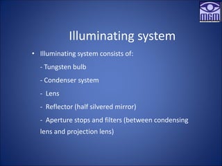
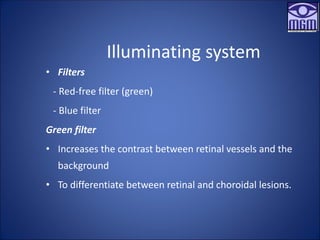
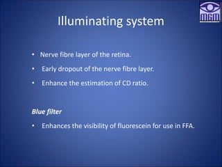
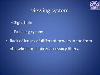
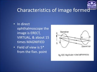
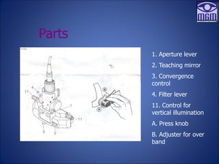
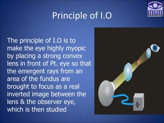
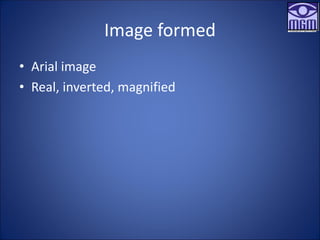
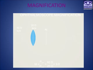
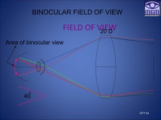
SlideShare a Scribd company logo
Direct & Indirect
Ophthalmoscope
Mahendrda Singh
PhD (Scholar)
Assistant Professor and Consultant Optometrist
CL Gupta Eye institute, Moradabad UP India
• Objective method of examining the posterior
segment of the eye.
• Presence or absence of abnormalities in the
posterior segment - structural or pathological.
• View of vascular & neurological structures of a living
organ in-situ.
Introduction
Types
• Can be divided into two types :
– Direct ophthalmoscope
– Indirect ophthalmoscope
Direct
Ophthalmoscope
History
1846- Charles Babbage, constructed
first ophthalmoscope.
1850’s- Von Helmholtz - Father of
ophthalmoscope.
PRINCIPLE
• Work on the principle of ANGULAR
Magnification.
Optics
• A convergent beam of
light is reflected into
the patient’s pupil.
• The emergent rays
from any point on the
patient’s fundus reach
the observer’s retina
through the viewing
hole in the
ophthalmoscope.
Optics (In Hyperopic patient’s)
• In a hypermetropic
patient’s, the emergent
ray from the illuminated
area of retina will be
divergent & thus can be
brought to focus on the
observer’s retina if the
latter accommodates,
or by the help of a
convex lens
Optics (In Myopic patient’s)
• In a myopic patient’s
the emergent rays will
be convergent & thus
can be brought to
focus on the observer
retina by the help of a
minus lens
Instrumentation
• Illuminating system
• viewing system
Illuminating system
• Illuminating system consists of:
- Tungsten bulb
- Condenser system
- Lens
- Reflector (half silvered mirror)
- Aperture stops and filters (between condensing
lens and projection lens)
Illuminating system
• Filters
- Red-free filter (green)
- Blue filter
Green filter
• Increases the contrast between retinal vessels and the
background
• To differentiate between retinal and choroidal lesions.
Illuminating system
• Nerve fibre layer of the retina.
• Early dropout of the nerve fibre layer.
• Enhance the estimation of CD ratio.
Blue filter
• Enhances the visibility of fluorescein for use in FFA.
viewing system
–Sight hole
–Focusing system
• Rack of lenses of different powers in the form
of a wheel or chain & accessory filters.
Lens track
chain
Eyebrow rest
Sight hole
viewing system
• It consists of 3 aperture stops :
1) Small - for macula
2) Intermediate - for viewing the fundus through
normal pupils
3) A large - to view through dilated fundus.
viewing system
• Slit diaphragm - observing elevated retinal
lesions.
• Half circle - reduces reflection by limiting the
illumination & observing certain fine retinal
details.
• Fixation star - to determine patients fixation in
strabismic amblyopia.
Half circle
Small aperture
Intermediate aperture
Fixation star
Slit aperture
Filter changing lever
Green filter
Large aperture
Sight hole
Blue filter
Aperture stop
Characteristics of image formed
• In direct
ophthalmoscope the
image is ERECT,
VIRTUAL, & about 15
times MAGNIFIED
• Field of view is 5*
from the fixn. point
Magnification
Depend on angular magnification,
Therefore A M is achieved by
AM=POWER OF THE EYE/4
M=60/4
=15X.
Field of vision
• Directly proportional to the size of the pupil of
observed eye
• Directly proportional to the axial length of the
observer’s eye
• Inversely proportional to the distance between the
observer’s & observed eye
• The smaller the sight hole of ophthalmoscope the
better the field of vision
Technique
• Patient seated in semi-dark room & looking straight
ahead
• Pt. right eye should be examined by the observer
with his right eye & left with the left
• Once the red reflex is seen, the observer should
move as close to the patient’s eye as possible
(theoretically at the antr. focal plane of the pt. eye
i.e. 15.4mm)
Clinical uses
• The homogeneity of cornea & lens may also
checked,
• Irregularities in the optical structures as black
shadow.
• Small hemorrhages or aneurysms, which can
easily locate.
• Used as distant direct ophthalmoscope.
• Used as BRUCKNER reflex test.
Advantages
• Magnification is about 15 x.
• Easier to use with small and undilated pupil.
• Easier mechanically.
• Portable.
Disadvantages
• Field of view is smaller
• Peripheral view of retina is not possible.
• It is not easy in viewing the fundus in cloudy
media.
• Stereopsis is not present.
• Limited Illumination.
Indirect
Ophthalmoscope
Introduction
• Introduced by Nagel in 1864
• Very popular method for examination of the
posterior segment
TYPES
• BINOCULAR INDIRECT OPHTHALMOSCOPE
• MONOCULAR INDIRECT OPHTHALMOSCOPE
Indirect Ophthalmoscope
1. Aperture lever
2. Teaching mirror
3. Convergence
control
4. Filter lever
11. Control for
vertical illumination
A. Press knob
B. Adjuster for over
band
Parts
Parts
5. Unscrew
knob
6. Control for
over band
7.Circumferen
ce adjuster
8.Height
adjuster
9.Over band
10.optics.
Parts
12.Brightness selection
13.Bulb connector
14.Bulb
15.Locating pin
16.Slit
17.Fibre optics cable
18.Cord socket
Accessories used
• 1.Condensing Lens
• 2.Indenter
Principle of I.O
The principle of I.O is to
make the eye highly myopic
by placing a strong convex
lens in front of Pt. eye so that
the emergent rays from an
area of the fundus are
brought to focus as a real
inverted image between the
lens & the observer eye,
which is then studied
Optical system of I.O
• Binocularity is
achieved by reducing
the observer I.P.D to
approx. 15mm by
prisms/mirror.
FIRST ATTEMPT AT BINOCULAR VIEW
Obs. L eye
Obs. R eye
S’s eye
Combine L and R eye views
Observer’s eyes have to be too close
IMAGE ORIENTATION
MAGNIFICATION
FIELD OF VIEW
Characteristics of image
IMAGE ORIENTATION
The emergent rays from
the illuminated area of
retina are parallel in
emmetropic Pt. & are
therefore brought to focus
by the condensing lens at
its principal focus, thus an
inverted image of the
retina is formed in the air
between the condensing
lens & the observer.
Image formed
• Arial image
• Real, inverted, magnified
MAGNIFICATION
20 D
lens
RI
60 D
eye
OPHTHALMOSCOPE MAGNIFICATION
Peye
Plens
=
60 D
20 D
= 3.0
M =
FIELD OF VIEW
20 D
40
Area of binocular view
BINOCULAR FIELD OF VIEW
GTT 04
Advantages
• Large field of view.
• Easy to view the extreme periphery.
• Easy to view in cloudy media
• Use of indenter becomes easy.
• Good illumination.
• Hand free for operative purpose.
Disadvantages
• Low magnification.
• It cannot be well perform in undilated pupil.
• Mechanically not easy.
• Not portable.
Monocular I O
Its virtue is to permits visualisation of the
fundus through an undilated pupil and the
image seen is errect rather than inverted and
sterioscopic visualisation is not possible.
Few difficulties to deal with
• Unwanted reflections of light which comes
from the anterior and posterior surface of the
convex lens and the patients cornea.
• In case of small pupil.
• In case of large pupils.
Comparison
Feature Direct Indirect (20 D)
Magnification 15x 3 x
Field diameter 2 DD 9 DD
Illumination Limited High
Depth of focus Small Large
Stereopsis Absent Present
Image
Orientation Upright reversed
Periphery view Limited Full
Working distance Close Arm’s length
Scleral indentation Difficult Easy
Colour Code for Fundus Chart
Color Code for Fundus Chart
Red
Attached retina
Arteries
Retinal breaks
Thin retina
Retinal hemorrhages (superficial
& deep)
Microaneurysms
Retinal new vessels (flat &
elevated)
Color Code for Fundus Chart
Blue
Detached retina
Retinoschisis
Veins
Outline of retinal breaks
Lattice degeneration
BROWN
CHOROIDAL DETACHMENT
Color Code for Fundus Chart
Yellow
Exudates, Drusen
Green
Media Opacities
(label specific lesion)
Black
Retino-choroidal pigmentation
Ora serrata
• Thank you

More Related Content

Similar to Direct & Indirect Ophthalmoloscope.pdf

Direct & indirect ophthalmoscopy
Direct & indirect ophthalmoscopyDirect & indirect ophthalmoscopy
Direct & indirect ophthalmoscopy
DR. ROHIT AGRAWAL
 
ophthalmoscopy.pptx
ophthalmoscopy.pptxophthalmoscopy.pptx
ophthalmoscopy.pptx
Divya785180
 
Slit lamp biomicroscopy
Slit lamp biomicroscopySlit lamp biomicroscopy
Slit lamp biomicroscopy
nafiz mahmood
 
Slitlamp bimicroscopy
Slitlamp bimicroscopy Slitlamp bimicroscopy
Slitlamp bimicroscopy
Obehi Osoata
 
Bio optos-8-21-2011
Bio optos-8-21-2011Bio optos-8-21-2011
Bio optos-8-21-2011
Aali Shree
 
INDIRECT OPHTHALMOSCOPY ppt
INDIRECT OPHTHALMOSCOPY pptINDIRECT OPHTHALMOSCOPY ppt
INDIRECT OPHTHALMOSCOPY ppt
SoundariyaAnbarasan
 
Slit Lamp Biomicroscopy.
Slit Lamp Biomicroscopy.Slit Lamp Biomicroscopy.
Slit Lamp Biomicroscopy.
Mystic Dider
 
Binocular Indirect Ophthalmoscopy
Binocular Indirect OphthalmoscopyBinocular Indirect Ophthalmoscopy
Binocular Indirect Ophthalmoscopy
abhishek ghelani
 
SLIT LAMP BIOMICROSCOPE MOPB 021.pptx
SLIT LAMP BIOMICROSCOPE MOPB 021.pptxSLIT LAMP BIOMICROSCOPE MOPB 021.pptx
SLIT LAMP BIOMICROSCOPE MOPB 021.pptx
bakanangemmahpholoan
 
directophthalmoscope-180730142501 (2).pdf
directophthalmoscope-180730142501 (2).pdfdirectophthalmoscope-180730142501 (2).pdf
directophthalmoscope-180730142501 (2).pdf
anju468752
 
Direct ophthalmoscope
Direct ophthalmoscopeDirect ophthalmoscope
Direct ophthalmoscope
Rasika Walpitagamage
 
ilovepdf_merged.pdf
ilovepdf_merged.pdfilovepdf_merged.pdf
ilovepdf_merged.pdf
anju468752
 
directophthalmoscope-180730142501 (2).pptx
directophthalmoscope-180730142501 (2).pptxdirectophthalmoscope-180730142501 (2).pptx
directophthalmoscope-180730142501 (2).pptx
anju468752
 
Ophthalmoscopy
OphthalmoscopyOphthalmoscopy
Ophthalmoscopy
SalalKhan5
 
Gonioscopy
GonioscopyGonioscopy
Gonioscopy
Simanta Borah
 
Slit lamp biomicroscope
Slit lamp biomicroscopeSlit lamp biomicroscope
Slit lamp biomicroscope
عيسى مشتهى
 
Indirect Ophthalmoscopy .pptx
Indirect Ophthalmoscopy .pptxIndirect Ophthalmoscopy .pptx
Indirect Ophthalmoscopy .pptx
Dr.Md.Ashfakur Rahaman Rayhan
 
Refraction in different refractive errors and their Management
Refraction in different refractive errors and their ManagementRefraction in different refractive errors and their Management
Refraction in different refractive errors and their Management
DrArvindMorya
 
Slit lamp biomicroscope
Slit lamp biomicroscopeSlit lamp biomicroscope
Slit lamp biomicroscope
Md. Nurul Islam
 
Retinoscopy @adi
Retinoscopy @adiRetinoscopy @adi
Retinoscopy @adi
Farhana Adi
 

Similar to Direct & Indirect Ophthalmoloscope.pdf (20)

Direct & indirect ophthalmoscopy
Direct & indirect ophthalmoscopyDirect & indirect ophthalmoscopy
Direct & indirect ophthalmoscopy
 
ophthalmoscopy.pptx
ophthalmoscopy.pptxophthalmoscopy.pptx
ophthalmoscopy.pptx
 
Slit lamp biomicroscopy
Slit lamp biomicroscopySlit lamp biomicroscopy
Slit lamp biomicroscopy
 
Slitlamp bimicroscopy
Slitlamp bimicroscopy Slitlamp bimicroscopy
Slitlamp bimicroscopy
 
Bio optos-8-21-2011
Bio optos-8-21-2011Bio optos-8-21-2011
Bio optos-8-21-2011
 
INDIRECT OPHTHALMOSCOPY ppt
INDIRECT OPHTHALMOSCOPY pptINDIRECT OPHTHALMOSCOPY ppt
INDIRECT OPHTHALMOSCOPY ppt
 
Slit Lamp Biomicroscopy.
Slit Lamp Biomicroscopy.Slit Lamp Biomicroscopy.
Slit Lamp Biomicroscopy.
 
Binocular Indirect Ophthalmoscopy
Binocular Indirect OphthalmoscopyBinocular Indirect Ophthalmoscopy
Binocular Indirect Ophthalmoscopy
 
SLIT LAMP BIOMICROSCOPE MOPB 021.pptx
SLIT LAMP BIOMICROSCOPE MOPB 021.pptxSLIT LAMP BIOMICROSCOPE MOPB 021.pptx
SLIT LAMP BIOMICROSCOPE MOPB 021.pptx
 
directophthalmoscope-180730142501 (2).pdf
directophthalmoscope-180730142501 (2).pdfdirectophthalmoscope-180730142501 (2).pdf
directophthalmoscope-180730142501 (2).pdf
 
Direct ophthalmoscope
Direct ophthalmoscopeDirect ophthalmoscope
Direct ophthalmoscope
 
ilovepdf_merged.pdf
ilovepdf_merged.pdfilovepdf_merged.pdf
ilovepdf_merged.pdf
 
directophthalmoscope-180730142501 (2).pptx
directophthalmoscope-180730142501 (2).pptxdirectophthalmoscope-180730142501 (2).pptx
directophthalmoscope-180730142501 (2).pptx
 
Ophthalmoscopy
OphthalmoscopyOphthalmoscopy
Ophthalmoscopy
 
Gonioscopy
GonioscopyGonioscopy
Gonioscopy
 
Slit lamp biomicroscope
Slit lamp biomicroscopeSlit lamp biomicroscope
Slit lamp biomicroscope
 
Indirect Ophthalmoscopy .pptx
Indirect Ophthalmoscopy .pptxIndirect Ophthalmoscopy .pptx
Indirect Ophthalmoscopy .pptx
 
Refraction in different refractive errors and their Management
Refraction in different refractive errors and their ManagementRefraction in different refractive errors and their Management
Refraction in different refractive errors and their Management
 
Slit lamp biomicroscope
Slit lamp biomicroscopeSlit lamp biomicroscope
Slit lamp biomicroscope
 
Retinoscopy @adi
Retinoscopy @adiRetinoscopy @adi
Retinoscopy @adi
 

More from mahendra singh

AVAILABLE_COMMUNITY_SERVICES_FOR_LOW_VISION_PATIENTS
AVAILABLE_COMMUNITY_SERVICES_FOR_LOW_VISION_PATIENTSAVAILABLE_COMMUNITY_SERVICES_FOR_LOW_VISION_PATIENTS
AVAILABLE_COMMUNITY_SERVICES_FOR_LOW_VISION_PATIENTS
mahendra singh
 
Low Vision Managment, Age Related Macular Degeneration ARMD
Low Vision Managment, Age Related Macular Degeneration ARMDLow Vision Managment, Age Related Macular Degeneration ARMD
Low Vision Managment, Age Related Macular Degeneration ARMD
mahendra singh
 
antisuppression exercises.ppt
antisuppression exercises.pptantisuppression exercises.ppt
antisuppression exercises.ppt
mahendra singh
 
Anomalies Of Convergence
Anomalies Of ConvergenceAnomalies Of Convergence
Anomalies Of Convergence
mahendra singh
 
Amblyopia.ppt
Amblyopia.pptAmblyopia.ppt
Amblyopia.ppt
mahendra singh
 
oculocutaneous albinism
oculocutaneous albinismoculocutaneous albinism
oculocutaneous albinism
mahendra singh
 
ageing and eye.ppt
ageing and eye.pptageing and eye.ppt
ageing and eye.ppt
mahendra singh
 
AC/A ratio
AC/A ratio AC/A ratio
AC/A ratio
mahendra singh
 
antisuppression exercises.ppt
antisuppression exercises.pptantisuppression exercises.ppt
antisuppression exercises.ppt
mahendra singh
 
Amblyopia.ppt
Amblyopia.pptAmblyopia.ppt
Amblyopia.ppt
mahendra singh
 
Diffrection of light.ppt.pptx
Diffrection of light.ppt.pptxDiffrection of light.ppt.pptx
Diffrection of light.ppt.pptx
mahendra singh
 
MAHENDRA SINGH FINAL PPT 27TH MARCH 2022.pptx
MAHENDRA SINGH FINAL PPT 27TH MARCH 2022.pptxMAHENDRA SINGH FINAL PPT 27TH MARCH 2022.pptx
MAHENDRA SINGH FINAL PPT 27TH MARCH 2022.pptx
mahendra singh
 
Lensometer.pptx
Lensometer.pptxLensometer.pptx
Lensometer.pptx
mahendra singh
 
Research Problem ppt.pptx
Research Problem ppt.pptxResearch Problem ppt.pptx
Research Problem ppt.pptx
mahendra singh
 
Retinal vein occulision
Retinal vein occulisionRetinal vein occulision
Retinal vein occulision
mahendra singh
 

More from mahendra singh (15)

AVAILABLE_COMMUNITY_SERVICES_FOR_LOW_VISION_PATIENTS
AVAILABLE_COMMUNITY_SERVICES_FOR_LOW_VISION_PATIENTSAVAILABLE_COMMUNITY_SERVICES_FOR_LOW_VISION_PATIENTS
AVAILABLE_COMMUNITY_SERVICES_FOR_LOW_VISION_PATIENTS
 
Low Vision Managment, Age Related Macular Degeneration ARMD
Low Vision Managment, Age Related Macular Degeneration ARMDLow Vision Managment, Age Related Macular Degeneration ARMD
Low Vision Managment, Age Related Macular Degeneration ARMD
 
antisuppression exercises.ppt
antisuppression exercises.pptantisuppression exercises.ppt
antisuppression exercises.ppt
 
Anomalies Of Convergence
Anomalies Of ConvergenceAnomalies Of Convergence
Anomalies Of Convergence
 
Amblyopia.ppt
Amblyopia.pptAmblyopia.ppt
Amblyopia.ppt
 
oculocutaneous albinism
oculocutaneous albinismoculocutaneous albinism
oculocutaneous albinism
 
ageing and eye.ppt
ageing and eye.pptageing and eye.ppt
ageing and eye.ppt
 
AC/A ratio
AC/A ratio AC/A ratio
AC/A ratio
 
antisuppression exercises.ppt
antisuppression exercises.pptantisuppression exercises.ppt
antisuppression exercises.ppt
 
Amblyopia.ppt
Amblyopia.pptAmblyopia.ppt
Amblyopia.ppt
 
Diffrection of light.ppt.pptx
Diffrection of light.ppt.pptxDiffrection of light.ppt.pptx
Diffrection of light.ppt.pptx
 
MAHENDRA SINGH FINAL PPT 27TH MARCH 2022.pptx
MAHENDRA SINGH FINAL PPT 27TH MARCH 2022.pptxMAHENDRA SINGH FINAL PPT 27TH MARCH 2022.pptx
MAHENDRA SINGH FINAL PPT 27TH MARCH 2022.pptx
 
Lensometer.pptx
Lensometer.pptxLensometer.pptx
Lensometer.pptx
 
Research Problem ppt.pptx
Research Problem ppt.pptxResearch Problem ppt.pptx
Research Problem ppt.pptx
 
Retinal vein occulision
Retinal vein occulisionRetinal vein occulision
Retinal vein occulision
 

Recently uploaded

PET CT beginners Guide covers some of the underrepresented topics in PET CT
PET CT  beginners Guide  covers some of the underrepresented topics  in PET CTPET CT  beginners Guide  covers some of the underrepresented topics  in PET CT
PET CT beginners Guide covers some of the underrepresented topics in PET CT
MiadAlsulami
 
Unlocking the Secrets to Safe Patient Handling.pdf
Unlocking the Secrets to Safe Patient Handling.pdfUnlocking the Secrets to Safe Patient Handling.pdf
Unlocking the Secrets to Safe Patient Handling.pdf
Lift Ability
 
Let's Talk About It: Breast Cancer (What is Mindset and Does it Really Matter?)
Let's Talk About It: Breast Cancer (What is Mindset and Does it Really Matter?)Let's Talk About It: Breast Cancer (What is Mindset and Does it Really Matter?)
Let's Talk About It: Breast Cancer (What is Mindset and Does it Really Matter?)
bkling
 
Tips for Pet Care in winters How to take care of pets.
Tips for Pet Care in winters How to take care of pets.Tips for Pet Care in winters How to take care of pets.
Tips for Pet Care in winters How to take care of pets.
Dinesh Chauhan
 
Pneumothorax and role of Physiotherapy in it.
Pneumothorax and role of Physiotherapy in it.Pneumothorax and role of Physiotherapy in it.
Pneumothorax and role of Physiotherapy in it.
Vishal kr Thakur
 
Innovative Minds France's Most Impactful Healthcare Leaders.pdf
Innovative Minds France's Most Impactful Healthcare Leaders.pdfInnovative Minds France's Most Impactful Healthcare Leaders.pdf
Innovative Minds France's Most Impactful Healthcare Leaders.pdf
eurohealthleaders
 
KEY Points of Leicester travel clinic In London doc.docx
KEY Points of Leicester travel clinic In London doc.docxKEY Points of Leicester travel clinic In London doc.docx
KEY Points of Leicester travel clinic In London doc.docx
NX Healthcare
 
TEST BANK FOR Health Assessment in Nursing 7th Edition by Weber Chapters 1 - ...
TEST BANK FOR Health Assessment in Nursing 7th Edition by Weber Chapters 1 - ...TEST BANK FOR Health Assessment in Nursing 7th Edition by Weber Chapters 1 - ...
TEST BANK FOR Health Assessment in Nursing 7th Edition by Weber Chapters 1 - ...
rightmanforbloodline
 
TEST BANK For Accounting Information Systems, 3rd Edition by Vernon Richardso...
TEST BANK For Accounting Information Systems, 3rd Edition by Vernon Richardso...TEST BANK For Accounting Information Systems, 3rd Edition by Vernon Richardso...
TEST BANK For Accounting Information Systems, 3rd Edition by Vernon Richardso...
rightmanforbloodline
 
DRAFT Ventilator Rapid Reference version 2.4.pdf
DRAFT Ventilator Rapid Reference  version  2.4.pdfDRAFT Ventilator Rapid Reference  version  2.4.pdf
DRAFT Ventilator Rapid Reference version 2.4.pdf
Robert Cole
 
Hypertension and it's role of physiotherapy in it.
Hypertension and it's role of physiotherapy in it.Hypertension and it's role of physiotherapy in it.
Hypertension and it's role of physiotherapy in it.
Vishal kr Thakur
 
Top Rated Massage Center In Ajman Chandrima Spa
Top Rated Massage Center In Ajman Chandrima SpaTop Rated Massage Center In Ajman Chandrima Spa
Top Rated Massage Center In Ajman Chandrima Spa
Chandrima Spa Ajman
 
PrudentRx: A Resource for Patient Education and Engagement
PrudentRx: A Resource for Patient Education and EngagementPrudentRx: A Resource for Patient Education and Engagement
PrudentRx: A Resource for Patient Education and Engagement
PrudentRx Program
 
Can Allopathy and Homeopathy Be Used Together in India.pdf
Can Allopathy and Homeopathy Be Used Together in India.pdfCan Allopathy and Homeopathy Be Used Together in India.pdf
Can Allopathy and Homeopathy Be Used Together in India.pdf
Dharma Homoeopathy
 
Luxurious Spa In Ajman Chandrima Massage Center
Luxurious Spa In Ajman Chandrima Massage CenterLuxurious Spa In Ajman Chandrima Massage Center
Luxurious Spa In Ajman Chandrima Massage Center
Chandrima Spa Ajman
 
MBC Support Group for Black Women – Insights in Genetic Testing.pdf
MBC Support Group for Black Women – Insights in Genetic Testing.pdfMBC Support Group for Black Women – Insights in Genetic Testing.pdf
MBC Support Group for Black Women – Insights in Genetic Testing.pdf
bkling
 
Time line.ppQAWSDRFTGYUIOPÑLKIUYTREWASDFTGY
Time line.ppQAWSDRFTGYUIOPÑLKIUYTREWASDFTGYTime line.ppQAWSDRFTGYUIOPÑLKIUYTREWASDFTGY
Time line.ppQAWSDRFTGYUIOPÑLKIUYTREWASDFTGY
DianaRodriguez639773
 
Trauma Outpatient Center .
Trauma Outpatient Center                       .Trauma Outpatient Center                       .
Trauma Outpatient Center .
TraumaOutpatientCent
 
Can coffee help me lose weight? Yes, 25,422 users in the USA use it for that ...
Can coffee help me lose weight? Yes, 25,422 users in the USA use it for that ...Can coffee help me lose weight? Yes, 25,422 users in the USA use it for that ...
Can coffee help me lose weight? Yes, 25,422 users in the USA use it for that ...
nirahealhty
 
Feeding plate for a newborn with Cleft Palate.pptx
Feeding plate for a newborn with Cleft Palate.pptxFeeding plate for a newborn with Cleft Palate.pptx
Feeding plate for a newborn with Cleft Palate.pptx
SatvikaPrasad
 

Recently uploaded (20)

PET CT beginners Guide covers some of the underrepresented topics in PET CT
PET CT  beginners Guide  covers some of the underrepresented topics  in PET CTPET CT  beginners Guide  covers some of the underrepresented topics  in PET CT
PET CT beginners Guide covers some of the underrepresented topics in PET CT
 
Unlocking the Secrets to Safe Patient Handling.pdf
Unlocking the Secrets to Safe Patient Handling.pdfUnlocking the Secrets to Safe Patient Handling.pdf
Unlocking the Secrets to Safe Patient Handling.pdf
 
Let's Talk About It: Breast Cancer (What is Mindset and Does it Really Matter?)
Let's Talk About It: Breast Cancer (What is Mindset and Does it Really Matter?)Let's Talk About It: Breast Cancer (What is Mindset and Does it Really Matter?)
Let's Talk About It: Breast Cancer (What is Mindset and Does it Really Matter?)
 
Tips for Pet Care in winters How to take care of pets.
Tips for Pet Care in winters How to take care of pets.Tips for Pet Care in winters How to take care of pets.
Tips for Pet Care in winters How to take care of pets.
 
Pneumothorax and role of Physiotherapy in it.
Pneumothorax and role of Physiotherapy in it.Pneumothorax and role of Physiotherapy in it.
Pneumothorax and role of Physiotherapy in it.
 
Innovative Minds France's Most Impactful Healthcare Leaders.pdf
Innovative Minds France's Most Impactful Healthcare Leaders.pdfInnovative Minds France's Most Impactful Healthcare Leaders.pdf
Innovative Minds France's Most Impactful Healthcare Leaders.pdf
 
KEY Points of Leicester travel clinic In London doc.docx
KEY Points of Leicester travel clinic In London doc.docxKEY Points of Leicester travel clinic In London doc.docx
KEY Points of Leicester travel clinic In London doc.docx
 
TEST BANK FOR Health Assessment in Nursing 7th Edition by Weber Chapters 1 - ...
TEST BANK FOR Health Assessment in Nursing 7th Edition by Weber Chapters 1 - ...TEST BANK FOR Health Assessment in Nursing 7th Edition by Weber Chapters 1 - ...
TEST BANK FOR Health Assessment in Nursing 7th Edition by Weber Chapters 1 - ...
 
TEST BANK For Accounting Information Systems, 3rd Edition by Vernon Richardso...
TEST BANK For Accounting Information Systems, 3rd Edition by Vernon Richardso...TEST BANK For Accounting Information Systems, 3rd Edition by Vernon Richardso...
TEST BANK For Accounting Information Systems, 3rd Edition by Vernon Richardso...
 
DRAFT Ventilator Rapid Reference version 2.4.pdf
DRAFT Ventilator Rapid Reference  version  2.4.pdfDRAFT Ventilator Rapid Reference  version  2.4.pdf
DRAFT Ventilator Rapid Reference version 2.4.pdf
 
Hypertension and it's role of physiotherapy in it.
Hypertension and it's role of physiotherapy in it.Hypertension and it's role of physiotherapy in it.
Hypertension and it's role of physiotherapy in it.
 
Top Rated Massage Center In Ajman Chandrima Spa
Top Rated Massage Center In Ajman Chandrima SpaTop Rated Massage Center In Ajman Chandrima Spa
Top Rated Massage Center In Ajman Chandrima Spa
 
PrudentRx: A Resource for Patient Education and Engagement
PrudentRx: A Resource for Patient Education and EngagementPrudentRx: A Resource for Patient Education and Engagement
PrudentRx: A Resource for Patient Education and Engagement
 
Can Allopathy and Homeopathy Be Used Together in India.pdf
Can Allopathy and Homeopathy Be Used Together in India.pdfCan Allopathy and Homeopathy Be Used Together in India.pdf
Can Allopathy and Homeopathy Be Used Together in India.pdf
 
Luxurious Spa In Ajman Chandrima Massage Center
Luxurious Spa In Ajman Chandrima Massage CenterLuxurious Spa In Ajman Chandrima Massage Center
Luxurious Spa In Ajman Chandrima Massage Center
 
MBC Support Group for Black Women – Insights in Genetic Testing.pdf
MBC Support Group for Black Women – Insights in Genetic Testing.pdfMBC Support Group for Black Women – Insights in Genetic Testing.pdf
MBC Support Group for Black Women – Insights in Genetic Testing.pdf
 
Time line.ppQAWSDRFTGYUIOPÑLKIUYTREWASDFTGY
Time line.ppQAWSDRFTGYUIOPÑLKIUYTREWASDFTGYTime line.ppQAWSDRFTGYUIOPÑLKIUYTREWASDFTGY
Time line.ppQAWSDRFTGYUIOPÑLKIUYTREWASDFTGY
 
Trauma Outpatient Center .
Trauma Outpatient Center                       .Trauma Outpatient Center                       .
Trauma Outpatient Center .
 
Can coffee help me lose weight? Yes, 25,422 users in the USA use it for that ...
Can coffee help me lose weight? Yes, 25,422 users in the USA use it for that ...Can coffee help me lose weight? Yes, 25,422 users in the USA use it for that ...
Can coffee help me lose weight? Yes, 25,422 users in the USA use it for that ...
 
Feeding plate for a newborn with Cleft Palate.pptx
Feeding plate for a newborn with Cleft Palate.pptxFeeding plate for a newborn with Cleft Palate.pptx
Feeding plate for a newborn with Cleft Palate.pptx
 

Direct & Indirect Ophthalmoloscope.pdf

  • 1. Direct & Indirect Ophthalmoscope Mahendrda Singh PhD (Scholar) Assistant Professor and Consultant Optometrist CL Gupta Eye institute, Moradabad UP India
  • 2. • Objective method of examining the posterior segment of the eye. • Presence or absence of abnormalities in the posterior segment - structural or pathological. • View of vascular & neurological structures of a living organ in-situ. Introduction
  • 3. Types • Can be divided into two types : – Direct ophthalmoscope – Indirect ophthalmoscope
  • 5. History 1846- Charles Babbage, constructed first ophthalmoscope. 1850’s- Von Helmholtz - Father of ophthalmoscope.
  • 6. PRINCIPLE • Work on the principle of ANGULAR Magnification.
  • 7. Optics • A convergent beam of light is reflected into the patient’s pupil. • The emergent rays from any point on the patient’s fundus reach the observer’s retina through the viewing hole in the ophthalmoscope.
  • 8. Optics (In Hyperopic patient’s) • In a hypermetropic patient’s, the emergent ray from the illuminated area of retina will be divergent & thus can be brought to focus on the observer’s retina if the latter accommodates, or by the help of a convex lens
  • 9. Optics (In Myopic patient’s) • In a myopic patient’s the emergent rays will be convergent & thus can be brought to focus on the observer retina by the help of a minus lens
  • 11. Illuminating system • Illuminating system consists of: - Tungsten bulb - Condenser system - Lens - Reflector (half silvered mirror) - Aperture stops and filters (between condensing lens and projection lens)
  • 12. Illuminating system • Filters - Red-free filter (green) - Blue filter Green filter • Increases the contrast between retinal vessels and the background • To differentiate between retinal and choroidal lesions.
  • 13. Illuminating system • Nerve fibre layer of the retina. • Early dropout of the nerve fibre layer. • Enhance the estimation of CD ratio. Blue filter • Enhances the visibility of fluorescein for use in FFA.
  • 14. viewing system –Sight hole –Focusing system • Rack of lenses of different powers in the form of a wheel or chain & accessory filters.
  • 16. viewing system • It consists of 3 aperture stops : 1) Small - for macula 2) Intermediate - for viewing the fundus through normal pupils 3) A large - to view through dilated fundus.
  • 17. viewing system • Slit diaphragm - observing elevated retinal lesions. • Half circle - reduces reflection by limiting the illumination & observing certain fine retinal details. • Fixation star - to determine patients fixation in strabismic amblyopia.
  • 18. Half circle Small aperture Intermediate aperture Fixation star Slit aperture Filter changing lever Green filter Large aperture Sight hole Blue filter Aperture stop
  • 19.
  • 20. Characteristics of image formed • In direct ophthalmoscope the image is ERECT, VIRTUAL, & about 15 times MAGNIFIED • Field of view is 5* from the fixn. point
  • 21. Magnification Depend on angular magnification, Therefore A M is achieved by AM=POWER OF THE EYE/4 M=60/4 =15X.
  • 22. Field of vision • Directly proportional to the size of the pupil of observed eye • Directly proportional to the axial length of the observer’s eye • Inversely proportional to the distance between the observer’s & observed eye • The smaller the sight hole of ophthalmoscope the better the field of vision
  • 23. Technique • Patient seated in semi-dark room & looking straight ahead • Pt. right eye should be examined by the observer with his right eye & left with the left • Once the red reflex is seen, the observer should move as close to the patient’s eye as possible (theoretically at the antr. focal plane of the pt. eye i.e. 15.4mm)
  • 24. Clinical uses • The homogeneity of cornea & lens may also checked, • Irregularities in the optical structures as black shadow. • Small hemorrhages or aneurysms, which can easily locate. • Used as distant direct ophthalmoscope. • Used as BRUCKNER reflex test.
  • 25. Advantages • Magnification is about 15 x. • Easier to use with small and undilated pupil. • Easier mechanically. • Portable.
  • 26. Disadvantages • Field of view is smaller • Peripheral view of retina is not possible. • It is not easy in viewing the fundus in cloudy media. • Stereopsis is not present. • Limited Illumination.
  • 28. Introduction • Introduced by Nagel in 1864 • Very popular method for examination of the posterior segment
  • 29. TYPES • BINOCULAR INDIRECT OPHTHALMOSCOPE • MONOCULAR INDIRECT OPHTHALMOSCOPE
  • 31.
  • 32.
  • 33.
  • 34.
  • 35. 1. Aperture lever 2. Teaching mirror 3. Convergence control 4. Filter lever 11. Control for vertical illumination A. Press knob B. Adjuster for over band Parts
  • 36. Parts 5. Unscrew knob 6. Control for over band 7.Circumferen ce adjuster 8.Height adjuster 9.Over band 10.optics.
  • 37. Parts 12.Brightness selection 13.Bulb connector 14.Bulb 15.Locating pin 16.Slit 17.Fibre optics cable 18.Cord socket
  • 38. Accessories used • 1.Condensing Lens • 2.Indenter
  • 39. Principle of I.O The principle of I.O is to make the eye highly myopic by placing a strong convex lens in front of Pt. eye so that the emergent rays from an area of the fundus are brought to focus as a real inverted image between the lens & the observer eye, which is then studied
  • 40. Optical system of I.O • Binocularity is achieved by reducing the observer I.P.D to approx. 15mm by prisms/mirror.
  • 41. FIRST ATTEMPT AT BINOCULAR VIEW Obs. L eye Obs. R eye S’s eye Combine L and R eye views Observer’s eyes have to be too close
  • 42. IMAGE ORIENTATION MAGNIFICATION FIELD OF VIEW Characteristics of image
  • 43. IMAGE ORIENTATION The emergent rays from the illuminated area of retina are parallel in emmetropic Pt. & are therefore brought to focus by the condensing lens at its principal focus, thus an inverted image of the retina is formed in the air between the condensing lens & the observer.
  • 44. Image formed • Arial image • Real, inverted, magnified
  • 45. MAGNIFICATION 20 D lens RI 60 D eye OPHTHALMOSCOPE MAGNIFICATION Peye Plens = 60 D 20 D = 3.0 M =
  • 46. FIELD OF VIEW 20 D 40 Area of binocular view BINOCULAR FIELD OF VIEW GTT 04
  • 47. Advantages • Large field of view. • Easy to view the extreme periphery. • Easy to view in cloudy media • Use of indenter becomes easy. • Good illumination. • Hand free for operative purpose.
  • 48. Disadvantages • Low magnification. • It cannot be well perform in undilated pupil. • Mechanically not easy. • Not portable.
  • 49. Monocular I O Its virtue is to permits visualisation of the fundus through an undilated pupil and the image seen is errect rather than inverted and sterioscopic visualisation is not possible.
  • 50. Few difficulties to deal with • Unwanted reflections of light which comes from the anterior and posterior surface of the convex lens and the patients cornea. • In case of small pupil. • In case of large pupils.
  • 51. Comparison Feature Direct Indirect (20 D) Magnification 15x 3 x Field diameter 2 DD 9 DD Illumination Limited High Depth of focus Small Large Stereopsis Absent Present Image Orientation Upright reversed Periphery view Limited Full Working distance Close Arm’s length Scleral indentation Difficult Easy
  • 52. Colour Code for Fundus Chart
  • 53. Color Code for Fundus Chart Red Attached retina Arteries Retinal breaks Thin retina Retinal hemorrhages (superficial & deep) Microaneurysms Retinal new vessels (flat & elevated)
  • 54. Color Code for Fundus Chart Blue Detached retina Retinoschisis Veins Outline of retinal breaks Lattice degeneration BROWN CHOROIDAL DETACHMENT
  • 55. Color Code for Fundus Chart Yellow Exudates, Drusen Green Media Opacities (label specific lesion) Black Retino-choroidal pigmentation Ora serrata