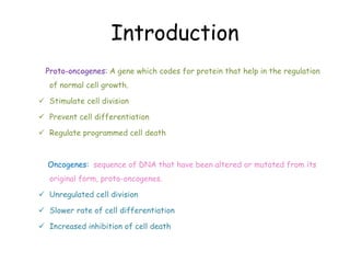
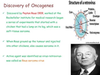
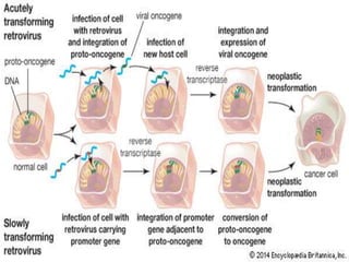
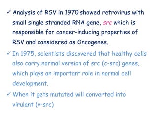
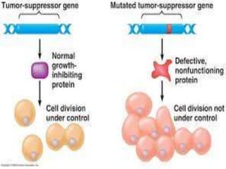
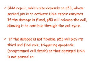
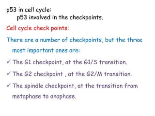
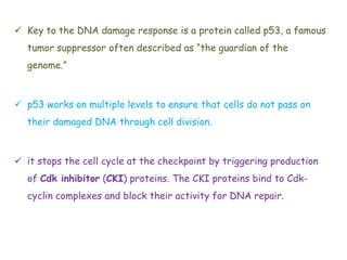
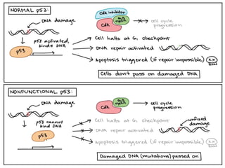
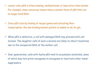
This document discusses proto-oncogenes, oncogenes, and tumor suppressor genes. Proto-oncogenes are normal genes that regulate cell growth, while oncogenes are mutated proto-oncogenes that contribute to uncontrolled cell growth. The discovery of oncogenes began with Rous sarcoma virus, which was found to contain the viral oncogene v-src. The tumor suppressor gene p53 plays a key role in DNA repair and apoptosis, preventing cancer formation. When mutated, p53 cannot regulate the cell cycle properly, allowing damaged DNA to be passed on and tumors to form.
An oncogene is a gene that has the potential to cause cancer. In tumor cells, they are mutated or expressed at high levels. Most normal cells undergo a programmed form of rapid cell death (apoptosis) when critical functions are altered.
Introduction
History
Tumor suppressor gene- pRB
- RB gene
- Role of RB in regulation of cell cycle
- Tumor associated with RB gene mutation
Tumor suppressor gene- p53
- What is p53 gene?
- Function of p53 gene
- How it regulates cell cycle
- What happen if p53 gene inactivated
- Cancer associated with p53 mutation
- Conclusion
- References
Just like the Dr. Henry Jekyll & Mr. Edward Hyde from the 'Strange Case of Dr. Jekyll & Mr. Hyde' written by R. L. Stevenson, in our body, there are several genes which behave exactly like them. When these genes are the good guys they are called Proto-Oncogenes and when they are the bad guys they are Oncogenes.
The p53 gene like the Rb gene, is a tumor suppressor gene, i.e., its activity stops the formation of tumors. If a person inherits only one functional copy
APOPTOSIS & CANCER
How is the initiator caspase frist activated in response to an apoptotic signal?
The role of pRB in controlling transcription of genes
The Role of p53: Guardian of the Genome
Mutations in the PI3K/Akt/mTOR Pathway Drive Cancer Cells to Grow
A detailed description of programmed cell death mechanism also called Apoptosis.
It explains about the factors, mechanism and pathways involved in the apoptosis.
Oncogenes, proto-oncogenes and tumor suppressor geneVaishnaviJanjal
An oncogene is a mutated gene that has the potential to cause cancer. Before an oncogene becomes mutated, it is called a proto-oncogene, and it plays a role in regulating normal cell division. Cancer can arise when a proto-oncogene is mutated, changing it into an oncogene and causing the cell to divide and multiply uncontrollably. Some oncogenes work like an accelerator pedal in a car, pushing a cell to divide again and again. Others work like a faulty brake in a car parked on a hill, also causing the cell to divide unchecked.
Tumor suppressor genes are normal genes that slow down cell division or tell cells to die at the right time (a process known
as apoptosis or programmed cell death). When tumor suppressor genes don't work properly, cells can grow out of control, which can lead to cancer.
Similar to oncogenes and tumour supressor genes (20)
This pdf is about the Schizophrenia.
For more details visit on YouTube; @SELF-EXPLANATORY;
https://www.youtube.com/channel/UCAiarMZDNhe1A3Rnpr_WkzA/videos
Thanks...!
THE IMPORTANCE OF MARTIAN ATMOSPHERE SAMPLE RETURN.Sérgio Sacani
The return of a sample of near-surface atmosphere from Mars would facilitate answers to several first-order science questions surrounding the formation and evolution of the planet. One of the important aspects of terrestrial planet formation in general is the role that primary atmospheres played in influencing the chemistry and structure of the planets and their antecedents. Studies of the martian atmosphere can be used to investigate the role of a primary atmosphere in its history. Atmosphere samples would also inform our understanding of the near-surface chemistry of the planet, and ultimately the prospects for life. High-precision isotopic analyses of constituent gases are needed to address these questions, requiring that the analyses are made on returned samples rather than in situ.
A brief information about the SCOP protein database used in bioinformatics.
The Structural Classification of Proteins (SCOP) database is a comprehensive and authoritative resource for the structural and evolutionary relationships of proteins. It provides a detailed and curated classification of protein structures, grouping them into families, superfamilies, and folds based on their structural and sequence similarities.
Earliest Galaxies in the JADES Origins Field: Luminosity Function and Cosmic ...Sérgio Sacani
We characterize the earliest galaxy population in the JADES Origins Field (JOF), the deepest
imaging field observed with JWST. We make use of the ancillary Hubble optical images (5 filters
spanning 0.4−0.9µm) and novel JWST images with 14 filters spanning 0.8−5µm, including 7 mediumband filters, and reaching total exposure times of up to 46 hours per filter. We combine all our data
at > 2.3µm to construct an ultradeep image, reaching as deep as ≈ 31.4 AB mag in the stack and
30.3-31.0 AB mag (5σ, r = 0.1” circular aperture) in individual filters. We measure photometric
redshifts and use robust selection criteria to identify a sample of eight galaxy candidates at redshifts
z = 11.5 − 15. These objects show compact half-light radii of R1/2 ∼ 50 − 200pc, stellar masses of
M⋆ ∼ 107−108M⊙, and star-formation rates of SFR ∼ 0.1−1 M⊙ yr−1
. Our search finds no candidates
at 15 < z < 20, placing upper limits at these redshifts. We develop a forward modeling approach to
infer the properties of the evolving luminosity function without binning in redshift or luminosity that
marginalizes over the photometric redshift uncertainty of our candidate galaxies and incorporates the
impact of non-detections. We find a z = 12 luminosity function in good agreement with prior results,
and that the luminosity function normalization and UV luminosity density decline by a factor of ∼ 2.5
from z = 12 to z = 14. We discuss the possible implications of our results in the context of theoretical
models for evolution of the dark matter halo mass function.
Introduction:
RNA interference (RNAi) or Post-Transcriptional Gene Silencing (PTGS) is an important biological process for modulating eukaryotic gene expression.
It is highly conserved process of posttranscriptional gene silencing by which double stranded RNA (dsRNA) causes sequence-specific degradation of mRNA sequences.
dsRNA-induced gene silencing (RNAi) is reported in a wide range of eukaryotes ranging from worms, insects, mammals and plants.
This process mediates resistance to both endogenous parasitic and exogenous pathogenic nucleic acids, and regulates the expression of protein-coding genes.
What are small ncRNAs?
micro RNA (miRNA)
short interfering RNA (siRNA)
Properties of small non-coding RNA:
Involved in silencing mRNA transcripts.
Called “small” because they are usually only about 21-24 nucleotides long.
Synthesized by first cutting up longer precursor sequences (like the 61nt one that Lee discovered).
Silence an mRNA by base pairing with some sequence on the mRNA.
Discovery of siRNA?
The first small RNA:
In 1993 Rosalind Lee (Victor Ambros lab) was studying a non- coding gene in C. elegans, lin-4, that was involved in silencing of another gene, lin-14, at the appropriate time in the
development of the worm C. elegans.
Two small transcripts of lin-4 (22nt and 61nt) were found to be complementary to a sequence in the 3' UTR of lin-14.
Because lin-4 encoded no protein, she deduced that it must be these transcripts that are causing the silencing by RNA-RNA interactions.
Types of RNAi ( non coding RNA)
MiRNA
Length (23-25 nt)
Trans acting
Binds with target MRNA in mismatch
Translation inhibition
Si RNA
Length 21 nt.
Cis acting
Bind with target Mrna in perfect complementary sequence
Piwi-RNA
Length ; 25 to 36 nt.
Expressed in Germ Cells
Regulates trnasposomes activity
MECHANISM OF RNAI:
First the double-stranded RNA teams up with a protein complex named Dicer, which cuts the long RNA into short pieces.
Then another protein complex called RISC (RNA-induced silencing complex) discards one of the two RNA strands.
The RISC-docked, single-stranded RNA then pairs with the homologous mRNA and destroys it.
THE RISC COMPLEX:
RISC is large(>500kD) RNA multi- protein Binding complex which triggers MRNA degradation in response to MRNA
Unwinding of double stranded Si RNA by ATP independent Helicase
Active component of RISC is Ago proteins( ENDONUCLEASE) which cleave target MRNA.
DICER: endonuclease (RNase Family III)
Argonaute: Central Component of the RNA-Induced Silencing Complex (RISC)
One strand of the dsRNA produced by Dicer is retained in the RISC complex in association with Argonaute
ARGONAUTE PROTEIN :
1.PAZ(PIWI/Argonaute/ Zwille)- Recognition of target MRNA
2.PIWI (p-element induced wimpy Testis)- breaks Phosphodiester bond of mRNA.)RNAse H activity.
MiRNA:
The Double-stranded RNAs are naturally produced in eukaryotic cells during development, and they have a key role in regulating gene expression .
Cancer cell metabolism: special Reference to Lactate PathwayAADYARAJPANDEY1
Normal Cell Metabolism:
Cellular respiration describes the series of steps that cells use to break down sugar and other chemicals to get the energy we need to function.
Energy is stored in the bonds of glucose and when glucose is broken down, much of that energy is released.
Cell utilize energy in the form of ATP.
The first step of respiration is called glycolysis. In a series of steps, glycolysis breaks glucose into two smaller molecules - a chemical called pyruvate. A small amount of ATP is formed during this process.
Most healthy cells continue the breakdown in a second process, called the Kreb's cycle. The Kreb's cycle allows cells to “burn” the pyruvates made in glycolysis to get more ATP.
The last step in the breakdown of glucose is called oxidative phosphorylation (Ox-Phos).
It takes place in specialized cell structures called mitochondria. This process produces a large amount of ATP. Importantly, cells need oxygen to complete oxidative phosphorylation.
If a cell completes only glycolysis, only 2 molecules of ATP are made per glucose. However, if the cell completes the entire respiration process (glycolysis - Kreb's - oxidative phosphorylation), about 36 molecules of ATP are created, giving it much more energy to use.
IN CANCER CELL:
Unlike healthy cells that "burn" the entire molecule of sugar to capture a large amount of energy as ATP, cancer cells are wasteful.
Cancer cells only partially break down sugar molecules. They overuse the first step of respiration, glycolysis. They frequently do not complete the second step, oxidative phosphorylation.
This results in only 2 molecules of ATP per each glucose molecule instead of the 36 or so ATPs healthy cells gain. As a result, cancer cells need to use a lot more sugar molecules to get enough energy to survive.
introduction to WARBERG PHENOMENA:
WARBURG EFFECT Usually, cancer cells are highly glycolytic (glucose addiction) and take up more glucose than do normal cells from outside.
Otto Heinrich Warburg (; 8 October 1883 – 1 August 1970) In 1931 was awarded the Nobel Prize in Physiology for his "discovery of the nature and mode of action of the respiratory enzyme.
WARNBURG EFFECT : cancer cells under aerobic (well-oxygenated) conditions to metabolize glucose to lactate (aerobic glycolysis) is known as the Warburg effect. Warburg made the observation that tumor slices consume glucose and secrete lactate at a higher rate than normal tissues.
What is greenhouse gasses and how many gasses are there to affect the Earth.moosaasad1975
What are greenhouse gasses how they affect the earth and its environment what is the future of the environment and earth how the weather and the climate effects.
Richard's aventures in two entangled wonderlandsRichard Gill
Since the loophole-free Bell experiments of 2020 and the Nobel prizes in physics of 2022, critics of Bell's work have retreated to the fortress of super-determinism. Now, super-determinism is a derogatory word - it just means "determinism". Palmer, Hance and Hossenfelder argue that quantum mechanics and determinism are not incompatible, using a sophisticated mathematical construction based on a subtle thinning of allowed states and measurements in quantum mechanics, such that what is left appears to make Bell's argument fail, without altering the empirical predictions of quantum mechanics. I think however that it is a smoke screen, and the slogan "lost in math" comes to my mind. I will discuss some other recent disproofs of Bell's theorem using the language of causality based on causal graphs. Causal thinking is also central to law and justice. I will mention surprising connections to my work on serial killer nurse cases, in particular the Dutch case of Lucia de Berk and the current UK case of Lucy Letby.
Professional air quality monitoring systems provide immediate, on-site data for analysis, compliance, and decision-making.
Monitor common gases, weather parameters, particulates.
2. Introduction
Proto-oncogenes: A gene which codes for protein that help in the regulation
of normal cell growth.
Stimulate cell division
Prevent cell differentiation
Regulate programmed cell death
Oncogenes: sequence of DNA that have been altered or mutated from its
original form, proto-oncogenes.
Unregulated cell division
Slower rate of cell differentiation
Increased inhibition of cell death
3. Discovery of Oncogenes
Discoverd by Peyton Rous 1909, worked at the
Rockefeller institute for medical research began
a series of experiments that started with a
chicken that had a lump on its leg, which was a
soft-tissue sarcoma.
When Rous ground up the tumour and injected
into other chickens, also causes sarcoma in it.
Active agent was identified as virus-retrovirus-
was called as Rous sarcoma virus
4.
5. Analysis of RSV in 1970 showed retrovirus with
small single stranded RNA gene, src which is
responsible for cancer-inducing properties of
RSV and considered as Oncogenes.
In 1975, scientists discovered that healthy cells
also carry normal version of src (c-src) genes,
which plays an important role in normal cell
development.
When it gets mutated will converted into
virulant (v-src)
6. SRC: Proto-oncogene tyrosine-protein kinase
Cell morphology, motility
Proliferation and survival
Structure:
14-c myristoyl group, SH3 andSH2 domain, protein
tyrosine kinase domain and c-terminal regulatory tail
Chief phosphorylation site of src include tyrosine
416 result in activaion from autophosphorylation and
tyrosine 527 result in inhibition from
phosphorylation by c-terminal src kinase.
7. Tyrosine kinase signalling and
oncogenesis
Normal cells receive and interpret external signals which
influence their growth, shape, metabolism and movement.
Complex interactions between molecules inside the cell
transmit these signals and initiate appropriate cellular
responses.
Protein phosphorylation on Tyrosine (Tyr) residues has
evolved as an important mechanism to coordinate this
intracellular signalling process in multicellular organisms.
8.
9. Inactivation:
Y416 is autophosphorylation site in the activation loop and
negative regulation in tyrosine site by phosphorylation at
Y527 in the c-terminal end.
c-src is auto inhibited through intramolecular association
between the SH2 and Y527, and SH3 and poly proline motif
in the linker.
Activation:
Activation is by either dephosphorylation of Y527 to
release the SH2 domain or by binding of peptide ligand to
recognise SH2 leading to autophosphorylation of Y416.
10. When Src goes wrong???
Under normal circumstances, Src is predominantly inactive in cells,
being switched on only at specific times.
fine balance between phosphorylation and dephosphorylation is
disrupted, changes can occur in Src activity with drastic results.
Several cancers, including colon and breast cancer, have been
associated with an increase in Src activity.
v-Src, from the transforming virus, Rous Sarcoma Virus. v-Src was
found to lack the region of the cellular protein (c-Src) that contains
Tyr527, making it continually active.
11. In late stage colon cancers, mutations have been reported in the src
gene that cause the loss of the region containing Tyr527, leading to
Src over-activity.
Proteins that regulate Src have also been found at abnormal levels
in cancer cells, including both those that activate and those that
inactivate Src.
Proteins such as PTPalpha, SHP-1 and PTP1B that activate Src by
dephosphorylating Tyr527 have been detected at elevated levels in
various cancer cells, including epidermal and breast carcinoma cells.
12. Therapeutic applications
Control over the metabolic alterations :
A decreased oxidative phosphorylation, increased glucose
consumubsequent lactate production are the remarkable features of
oncogenic activation .
Over the last decade, evidence has accumulated that the oncogenes myc,
AKT, nuclear factor kB (NF-kB) and the tyrosine kinase receptors
(insulin-like growth factor 1, epidermal growth factor, EGF; IGF-1; Her-2;
etc.), which turn on phosphatidylinositol 3-kinases (PI3Ks), mammalian
target of rapamycin (mTOR) pathway and the Ras, RAF–Mitogen-
Activated Protein Kinase (MAP kinase) along with hypoxia induced factor
(HIF), can activate the transcription of numerous genes that encode the
proteins that mediate the various metabolic pathways .
13. Myc and HIF-1 transcription factors
enhances the rate of transcription of some
of the GLUT transporters and hexokinase
2, increasing both glucose uptake and its
retention in the cell .
Increases the oxidative phosphorylation
and regulate the metabolic pathway.
14. Tumor suppressor gene:
A protective gene that normally limits the growth of tumors. When a tumor
suppressor gene is mutated (altered), it may fail to keep a cancer from growing.
The first insight into the activity of tumor suppressor genes came from somatic cell
hybridization experiments, initiated by Henry Harris and his colleagues in 1969.
The fusion of normal cells with tumor cells yielded hybrid cells containing chromosomes
from both parents.
The first tumor suppressor gene was identified by studies of retinoblastoma, a rare
childhood eye tumor.
15. Protein that halts cell cycle progression in response to
DNA damage may no longer sense damage or trigger a
response. Genes that normally block cell cycle progression
are known as tumor suppressors.
Tumor suppressors prevent the formation of cancerous
tumors when they are working correctly, and tumors may
form when they mutate so they no longer work.
One of the most important tumour suppressor gene is p53,
which plays a key role in the cellular response to DNA
damage.
18. DNA repair, which also depends on p53, whose
second job is to activate DNA repair enzymes.
If the damage is fixed, p53 will release the cell,
allowing it to continue through the cell cycle.
If the damage is not fixable, p53 will play its
third and final role: triggering apoptosis
(programmed cell death) so that damaged DNA
is not passed on.
19. p53 in cell cycle:
p53 involved in the checkpoints.
Cell cycle check points:
There are a number of checkpoints, but the three
most important ones are:
The G1 checkpoint, at the G1/S transition.
The G2 checkpoint , at the G2/M transition.
The spindle checkpoint, at the transition from
metaphase to anaphase.
20. Key to the DNA damage response is a protein called p53, a famous
tumor suppressor often described as “the guardian of the
genome.”
p53 works on multiple levels to ensure that cells do not pass on
their damaged DNA through cell division.
it stops the cell cycle at the checkpoint by triggering production
of Cdk inhibitor (CKI) proteins. The CKI proteins bind to Cdk-
cyclin complexes and block their activity for DNA repair.
21.
22. cancer cells, p53 is often missing, nonfunctional, or less active than normal.
For example, many cancerous tumors have a mutant form of p53 that can
no longer bind DNA.
Since p53 acts by binding to target genes and activating their
transcription, the non-binding mutant protein is unable to do its job.
When p53 is defective, a cell with damaged DNA may proceed with cell
division. The daughter cells of such a division are likely to inherit mutations
due to the unrepaired DNA of the mother cell.
Over generations, cells with faulty p53 tend to accumulate mutations, some
of which may turn proto-oncogenes to oncogenes or inactivate other tumor
suppressors.
23. Apoptosis
The cells between your embryonic fingers died in
a process called apoptosis, a common form of
programmed cell death.
In programmed cell death, cells undergo
“cellular suicide” when they receive certain cues.
Apoptosis involves the death of a cell, but it
benefits the organism as a whole.
24. Cells that undergo apoptosis go through a
different and much more orderly process.
They shrink and develop bubble-like
protrusions (technical name: “blebs”) on
their surface.
The DNA in the nucleus gets chopped up
into small pieces, and some organelles of
the cell, such as the endoplasmic reticulum,
break down into fragments.
25. They release signals that attract debris-eating
(phagocytic) immune cells, such as macrophages.
Also, the fragments of the dying cell display a
lipid molecule called phosphatidylserine on their
surface.
Phosphatidyserine is usually hidden on the inside
of the membrane, and when it is on the outside,
it lets the phagocytes bind and "eat" the cell
fragments.
26. Pathways :
Intrinsic and extrinsic pathways
Extrinsic pathway:
In this pathway, phosphatidyserine is the death signal, which
activates the pro-caspase 8(inactive) to caspase 8.
Activated caspase 8 , which activated the procaspase 3 to
caspase3.
Intrinsic pathway:
p53, which activates the bax, and bax activates cyt C, binds
to apaf1 to form apoptosome.
Activation of apoptosome activates procaspase 9 to active
form of caspase 9
27.
28. Applications of p53-Based Cancer
Therapy
Reactivating Mutant p53:
PhiKan083 is a carbazole derivative found from in silico screening of
the crystal structure of p53.
By binding mutated p53, PhiKan083 raises the melting temperature of
mutated p53, which results in the reactivation of its function.
PRIMA-1 -Restored sequence-specific DNA binding and active the
conformation.
CP-31398- Restore the protein folding of mutated p53 to a more
natural conformation that permits a wild-type function
29. p53 Stabilization:
MDM2 is an E3 ubiquitin ligase which controls p53
degradation. Many tumors overexpress MDM2, even
tumors without p53 mutations.
Nutlins are cis-imidazoline compounds that act as
antagonists of the MDM2-p53 interaction.
MI-219 is another small molecule that inhibits the
MDM2-p53 interaction. MI-219 also activates the
p53 pathway in cells with wild-type p53. Apoptosis
and cell cycle arrest were observed in xenograft
tumors which resulted in tumor regression .
30. Classes of Drugs for p53 Stabilization:
Tenovin was found by a cell-based drug screen to
activate p53. Tenovin acts as an inhibitor of the
protein-deacetylating activities of SirT1 and
SirT2.
The intraperitoneal administration of tenovin-6
has been demonstrated to induce a regression of
xenograft tumors in a mouse mode
31. p53 Gene Therapy:
p53-based gene therapy was reported in 1996. A
retroviral vector containing the wild-type p53 gene
under the control of an actin promoter was injected
directly into tumors of nonsmall cell lung cancer
patients.
After development of a replication-defective
recombinant p53 virus (Ad5CMV-p53) , many clinical
trials have been performed, including one in
esophageal cancer patients .
32. Conclusion
Cytoplasmic p53 can activate a transcription-
independent apoptotic program. The next generation
of p53-based cancer therapeutic approaches should
therefore be developed to take advantage of this
cytosolic function.
This may be safer than regulating the transcription
modulation of wild-type p53, which can induce both
prosurvival and proapoptotic effects in tumor cells.
33. Recent reports have showed that p53 regulates the process of
self-renewal of neural stem cells and hematopoietic stem cells.
The cancer stem model insists that tumors are maintained by a
small population of cancer stem cells that can divide both
symmetrically and asymmetrically.
Mammary stem cells with the targeted mutation of p53 have been
reported to show the same properties as cancer stem cells. The
reactivation of p53 restored the asymmetric cell division of
cancer stem cells and induced tumor growth inhibition .
34. References
T. Soussi and C. Béroud, “Assessing TP53 status in human tumours
to evaluate clinical outcome,” Nature Reviews Cancer, vol. 1, no. 3, pp.
233–240, 2001. View at Google Scholar · View at Scopus
K. H. Vousden and C. Prives, “Blinded by the light: the growing
complexity of p53,” Cell, vol. 137, no. 3, pp. 413–431, 2009. View at
Publisher · View at Google Scholar · View at Scopus
T. Riley, E. Sontag, P. Chen, and A. Levine, “Transcriptional control
of human p53-regulated genes,” Nature Reviews Molecular Cell
Biology, vol. 9, no. 5, pp. 402–412, 2008. View at Publisher · View at
Google Scholar · View at Scopus
35. Bjorn MJ, Groetsema G, Scalapino L. Antibody-Pseudomonas exotoxin A conjugates
cytotoxic to human breast cancer cells in vitro. Cancer Res 46:3262, 1986.PubMed
Cline M, Slamon DJ, Lipsick JS. Oncogenes: implications for the diagnosis and
treatment of cancer. Ann Intern Med 101:223, 1984.PubMed
Cote RJ, Houghton AN. The generation of human monoclonal antibodies and their
use in the analysis of the humoral immune response to cancer. In: Engleman EG,
Foung SKH, Larrick JW, Raubitschek A (eds), Human Hybridomas and Monoclonal
Antibodies. New York: Plenum Press, 1985, p. 189.
Douillard JY, Lehur PA, Aillet G, et al. Immunohistochemical antigenic expression
and in vivo tumor uptake of monoclonal antibodies with specificity for tumors of
the gastrointestinal tract. Cancer Res 46:4221, 1986.