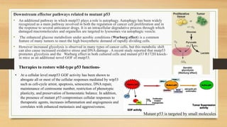
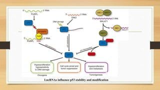
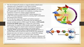
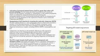
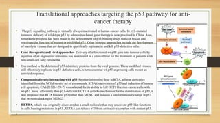
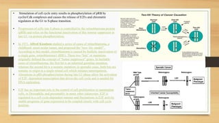
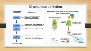
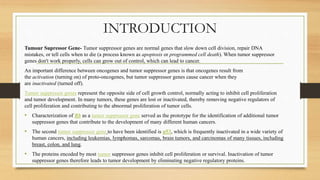
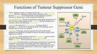
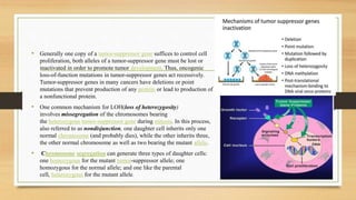
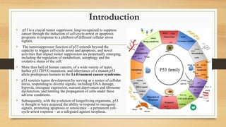
The document discusses the structure, function, and mechanisms of action of the tumor suppressor genes p53 and Rb, highlighting their roles in regulating cell cycle, apoptosis, and DNA repair. It details how the inactivation of these genes contributes to cancer development and outlines the genetic and epigenetic alterations associated with p53 mutations in various tumors. Additionally, the document explores therapeutic approaches targeting p53 pathways to improve cancer treatment outcomes.
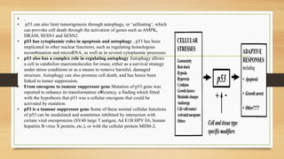
![• p53 restricts tumor development by serving as a sensor of cellular
stress, responding to diverse signals, including DNA damage,
hypoxia, oncogene expression, nutrient deprivation and ribosome
dysfunction, and limiting the propagation of cells under these adverse
conditions.
• p53 is thought to have acquired the ability to respond to oncogenic
signals, promoting apoptosis or senescence – a permanent cell-cycle-
arrest response – as a safeguard against neoplasia.
• Alternatively, under conditions of low-level stress, p53 elicits
protective, pro-survival responses, such as temporary cell-cycle arrest,
DNA repair and antioxidant protein production, to maintain genome
integrity and viability in cells that sustain limited, reparable damage.
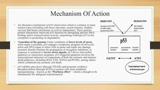
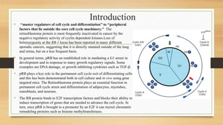
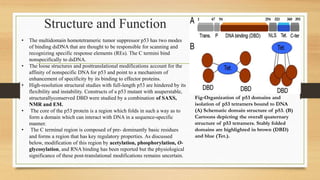
• p53 has protein domains typical of transcriptional activators
Similarly to other transcription factors, p53 has a modular protein
domain structure.
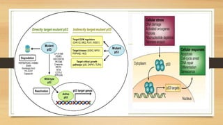
• The six most common p53 amino acid residues altered in cancer –
known as ‘hotspots’ – are R175, G245, R248, R249, R273 and R282.
In addition to disrupting DNA binding, these mutations can confer
gain-of-function capabilities on p53, and have been linked to
increased invasiveness and metastasis of tumors.
• p53 induces a host of transcriptional programs involved in
different responses Once active p53 is bound to DNA, it can
stimulate the transcription of many proteincoding and non-protein-
coding genes [e.g. microRNAs or large intergenic non-coding RNAs
(lincRNAs)], which is a function of fundamental importance for all
p53-mediated responses.](https://image.slidesharecdn.com/animalcellppt-200711083344/85/Tumour-supressor-genes-8-320.jpg)