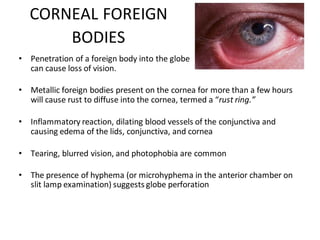
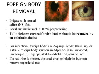
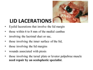
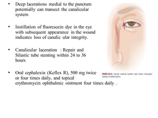
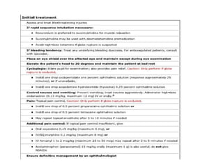
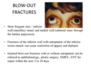
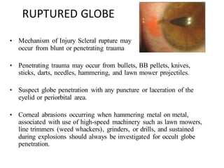
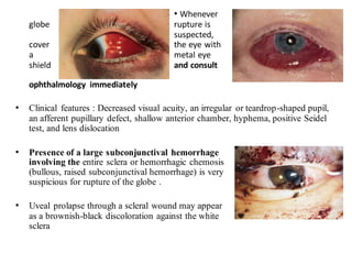
The document discusses various types of ocular injuries including open globe injuries, closed globe injuries, and periocular injuries. Open globe injuries involve a full thickness break of the eye wall, while closed globe injuries do not penetrate the full thickness. Periocular injuries involve structures surrounding the eye such as the eyelids. Specific injuries like corneal abrasions, hyphema, and lid lacerations are described along with their typical presentations, evaluations, and treatments. Ocular trauma requires careful examination and many cases need referral to an ophthalmologist.
The document discusses slit lamp examinations, which use a high-intensity light source focused as a slit and viewed through a microscope to examine the anterior segment of the eye. It describes the basic components and principles of the slit lamp biomicroscope, various illumination techniques used to examine different ocular structures, and historical developments of the slit lamp.
Cycloplegia involves paralyzing the ciliary muscle through the use of cycloplegic drugs like atropine and cyclopentolate. This allows for an objective determination of refractive error by eliminating accommodation. Cyclorefraction is important for accurately diagnosing latent hyperopia in children, which if left uncorrected can cause vision problems. Cyclopentolate is generally the drug of choice for routine cyclorefraction due to its fast onset and relatively short duration of action. Completion of cycloplegia is determined through push-up testing to ensure no residual accommodation remains before performing refraction.
1) The Holladay 2 formula is considered one of the most accurate IOL calculation formulas today. It was developed in 1993 by Dr. Holladay based on a study of over 34,000 eyes to determine the key variables that predict effective lens position.
2) The Holladay 2 formula utilizes 7 parameters - axial length, keratometry, white-to-white, lens thickness, and anterior chamber depth - to calculate the effective lens position and appropriate IOL power.
3) The Holladay 2 formula is recommended for its high accuracy and predictability. The IOLMaster 500 is currently the only device that has the Holladay 2 formula integrated to automatically calculate the IOL
This document discusses the evaluation of the nasolacrimal system. It covers the history and anatomy of the system, classification of epiphora, diagnostic tests used to evaluate the system including anatomical tests like syringing and probing, functional tests like dye disappearance tests, and secretory tests like Schirmer's test. The document also discusses differentiating anatomical obstruction from functional/physiological causes of excessive tearing and localization of blockages in the system.
Ophthalmic viscosurgical devices (OVDs) are used in eye surgeries to protect tissues and maintain spaces. Common OVDs include sodium hyaluronate, chondroitin sulfate, and hydroxypropyl methylcellulose. OVDs can be classified as cohesive, dispersive, or viscoadaptive based on their rheological properties such as viscosity and elasticity. Cohesive OVDs are highly viscous and stick together, while dispersive OVDs coat tissues well but are less viscous. OVDs are used in cataract surgery for tasks like protecting the endothelium, maintaining the anterior chamber, and implanting IOLs. Complications can include increased intraocular
This document discusses the process of subjective refraction to determine a patient's prescription. It involves 5 main steps: 1) determining the best vision sphere for each eye, 2) using a Jackson Cross Cylinder to find the cylindrical axis and power, 3) refining the results, 4) binocular balancing to account for any differences between the eyes, and 5) determining the binocular best sphere. Fogging and duochrome tests are used to achieve the best vision sphere. Near additions are also considered for presbyopic patients based on their habitual reading distance and age. Trial lens sets and phoropters are the main instruments used.
This document discusses scleral buckling surgery for retinal detachment. It describes scleral buckling as an old technique that uses scleral implants or explants like an encircling band or local explant. The document highlights key steps in scleral buckling surgery including preoperative assessment to locate all retinal breaks, retinal drawing, localization of breaks intraoperatively, isolation of the recti muscles, retinopexy using methods like cryotherapy, and postoperative considerations. Scleral buckling is presented as an option for primary retinal detachment repair, especially in phakic patients, though the document also notes evidence supporting primary vitrectomy in some cases.
A surgical procedure featuring a partial thickness scleral flap that creates a fistula between AC and subconjunctival space for filtration of aqueous and creation of conjunctival bleb in an effort to lower IOP
Collagen cross linking (CXL) aims to strengthen the cornea through the formation of covalent bonds between collagen fibers. The standard Dresden protocol uses riboflavin and UV light over 30 minutes. Newer accelerated protocols reduce treatment time but increase UV intensity. Trans-epithelial CXL leaves the epithelium intact but appears less effective. Combining CXL with refractive treatments like PRK can improve outcomes for keratoconus. Future areas of research include techniques to speed up CXL like flashlinking and methods to enhance riboflavin penetration such as iontophoresis.
This document discusses the embryology and anatomy of the cornea. It describes how the cornea develops from surface ectoderm in the 4th-5th week of gestation, with mesenchymal cells forming the stroma and endothelium. The cornea continues developing through the fetal period, with layers such as Bowman's membrane forming between 12-26 weeks. The document also discusses the cellular components, functions, and common congenital anomalies of the cornea, including microcornea, megalocornea, cornea plana, keratoconus, and others.
This document provides information about fundus fluorescein angiography (FFA). It begins with basic principles of FFA and the dyes used, including sodium fluorescein and indocyanine green. The purpose, indications, contraindications, technique, phases, and interpretation of FFA are described. Abnormal fluorescence patterns like hyperfluorescence and hypofluorescence are discussed. Recent advances in wide-field imaging and indocyanine green angiography are also summarized.
This document discusses objective refraction techniques, primarily retinoscopy. It begins by explaining the principles of retinoscopy, including far point concept and how different ametropias affect the far point. It then describes the components and optics of the retinoscope, how it works, and retinoscopy techniques. Key aspects covered include neutralization, prerequisites for retinoscopy, and problems that can occur. Autorefractometry is also briefly discussed. In under 3 sentences:
Retinoscopy is the primary objective refraction technique discussed, which uses a retinoscope to illuminate the retina and observe the movement of the red reflex to determine the refractive error, neutralizing with trial lenses. The document covers the optics
The document summarizes the history and generations of intraocular lenses (IOLs). It discusses the first IOL implanted by Sir Harold Ridley, made of polymethyl methacrylate (PMMA). Later generations included foldable materials, multifocal IOLs, and accommodating IOLs. Premium IOLs now offer features like toric designs to correct astigmatism and aspheric optics for better vision quality. While multifocal IOLs provide simultaneous distance and near vision without glasses, they can cause visual side effects like halos and reduced contrast sensitivity. Accommodating IOLs aim to restore a degree of natural accommodation but may not provide fully unaided near vision for small print.
This document provides an overview of high myopia, including its classification, causes, symptoms, complications, and management approaches. High myopia is defined as a refractive error greater than -6.00 diopters or an axial length over 26.5mm. It can be caused by genetic factors or the general growth process. Symptoms include defective vision, closer working distance, and night blindness. Complications involve retinal degeneration and detachment. Management includes high-powered spectacles, contact lenses, and refractive surgeries such as LASIK or clear lens extraction.
1) Trabeculectomy is a glaucoma surgery that creates an opening in the eye to drain fluid from the anterior chamber and reduce intraocular pressure.
2) It involves making a partial thickness scleral flap, removing a block of tissue underneath, and suturing the flap loosely to allow fluid drainage.
3) Antifibrotic agents like mitomycin C or 5-fluorouracil are often applied to reduce scarring and improve surgical success rates.
An intraocular lens (IOL) is an artificial lens implanted in the eye during cataract surgery or refractive lens exchange to replace the natural crystalline lens and correct vision. IOLs have evolved over generations from rigid PMMA lenses to modern foldable designs made of silicone, acrylic, or hydrogel materials. IOLs are either placed in the posterior chamber behind the iris or in the anterior chamber in front of the iris. Newer multifocal and accommodating IOL designs aim to provide both distance and near vision. Precise biometry is required to calculate the optimal IOL power to achieve the desired postoperative refraction. Potential complications include dislocation, opacification, deposits, and inflammation.
This document defines and describes the anatomy and physiology of the lacrimal apparatus, which is responsible for tear formation and drainage in the eye. It discusses how lacrimal syringing is used to test the structural integrity of the lacrimal drainage system by passing fluid through the puncta and observing for any blockages or abnormalities. The document also outlines the equipment needed for lacrimal syringing and provides interpretations of different results that could indicate where in the drainage system a blockage may be located.
This document discusses fluorescein angiography (FA), a technique used to examine the circulation of the retina and choroid. FA involves injecting a fluorescent dye called sodium fluorescein and then taking photographs of the retina as the dye circulates through the eye. The dye is absorbed by the blood vessels and any leaks or blockages are visible. Precautions are taken and potential side effects explained to the patient. The sequence of FA images provides information about blood flow and any pathological conditions.
This document discusses challenges and new technologies for accommodating intraocular lenses to treat presbyopia. It summarizes several hinged and dual-optic accommodating IOL designs currently in development or clinical trials, including their mean accommodative amplitudes. However, it notes that fibrosis and capsular contraction often reduce accommodative abilities over time for hinged lenses. Other challenges include determining optimal lens powers and preventing complications. The document explores several experimental technologies as well, but notes many hurdles remain to restoring full accommodation.
BASIC INFO ON FUDUS FLORESCENCE ANGIOGRAPHYNalin Nayan
The document discusses fundus fluorescein angiography (FFA). FFA involves injecting a fluorescent dye called fluorescein and using a retinal camera to take photos of the retina and choroid as the dye circulates. It describes the five phases seen in FFA - choroidal, arterial, capillary, venous, and late phases. Abnormalities that may appear as hyperfluorescence or hypofluorescence on FFA are also outlined.
The document provides an overview of the anatomy and physiology of the cornea, beginning with its embryology and dimensions. It discusses the histology of the cornea's layers - epithelium, Bowman's layer, stroma, Descemet's membrane, and endothelium. For each layer, its composition, thickness, cell types, and clinical significance are described. The cornea's vascular supply, nerve supply, physiology, and various clinical conditions are also summarized. The document aims to provide applied and clinical relevance for understanding the structure and function of the cornea.
The document discusses the history and techniques of penetrating keratoplasty (PK). It notes that the first successful PK was performed in 1905. Common indications for PK include corneal opacities, dystrophies, trauma, and graft failure. The preoperative evaluation, surgical steps such as trephination and suturing, and postoperative care are described. Complications during and after PK can include perforation, bleeding, and graft rejection. Tight control of inflammation and immunosuppression are important for managing high-risk grafts.
Gonioscopy refers to techniques for examining the anterior chamber angle of the eye to evaluate and classify normal and abnormal angle structures. It involves using specialized lenses and lights to view the iridocorneal angle. There are two main methods: direct gonioscopy uses large contact lenses requiring saline, while indirect uses smaller lenses with mirrors or prisms that utilize tear film. Gonioscopy is used to diagnose angle-closure glaucoma and other conditions by allowing visualization of the trabecular meshwork, scleral spur, and other angle structures. Indentation gonioscopy, where the lens is pressed against the cornea, can differentiate appositional from synechial angle closure. Proper technique and
The document discusses recent advances in the management of obstruction of the lacrimal drainage system. It describes the history and evolution of various surgical techniques for treating this obstruction, including external dacryocystorhinostomy (DCR), endoscopic endonasal DCR, ultrasonic endoscopic DCR, non-laser endonasal DCR, endocanalicular laser DCR, and balloon-assisted DCR. It then provides details on the surgical procedure for external DCR, including patient preparation, instrumentation, osteotomy creation, flap formation, flap anastomosis, wound closure, and post-operative management. Potential complications are also briefly mentioned.
This document discusses potential complications of trabeculectomy, both intraoperative and postoperative. Intraoperative complications include buttonholing of the conjunctiva, scleral flap tears, lens injury, vitreous prolapse, hyphema, and suprachoroidal hemorrhage. Postoperative complications can be early such as hypotony, elevated intraocular pressure, choroidal effusions or late such as thin blebs, infections, and cataracts. Management strategies are provided for addressing complications depending on the specific issue.
The document summarizes management of ocular trauma from blunt injuries. It describes mechanisms of injury including kinetic energy transfer and oscillatory tissue damage. Treatment depends on injury severity and location, and may involve repair of open globe injuries, removal of intraocular foreign bodies, management of hyphema, lens subluxation or dislocation, and vitrectomy. The goal is to restore ocular integrity and address complications while minimizing further trauma from intervention.
The document discusses various types of penetrating ocular injuries including penetrating injuries, perforating injuries, and intraocular foreign bodies. It describes the common causes and effects of these injuries which can include laceration, vitreous hemorrhage, retinal tears and detachments. Diagnostic procedures like slit lamp examination and ultrasound are used to evaluate the injuries. Specific injuries like corneal lacerations, conjunctival lacerations, globe ruptures, and retinal detachments are also summarized. The treatment and management of different ocular injuries is covered as well.
Retinal detachment occurs when the retina separates from its underlying layers, which can cause vision loss if not treated. It is often caused by breaks or tears in the retina that allow fluid to enter and detach the retina. Diagnosis involves examining the eye through various tests like ophthalmoscopy, ultrasound and angiography. Treatment options aim to reattach the retina through procedures like laser photocoagulation, cryotherapy, scleral buckling surgery, pneumatic retinopexy or vitrectomy. These work by sealing retinal breaks or reducing fluid flow to allow the retina to reattach.
1. Corneal injuries can occur due to trauma, infection, or non-infectious causes. Trauma can cause lacerations or perforations, which are diagnosed using tests like Seidel's test. Small wounds may be treated medically but larger ones may require surgery.
2. Perforating eye injuries caused by intraocular foreign bodies often occur in occupational settings. Metallic foreign bodies carry less risk than organic materials. Location and size of the object impacts damage. Examination looks for entry sites and potential endophthalmitis.
3. Blunt eye injuries from impacts can cause globe rupture or retrobulbar hematoma, diagnosed using imaging and symptoms like proptosis. Immediate decomp
This document discusses various types of eye trauma including ruptures, lacerations, penetrating and perforating injuries of the eyewall. It also discusses principles of repair for conjunctival lacerations, corneal lacerations, full thickness tears, scleral lacerations, iris reconstruction using McCannel and Siepser techniques, iridodialysis repair, pupillary cerclage, and indications for surgery in cases of traumatic vitreous hemorrhage. Surgical principles emphasized include using fine sutures, clearing debris, preventing gape and prolapsing tissues, and burying knots towards the limbus.
This document provides information on retinal detachment including:
1. It describes the three main types of retinal detachment - rhegmatogenous, tractional, and exudative - and their causes.
2. Risk factors, clinical manifestations, diagnosis, and surgical management options like cryopexy, laser photocoagulation, scleral buckling, pneumatic retinopexy, and vitrectomy are summarized.
3. Post-operative care involves positioning, monitoring for complications, restricting activities that raise intraocular pressure, and reporting any new visual issues or pain immediately.
Retinal detachment is a disorder of the eye in which the retina separates from the layer underneath. Symptoms include an increase in the number of floaters, flashes of light, and worsening of the outer part of the visual field
This document summarizes key information about treating various ocular emergencies, including eyelid lacerations, globe proptosis, corneal ulcers, corneal lacerations, and glaucoma. It provides details on surgical and medical management for each condition, including antibiotic and pain medication protocols, suturing techniques, and postoperative care instructions. Clinical signs, diagnostic approaches, and classifications of ophthalmic antibiotics are also reviewed for certain conditions.
This document discusses the anatomy and management of the anophthalmic socket. It begins by defining an anophthalmic socket as an orbit lacking an eye globe, usually due to enucleation. True anophthalmia can be congenital or acquired. The most common causes of acquired anophthalmia are enucleation, exenteration, or evisceration performed to treat painful, blind eyes or remove tumors. The document outlines techniques for each procedure and discusses complications that can arise like socket contracture and management strategies like grafting. It emphasizes the importance of socket maintenance with conformers and prosthetics to prevent complications. In summary, this document provides an overview of anophthalmic socket anatomy,
Retinal detachment occurs when the retina separates from the underlying layers of the eye. It is usually caused by a tear or hole in the retina that allows fluid to enter and separate the retina. There are three main types of retinal detachment: rhegmatogenous, tractional, and exudative. Risk factors include high myopia, aging, trauma, and prior eye surgeries. Treatment options include laser photocoagulation, cryotherapy, scleral buckling surgery, vitrectomy, or pneumatic retinopexy to reattach the retina. Post-operative care focuses on rest, positioning, avoiding strain on the eyes, and follow up exams to monitor healing and prevent recurrence.
The document discusses anophthalmia, which refers to the complete absence of the eye and orbital tissues. Anophthalmia can be congenital or acquired through procedures like enucleation, evisceration, and exenteration. These procedures require orbital implants, conformers, and prosthetics to rehabilitate the socket. Long-term complications can include socket contracture, eyelid malpositions, and implant exposure or migration. Proper surgical planning and techniques aim to minimize complications and provide stable, comfortable sockets that allow for good prosthetic motility and cosmesis.
This document provides information on ocular trauma, including:
1. It defines various types of ocular injuries such as closed globe, open globe, contusion, laceration, and penetrating injuries.
2. It discusses classifications for open and closed globe injuries based on factors like visual acuity, zone of injury, and presence of RAPD.
3. It outlines the evaluation, history taking, examination and investigations for patients presenting with ocular trauma, as well as management approaches for different types of injuries like hyphema, intraocular foreign bodies, and open globe injuries.
The document discusses ocular trauma, providing classifications and guidelines for clinical evaluation and management. It describes the epidemiology of eye injuries and classifications for closed and open globe injuries. Evaluation involves history, exam, and potential investigations. Closed injuries include contusions while open injuries include ruptures and lacerations. Management depends on the injury type and aims to repair anatomy and maximize vision. Chemical burns require copious irrigation and treatments to normalize pH.
This document provides information on angle closure glaucoma, including its causes, risk factors, stages, clinical presentation, diagnostic tests, and treatment options. Angle closure glaucoma results from obstruction of the aqueous outflow pathway due to apposition or adhesion of the iris to the trabecular meshwork. It is more common in individuals with anatomically narrow anterior chamber angles and certain ethnic groups. Treatment involves lowering intraocular pressure through medications, laser procedures such as peripheral iridotomy or iridoplasty, or incisional surgeries like goniosynechialysis or trabeculectomy if needed.
Mechanical ocular trauma can cause a wide range of eye injuries from relatively minor to vision threatening. The document defines standard terminology for different types of eye injuries using the Birmingham Eye Trauma Terminology (BETT) system. It describes closed globe injuries which involve no penetration of the eyewall, open globe injuries which involve penetration of the eyewall, and different types of open globe injuries including globe rupture, penetrating injuries, and perforating injuries. It provides details on mechanisms of injury, clinical findings, examination techniques, and treatment approaches for different injury types.
This document provides information on various ophthalmology topics including:
1. Optic nerve swelling and papilledema are summarized.
2. Instruments used in ophthalmology such as the ophthalmoscope, slit lamp, and tonometers are described.
3. Common eye conditions like retinal detachment, glaucoma, and trauma are discussed in terms of signs, diagnosis, and management by general physicians.
This document summarizes various ocular emergencies commonly seen in emergency departments. It discusses trauma-related emergencies like penetrating injuries, blunt injuries, burns, as well as non-trauma emergencies such as infections, glaucoma, retinal vessel occlusions, retinal detachment, and ruptured globes. It also provides details on management of specific conditions like chemical injuries, acute angle closure glaucoma, foreign bodies, orbital fractures, and ocular infections.
Traumatic chorioretinal pathologies can result from both penetrating and non-penetrating ocular injuries. Males under 40 are most commonly affected. Closed globe injuries may cause commotio retinae, while open globe injuries risk choroidal rupture. Indirect injuries like Valsalva retinopathy or Terson syndrome can also cause intraocular bleeding. Traumatic macular holes and retinal detachments require surgical repair like vitrectomy. Overall ocular trauma remains a major cause of blindness worldwide.
Retinal detachment occurs when the retina separates from the underlying layer of the eye. It can cause vision loss if not treated. Risk factors include severe nearsightedness, prior eye surgery or injury, family history, and activities that increase intraocular pressure. Symptoms include flashes of light, floaters, and blurred or lost vision. Diagnosis involves eye exams like ultrasound or ophthalmoscopy. Treatment depends on the type of detachment but may include cryotherapy, laser photocoagulation, scleral buckling surgery, pneumatic retinopexy, or vitrectomy. With treatment, retinal detachment can be successfully repaired in 85-95% of cases to restore vision.
Retinal detachment is a serious condition where the retina peels away from its underlying supportive tissue layer. It can cause permanent vision loss if not repaired soon. The summary examines the risk factors, types, signs and symptoms, diagnosis, and treatment options for retinal detachment. Surgical methods like scleral buckling, cryopexy, laser photocoagulation, pneumatic retinopexy and vitrectomy aim to reattach the retina. Prompt treatment usually leads to vision improvement, though the prognosis depends on factors like macula involvement and presence of proliferative vitreoretinopathy. Post-operative care and home management are important to monitor for complications and aid recovery.
The document discusses the extraocular muscles (EOM) that control eye and eyelid movement. It describes the six muscles responsible for eye movement - the four rectus muscles (superior, inferior, medial, lateral) and two oblique muscles (superior, inferior). It also discusses the levator palpebrae superioris muscle responsible for raising the upper eyelid. The origins, insertions, innervations and actions of each muscle are defined.
The document discusses image formation using convex mirrors. It notes that convex mirrors always produce virtual, upright images that are located behind the mirror and are smaller than the object. As the object distance decreases, both the image distance and size increase. So as an object approaches a convex mirror, its virtual image also approaches the mirror and becomes larger. Additionally, the relationship between object and image distances is such that the distance between the image and mirror is always less than between the object and mirror for convex mirrors.
The document discusses the characteristics of images formed by a concave mirror based on the location of the object. It divides object locations into 5 cases: (1) beyond the center of curvature, (2) at the center of curvature, (3) between the center of curvature and focal point, (4) at the focal point, (5) in front of the focal point. For each case, it describes whether the image is real or virtual, upright or inverted, magnified or reduced compared to the object. Key factors that determine the image characteristics are the object's position relative to the focal point and center of curvature.
Optics is the study of light, including its interactions with matter. There are three main subfields: geometrical optics studies light as rays, physical optics studies light as waves, and quantum optics studies light as particles. Mirrors form images through the reflection of light rays according to specific rules. Plane mirrors form virtual upright images that are laterally inverted. Spherical mirrors can be concave or convex and form images using rules for tracing the path of light rays. Constructing ray diagrams involves using rays that pass through the center of curvature, focal point, or pole to locate the image point.
The nervous system is composed of the central nervous system (CNS), which includes the brain and spinal cord, and the peripheral nervous system (PNS), which includes nerves and ganglia. The CNS is protected by meninges and cerebrospinal fluid. The brain and spinal cord contain neurons and neuroglia cells. Neurons have dendrites, a cell body, and an axon and transmit nerve impulses. Neuroglia cells provide support and protection for neurons. The PNS transmits signals between the CNS and other body systems.
The document summarizes the gross anatomy of the head and neck. It describes the bones that make up the neurocranium (skull vault) and viscerocranium (facial skeleton). Key bones include the frontal, parietal, occipital, temporal, maxilla, mandible, zygomatic and nasal bones. It notes differences in a newborn skull, such as fontanelles between unfused bone plates that close during infancy. Clinical implications of skull fractures and suture obliteration with age are also discussed.
The document provides an introduction to the muscular system. It discusses that the muscular system is composed of muscle fibers which are specialized cells that are contractile. Muscles are responsible for movement when they are attached to bones or internal organs. There are three main types of muscle - skeletal, smooth, and cardiac muscle. The document then goes on to describe the structure of skeletal muscle and provides examples of muscle groups in the head, neck, trunk, upper extremities and more.
Histology is the study of tissues and organs under the microscope. It involves using light or electron microscopes to examine the microscopic anatomy of cells, tissues, and organs. The document provides an introduction to histology, describing the parts of light and electron microscopes. It also discusses the basic tissues - epithelial, connective, muscular and nervous tissue - and explains how histology can be classified into general and systemic histology. Histological techniques like staining and the structures of cells and their organelles are also outlined.
Epidemiological trends and health care implicationsania aslam
Epidemiology is the study of patterns, causes, and effects of health and disease conditions in defined populations. It informs public health policy and evidence-based medicine by identifying risk factors and targets for prevention. Epidemiologists conduct descriptive and analytical studies to understand disease distribution and determinants. They help with study design, data analysis, and dissemination of results. The bureau of epidemiology conducts epidemiological studies to protect public health from infectious and environmental diseases. It investigates outbreaks, conducts disease surveillance, and develops prevention strategies.
Temple of Asclepius in Thrace. Excavation resultsKrassimira Luka
The temple and the sanctuary around were dedicated to Asklepios Zmidrenus. This name has been known since 1875 when an inscription dedicated to him was discovered in Rome. The inscription is dated in 227 AD and was left by soldiers originating from the city of Philippopolis (modern Plovdiv).
Elevate Your Nonprofit's Online Presence_ A Guide to Effective SEO Strategies...TechSoup
Whether you're new to SEO or looking to refine your existing strategies, this webinar will provide you with actionable insights and practical tips to elevate your nonprofit's online presence.
🔥🔥🔥🔥🔥🔥🔥🔥🔥
إضغ بين إيديكم من أقوى الملازم التي صممتها
ملزمة تشريح الجهاز الهيكلي (نظري 3)
💀💀💀💀💀💀💀💀💀💀
تتميز هذهِ الملزمة بعِدة مُميزات :
1- مُترجمة ترجمة تُناسب جميع المستويات
2- تحتوي على 78 رسم توضيحي لكل كلمة موجودة بالملزمة (لكل كلمة !!!!)
#فهم_ماكو_درخ
3- دقة الكتابة والصور عالية جداً جداً جداً
4- هُنالك بعض المعلومات تم توضيحها بشكل تفصيلي جداً (تُعتبر لدى الطالب أو الطالبة بإنها معلومات مُبهمة ومع ذلك تم توضيح هذهِ المعلومات المُبهمة بشكل تفصيلي جداً
5- الملزمة تشرح نفسها ب نفسها بس تكلك تعال اقراني
6- تحتوي الملزمة في اول سلايد على خارطة تتضمن جميع تفرُعات معلومات الجهاز الهيكلي المذكورة في هذهِ الملزمة
واخيراً هذهِ الملزمة حلالٌ عليكم وإتمنى منكم إن تدعولي بالخير والصحة والعافية فقط
كل التوفيق زملائي وزميلاتي ، زميلكم محمد الذهبي 💊💊
🔥🔥🔥🔥🔥🔥🔥🔥🔥
Leveraging Generative AI to Drive Nonprofit InnovationTechSoup
In this webinar, participants learned how to utilize Generative AI to streamline operations and elevate member engagement. Amazon Web Service experts provided a customer specific use cases and dived into low/no-code tools that are quick and easy to deploy through Amazon Web Service (AWS.)
This document provides an overview of wound healing, its functions, stages, mechanisms, factors affecting it, and complications.
A wound is a break in the integrity of the skin or tissues, which may be associated with disruption of the structure and function.
Healing is the body’s response to injury in an attempt to restore normal structure and functions.
Healing can occur in two ways: Regeneration and Repair
There are 4 phases of wound healing: hemostasis, inflammation, proliferation, and remodeling. This document also describes the mechanism of wound healing. Factors that affect healing include infection, uncontrolled diabetes, poor nutrition, age, anemia, the presence of foreign bodies, etc.
Complications of wound healing like infection, hyperpigmentation of scar, contractures, and keloid formation.
Chapter wise All Notes of First year Basic Civil Engineering.pptxDenish Jangid
Chapter wise All Notes of First year Basic Civil Engineering
Syllabus
Chapter-1
Introduction to objective, scope and outcome the subject
Chapter 2
Introduction: Scope and Specialization of Civil Engineering, Role of civil Engineer in Society, Impact of infrastructural development on economy of country.
Chapter 3
Surveying: Object Principles & Types of Surveying; Site Plans, Plans & Maps; Scales & Unit of different Measurements.
Linear Measurements: Instruments used. Linear Measurement by Tape, Ranging out Survey Lines and overcoming Obstructions; Measurements on sloping ground; Tape corrections, conventional symbols. Angular Measurements: Instruments used; Introduction to Compass Surveying, Bearings and Longitude & Latitude of a Line, Introduction to total station.
Levelling: Instrument used Object of levelling, Methods of levelling in brief, and Contour maps.
Chapter 4
Buildings: Selection of site for Buildings, Layout of Building Plan, Types of buildings, Plinth area, carpet area, floor space index, Introduction to building byelaws, concept of sun light & ventilation. Components of Buildings & their functions, Basic concept of R.C.C., Introduction to types of foundation
Chapter 5
Transportation: Introduction to Transportation Engineering; Traffic and Road Safety: Types and Characteristics of Various Modes of Transportation; Various Road Traffic Signs, Causes of Accidents and Road Safety Measures.
Chapter 6
Environmental Engineering: Environmental Pollution, Environmental Acts and Regulations, Functional Concepts of Ecology, Basics of Species, Biodiversity, Ecosystem, Hydrological Cycle; Chemical Cycles: Carbon, Nitrogen & Phosphorus; Energy Flow in Ecosystems.
Water Pollution: Water Quality standards, Introduction to Treatment & Disposal of Waste Water. Reuse and Saving of Water, Rain Water Harvesting. Solid Waste Management: Classification of Solid Waste, Collection, Transportation and Disposal of Solid. Recycling of Solid Waste: Energy Recovery, Sanitary Landfill, On-Site Sanitation. Air & Noise Pollution: Primary and Secondary air pollutants, Harmful effects of Air Pollution, Control of Air Pollution. . Noise Pollution Harmful Effects of noise pollution, control of noise pollution, Global warming & Climate Change, Ozone depletion, Greenhouse effect
Text Books:
1. Palancharmy, Basic Civil Engineering, McGraw Hill publishers.
2. Satheesh Gopi, Basic Civil Engineering, Pearson Publishers.
3. Ketki Rangwala Dalal, Essentials of Civil Engineering, Charotar Publishing House.
4. BCP, Surveying volume 1
2. What is ocular trauma?
Damage or trauma inflicted to the eye by external means. The
concept includes both surface injuries and intraocular injuries.
During trauma soft tissues and bony structures around the eye
maybe involved.
3. • Ocular injuries are classified as open globe, closed globe, and
periocular .
• Open globe — Open globe injuries have a full thickness break of the
eye wall, which is composed of the sclera and the cornea.
• These injuries are further described as follows:
• ●Open globe ruptures – Full thickness eye injuries caused by blunt
trauma
• ●Open globe lacerations – Full thickness eye injuries caused by
sharp objects
Closed globe injuries
• Closed globe — Closed globe injuries do not have full thickness
breaks of the eye wall. They are further divided into lamellar
lacerations (partial thickness wound to the eye wall) or
contusions (no eye wall wound).
4. • Thus, the following injuries are also lamellar lacerations:
• ●Conjunctival laceration – Full thickness break of the
conjunctiva
• ●Partial thickness scleral laceration – Incomplete scleral break
not to the level of the choroid
• ●Partial thickness corneal laceration – Incomplete corneal
break without loss of aqueous humor
• Other closed globe injuries include:
• ●Conjunctival abrasion – Injury to the epithelium of the conjunctiva
• ●Corneal abrasion – Injury to the epithelium of the cornea
• ●Hyphema – Blood in the anterior chamber of the eye
5. • ●Traumatic iritis – Inflammation in the anterior chamber resulting from
trauma
• ●Traumatic mydriasis – Chronic pupil dilation usually from iris sphincter
damage
• ●Lens dislocation – Native or artificial lens implant displacement from its
original location
• ●Vitreous hemorrhage – Bleeding into the vitreous cavity
• ●Commotio retinae – Retinal whitening due to trauma-associated retinal
edema
• ●Retinal detachment – Separation of the retina from the underlying choroid
and sclera
Periocular injuries
• The orbital septum divides the pre-septal tissues (eyelids, meibomian
glands, lashes, nasolacrimal system , and the orbicularis oculi muscle) from
the deeper orbital components (orbital bones, fat, extraocular muscles,
nerves, and vessels).
6. • Periocular injuries include:
• ●Pre-septal:
• •Eyelid abrasions – Superficial skin injury not requiring surgical repair
• •Eyelid lacerations – Full thickness skin injury usually requiring surgical
repair
• •Canalicular lacerations – Full thickness eyelid skin injury, which includes
the lacrimal drainage system; usually requires nasolacrimal cannulation for
repair
• •Periocular ecchymoses – Skin bruising, which may indicate more serious
underlying injury
Orbital :
• •Eyelid lacerations with fat prolapse – Full thickness eyelid skin
injury with penetration through orbital septum ; requires complex
repair by ophthalmology
7. • •Eyelid lacerations with levator involvement – Full thickness
eyelid skin injury, which may lead to ptosis if not repaired; requires
ophthalmology consultation
• •Orbital fractures – Fracture of any of the bones making up the
socket and surrounding the eye
• •Extraocular muscle entrapment – Prolapse of extraocular
muscle(s) into the defect created by a fractured orbital bone; may
induce muscle ischemia requiring more urgent repair
• •Orbital compartmentsyndrome (OCS) – Elevated intraorbital pressure from
infection, bleeding or inflammation causing poor motility and possible ocular ischemia
• •Orbital foreign bodies – Any post-septal foreign body present in the eye socket but
outside the globe
• •Traumatic optic neuropathy – Acute vision loss and afferent pupil defect from
trauma-induced optic nerve damage.
8. • •Optic nerve avulsion – Acute vision loss and afferent pupil defect from traumatic
transection of the optic nerve
• •Ophthalmic artery injuries – Ocular ischemia from compression or laceration of the
CONJUCTIVAL ABRASION
• Conjunctiva has less innervation than the cornea
• Less symptomatic than corneal abrasions.
ophthalmic artery
9. • Foreign body sensation, mild pain, tearing, photophobia
• Conjunctival abrasion may be invisible without fluorescein
staining.
CONJUCTIVAL
LACERATION
• Seidel test to exclude perforation of the globe (can be negative
if a full-thickness laceration is small or has spontaneously
closed)
• Inspect the conjunctiva for a foreign body.
10. • Conjunctival foreign bodies usually can be removed with a
moistened, cotton tipped applicator .
• Erythromycin ophthalmic ointment 0.5% four times a day for 2
to 3 days
CORNEAL ABRASION
Cornea is richly innervated painful
Corneal epithelium regenerates quickly, heals
within 24 to 48 hours.
11. Causes : Contact lens wear, fingernails makeup
brushes, foreign objects blown into eyes while
driving or on windy days, or which drop into the
eye while working overhead
(construction) or under a car (mechanics).
• Symptoms : Intense pain, foreign body
sensation, photophobia, and tearing
• Inspection : conjunctival injection, tearing, and
lid swelling
• Slit lamp : flare and cells from iritis if the abrasion is large and >24 hours
old, but there is no corneal infiltrate
• The abrasion usually appears as a superficial, irregular corneal defect
appearing bright green under the cobalt bluelight after instillation of
fluorescein
12. • Key to pain control is adequate and persistent cycloplegia.
• If an abrasion is >2 mm or very painful, instill a cycloplegic
agent (cyclopentolate 1% or homatropine 5%) one drop every 6
to 8 hours
• Avoid atropine because the effect lasts for approximately 2
weeks.
13. • Patients with large abrasions or abrasions in the central visual
axis should follow up with ophthalmology in 24 hours, others
at 48 to 72 hours.
• Patching the eye does not promote healing, but some patients
feel better with the eye patched
14. CORNEAL
LACERATIONS
• Misshapen iris, macro- or microhyphema, decrease in visual acuity,
and shallow anterior chamber.
• The Seidel test will be positive.
• Causes : sharp sticks, fingernails, thorns, broken glass, or sharp
toys
• Pain out of proportion to physical findings, decrease in visual
acuity, or other unexplained ocular symptoms.
15. • CT of the orbit to identify changes in globe anatomy or contour or a
foreign body within the globe, and consult ophthalmology.
ULTRAVIOLET KERATITIS
Light in the UV range can cause death of corneal epithelial cells. Classically described as “snow
blindness”
Unprotected exposure to arc welders and tanning beds, and is often called “welder’s flash.”
Blepharospasm, conjunctival injection, and prominent tearing.
Slit lamp : diffuse punctate corneal edema, and instillation of fluorescein reveals diffuse punctate
corneal abrasions.
Treatment may include double patching of both eyes if the patient requests, use of cycloplegics,
topical antibiotics (erythromycin), and oral analgesics. Healing occurs in 24 to 36 hours
16. CORNEAL FOREIGN
BODIES
• Penetration of a foreign body into the globe
can cause loss of vision.
• Metallic foreign bodies present on the cornea for more than a few hours
will cause rust to diffuse into the cornea, termed a “rust ring.”
• Inflammatory reaction, dilating blood vessels of the conjunctiva and
causing edema of the lids, conjunctiva, and cornea
• Tearing, blurred vision, and photophobia are common
• The presence of hyphema (or microhyphema in the anterior chamber on
slit lamp examination) suggests globe perforation
17. FOREIGN BODY
REMOVAL
• Irrigate with normal
saline (NS) first
• Local anesthetic such as 0.5% proparacaine
• Full-thickness corneal foreign bodies should be removed by
an ophthalmologist
• For superficial foreign bodies, a 25-gauge needle (bevel up) or
a sterile foreign body spud on an Alger brush (a low-speed,
low-torque, battery operated hand-held drill) can be used
• If a rust ring is present, the spud or an ophthalmic burr can
remove superficial rust
18. LID LACERATIONS
• Eyelid lacerations that involve the lid margin
• those within 6 to 8 mm of the medial canthus
• involving the lacrimal duct or sac,
• those involving the inner surface of the lid,
• those involving the lid margins
• wounds associated with ptosis
• those involving the tarsal plate or levator palpebrae muscle
need repair by an oculoplastic specialist.
19. • Deep lacerations medial to the punctum
potentially can transect the canalicular
system
• Instillation of fluorescein dye in the eye
with subsequent appearance in the wound
indicates loss of canalic ular integrity.
• Canalicular laceration : Repair and
Silastic tube stenting within 24 to 36
hours
• Oral cephalexin (Keflex R), 500 mg twice
or four times daily, and topical
erythromycin ophthalmic ointment four times daily .
20. • Very small lacerations (<1 mm) at the lid edge only do not need
suturing and can heal spontaneously.
• Consider the possibility of corneal laceration and globe rupture
in all full-thickness lid lacerations.
• Partial-thickness lid lacerations usually be repaired in the ED, with
referral for ophthalmologic evaluation in 2 to 3 days.
• Use a soft, absorbable or nonabsorbable 6-0 or 7-0 suture.
• Have the suture ends closest to the cornea tucked under more distant
sutures to avoid corneal irritation
21. BLUNT EYE TRAUMA
• Assessment of the visual acuity, anterior chamber, and integrity of the globe.
• If the anterior chamber is flat, a ruptured globe is certain
• Restricted upgaze or lateral gaze suggests a blow-out fracture with entrapment
and a CT scan of facial bones should be obtained.
• Feel the orbital rim above and below for step-off deformities.
• Test for cutaneous sensation along the distribution of the inferior orbital nerve
(below the eye and ipsilateral side of the nose).
• Perform a slit lamp examination with fluorescein staining to check for
abrasions, lacerations, foreign bodies, hyphema, iritis, and lens dislocation.
• If vision and ocular anatomy and function are preserved, outpatient
follow-up by an ophthalmologist in the next 48 hours.
22. • If a ruptured globe is suspected due to loss of visual acuity, flat
anterior chamber, obvious full-thickness laceration, or intraocular
foreign body, do not manipulate the eye or measure intraocular
pressure.
• Consult ophthalmology immediately.
23.
24.
25. HYPHEMA
• Blood or blood clotsin the anterior chamber are referred to as a
hyphema
• Traumatic hyphema usually: bleedingfrom a ruptured iris root vessel.
• Spontaneoushyphemas : sickle cell disease
• Treatment: Elevate the patient’s head to promote settling of suspended
RBCs inferiorly to prevent occlusion of the trabecularmeshwork.
• After consultationwith the ophthalmologist, dilate the pupil to avoid
“pupillaryplay” (constriction and dilation movements of the iris in
26. response to changing lighting conditions), which can stretch the
involved iris vessel, causing additionalbleeding
• Control of intraocular pressure consists of topical β-
blockers, IV mannitol, topical α-adrenergic agonists
(apraclonidine), and oral, topical, or IV carbonic anhydrase
inhibitors (CAIs) such as Diamox.
27. • Do not give CAIs to patients with sickle cell disease.
• patients with hyphemas occupying one third or less of the
anterior chamber can be followed closely as outpatients
28.
29. BLOW-OUT
FRACTURES
• Most frequent sites : inferior
wall (maxillary sinus) and medial wall (ethmoid sinus through
the lamina papyracea).
• Fractures of the inferior wall with entrapment of the inferior
rectus muscle can cause restriction of upgaze and diplopia
• Isolated blow-out fractures with or without entrapment can be
referred to ophthalmology, plastic surgery, OMFS , ENT for
repair within the next 3 to 10 days.
30. • A Waters view x-ray : cloudy
maxillary sinus representing blood,
fluid, and tissue on the side of the
trauma.
• Oral antibiotics (cephalexin, 250 to
500 milligrams PO four times daily
for 10 days).
• Refer to an ophthalmologist for
an outpatient full dilated
examination to rule out any
unidentified retinal tears or
detachments.
31. RUPTURED GLOBE
• Mechanism of Injury Scleral rupture may
occur from blunt or penetrating trauma
• Penetrating trauma may occur from bullets, BB pellets, knives,
sticks, darts, needles, hammering, and lawn mower projectiles.
• Suspect globe penetration with any puncture or laceration of the
eyelid or periorbital area.
• Corneal abrasions occurring when hammering metal on metal,
associated with use of high-speed machinery such as lawn mowers,
line trimmers (weed whackers), grinders, or drills, and sustained
during explosions should always be investigated for occult globe
penetration.
32. • Whenever
globe rupture is
suspected,
cover the eye with
a metal eye
shield and consult
ophthalmology immediately
• Clinical features : Decreased visual acuity, an irregular or teardrop-shaped pupil,
an afferent pupillary defect, shallow anterior chamber, hyphema, positive Seidel
test, and lens dislocation
• Presence of a large subconjunctival hemorrhage
involving the entire sclera or hemorrhagic chemosis
(bullous, raised subconjunctival hemorrhage) is very
suspicious for rupture of the globe .
• Uveal prolapse through a scleral wound may appear
as a brownish-black discoloration against the white
sclera
33. • US as well as both direct and indirect ophthalmoscopy helpful
in locating orbital and intraocular foreign bodies.
• Immediate ophthalmologic consultation
• Broad-spectrum IV antibiotics and TT administered
• Provide sedation and analgesia
• Administer antiemetic agents to prevent increased IOP and
extrusion of intraocular contents from vomiting or retching.
• The patient should be maintained NPO, anticipating surgery
34. RETROBULBAR HEMATOMA
• Severe blunt trauma to the orbit can occasionally cause a retrobulbar
hematoma.
• Abrupt increase in IOP cause decreased blood flow to the optic nerve
and loss of vision.
• Pain, proptosis, and decreasing vision.
• A CT scan of the orbit will demonstrate retrobulbar hemorrhage.
• An intraocular pressure >40 mm Hg is a consideration for emergency
canthotomy.
35. CHEMICAL
OCULAR
INJURY
• Complications of chemical burns to the eye include scarring of
the cornea with permanent loss of vision and loss of the eye due
to corneal perforation.
• Irrigation of the eyes with 1 to 2 L of NS must be done
immediately and before any examination, including testing of
vision.
36.
37. ALKALI AND ACID INJURIES
• The most serious alkali injuries are associated with
ammonia found in many household cleaners, and
lye, a common ingredient in drain cleaners.
• Lye is also a component of concrete.
• Alkali injuries cause a liquefaction necrosis,
characterized by denaturing of proteins and
saponification of fats, allowing deep penetration
into tissue.
• Acid causes coagulation necrosis, with denaturing
of protein forming a coagulum that acts as a barrier
to further tissue penetration
38. • Irrigation should be with sterile NS or
other isotonic solution and may be
instilled into the eye via a Morgan Lens
• If the pH is >7.4, continue irrigation
until the pH remains neutral 30
minutes after the last irrigation
• Conjunctival injection and chemosis, scleral whitening,
secondary to ischemia and blood vessel injury
• Patients with chemosis (edema of the bulbar conjunctiva
overlying the white sclera) and no corneal or anterior
39. chamber findings should be treated after irrigation with
erythromycin ointment four times daily
• Refer for an ophthalmologic examination in 24 to 48 hours.
• These patients are considered to have “chemical
conjunctivitis.”
• A topical cycloplegic agent should be used three times daily
for pain reduction if an epithelial defect is present.
40. CYANOACRYLATE (SUPER
GLUE/CRAZY GLUE)
• Accidental instillation into the eye and adnexa can cause
adherence of the lids and clumps of adhesive to form on the
cornea.
• To remove crazy glue, instill generous amounts of
erythromycin ointment onto the eye and on the surface of the
eyelids to moisten, lubricate, and provide antibiotic coverage
41. • The glue will loosen and become easier to remove in a few
days.
• Refer to an ophthalmologist within 24 hours for complete
removal.