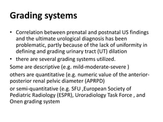
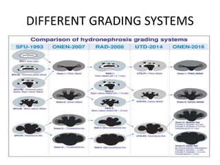
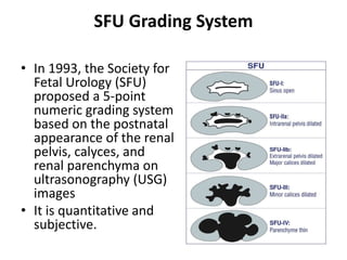
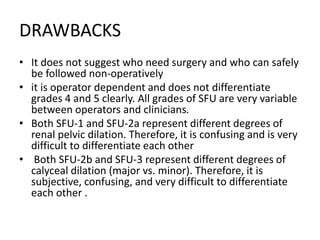
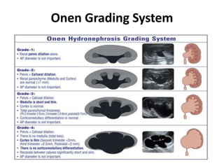
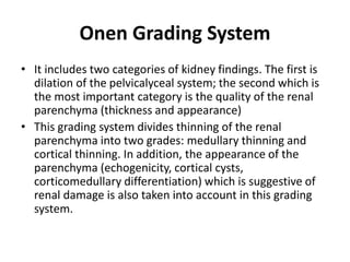
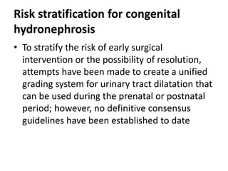
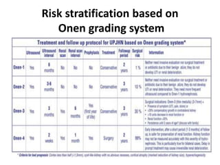
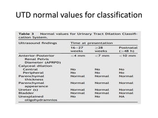
Different systems for grading and
risk stratification for congenital
hydronephrosis
BY/ Mohamed Elwany
CONGENITAL HYDRONEPHROPSIS
• Hydronephrosis refers to
dilatation of the renal
collecting system due to a
build-up of urine resulting
from drainage problems.
• Congenital hydronephrosis
occurs in up to 1%–5% of
all pregnancies
ULTRASOUND
• Urinary ultrasound (US) is the best
we have for the diagnosis and
follow-up of both prenatal and
post-natal hydronephrosis
• It is non-invasive, easily available,
fast, and low-cost; can be
performed directly in bedside
manner; and does not involve
radiation.
• It shows the size of kidneys,
thickness, and appearance of
parenchyma (echogenicity,
corticomedullary differentiation,
cortical cysts), severity of
hydronephrosis, ureteral dilation,
and bladder anatomy
Grading systems
• Correlation between prenatal and postnatal US findings
and the ultimate urological diagnosis has been
problematic, partly because of the lack of uniformity in
defining and grading urinary tract (UT) dilation
• there are several grading systems utilized.
Some are descriptive (e.g. mild-moderate-severe )
others are quantitative (e.g. numeric value of the anterior-
posterior renal pelvic diameter (APRPD)
or semi-quantitative (e.g. SFU ,European Society of
Pediatric Radiology (ESPR), Uroradiology Task Force , and
Onen grading system
DIFFERENT GRADING SYSTEMS
Anterior–Posterior (AP) Diameter of Renal
Pelvis (APDRP)
• The measurement of the AP diameter of the renal pelvis
is not standardized between different
disciplines, Unfortunately, it is significantly operator
dependent.
• The renal pelvis and AP diameter is very dynamic; its
measurement changes significantly depending on
hydration, bladder filling, position (supine or prone), and
respiration
• More importantly, its measurement is very variable and
misleading due to different renal pelvic configurations (
extrarenal and intrarenal pelvis )
SFU Grading System
• In 1993, the Society for
Fetal Urology (SFU)
proposed a 5-point
numeric grading system
based on the postnatal
appearance of the renal
pelvis, calyces, and
renal parenchyma on
ultrasonography (USG)
images
• It is quantitative and
subjective.
DRAWBACKS
• It does not suggest who need surgery and who can safely
be followed non-operatively
• it is operator dependent and does not differentiate
grades 4 and 5 clearly. All grades of SFU are very variable
between operators and clinicians.
• Both SFU-1 and SFU-2a represent different degrees of
renal pelvic dilation. Therefore, it is confusing and is very
difficult to differentiate each other
• Both SFU-2b and SFU-3 represent different degrees of
calyceal dilation (major vs. minor). Therefore, it is
subjective, confusing, and very difficult to differentiate
each other .
Radiology Grading System
• The radiology grading system
has partially been modified from
SFU for post-natal use
• It has the same grades 1, 2, and
3 as the SFU grading system
• In addition, it includes AP
diameter for the grades 1, 2, and
3.
• This grading system divides
parenchymal loss into two
different grades, suggesting the
importance of the renal
parenchyma to determine the
severity of hydronephrosis
UTD Classification
• This classification
suggests the general
term “urinary tract
dilation” to indicate
ultrasound findings that
include all ureteral and
kidney dilations
• UTD has been created
retrospectively based
on reviewing,
combining, and
summarizing the
current literature .
DRAWBACKS
• Central and peripheral calices: It is very operator
dependent to differentiate the dilation of peripheral
(minor) calices from those of central (major) calices
due to a high discrepancy within and between raters
for interpretation of the two types of calyceal dilation
Therefore, it is subjective and confusing and is very
difficult to differentiate each other
• The wide definition of UTD-P3 fails to demonstrate
accurately the severity of hydronephrosis and thus
significant misleads from prompt treatment. It does
not suggest who need surgical treatment and who can
safely be followed non-operatively.
Onen Grading System
• This grading system has been developed for
both prenatal and post-natal HN
• The Onen grading system is terminologically
simple and clear. Therefore, all disciplines
including radiology, perinatology, pediatric
nephrology, and pediatric urology can easily use
not only for clinical practice but also for future
researches.
• This grading system is beneficial in determining
the possible risk of renal damage, surgical
necessity, and prognosis in infants with HN.
Onen Grading System
Onen Grading System
• It includes two categories of kidney findings. The first is
dilation of the pelvicalyceal system; the second which is
the most important category is the quality of the renal
parenchyma (thickness and appearance)
• This grading system divides thinning of the renal
parenchyma into two grades: medullary thinning and
cortical thinning. In addition, the appearance of the
parenchyma (echogenicity, cortical cysts,
corticomedullary differentiation) which is suggestive of
renal damage is also taken into account in this grading
system.
Risk stratification for congenital
hydronephrosis
• To stratify the risk of early surgical
intervention or the possibility of resolution,
attempts have been made to create a unified
grading system for urinary tract dilatation that
can be used during the prenatal or postnatal
period; however, no definitive consensus
guidelines have been established to date
Risk stratification based on
Onen grading system
UTD risk stratification
UTD normal values for classification
Prenatal presentation
Postnatal presentation for UTD
RISK BASED MANAGEMENT

congenital HN.pptx

  • 1.
    Different systems forgrading and risk stratification for congenital hydronephrosis BY/ Mohamed Elwany
  • 2.
    CONGENITAL HYDRONEPHROPSIS • Hydronephrosisrefers to dilatation of the renal collecting system due to a build-up of urine resulting from drainage problems. • Congenital hydronephrosis occurs in up to 1%–5% of all pregnancies
  • 3.
    ULTRASOUND • Urinary ultrasound(US) is the best we have for the diagnosis and follow-up of both prenatal and post-natal hydronephrosis • It is non-invasive, easily available, fast, and low-cost; can be performed directly in bedside manner; and does not involve radiation. • It shows the size of kidneys, thickness, and appearance of parenchyma (echogenicity, corticomedullary differentiation, cortical cysts), severity of hydronephrosis, ureteral dilation, and bladder anatomy
  • 4.
    Grading systems • Correlationbetween prenatal and postnatal US findings and the ultimate urological diagnosis has been problematic, partly because of the lack of uniformity in defining and grading urinary tract (UT) dilation • there are several grading systems utilized. Some are descriptive (e.g. mild-moderate-severe ) others are quantitative (e.g. numeric value of the anterior- posterior renal pelvic diameter (APRPD) or semi-quantitative (e.g. SFU ,European Society of Pediatric Radiology (ESPR), Uroradiology Task Force , and Onen grading system
  • 5.
  • 6.
    Anterior–Posterior (AP) Diameterof Renal Pelvis (APDRP) • The measurement of the AP diameter of the renal pelvis is not standardized between different disciplines, Unfortunately, it is significantly operator dependent. • The renal pelvis and AP diameter is very dynamic; its measurement changes significantly depending on hydration, bladder filling, position (supine or prone), and respiration • More importantly, its measurement is very variable and misleading due to different renal pelvic configurations ( extrarenal and intrarenal pelvis )
  • 7.
    SFU Grading System •In 1993, the Society for Fetal Urology (SFU) proposed a 5-point numeric grading system based on the postnatal appearance of the renal pelvis, calyces, and renal parenchyma on ultrasonography (USG) images • It is quantitative and subjective.
  • 8.
    DRAWBACKS • It doesnot suggest who need surgery and who can safely be followed non-operatively • it is operator dependent and does not differentiate grades 4 and 5 clearly. All grades of SFU are very variable between operators and clinicians. • Both SFU-1 and SFU-2a represent different degrees of renal pelvic dilation. Therefore, it is confusing and is very difficult to differentiate each other • Both SFU-2b and SFU-3 represent different degrees of calyceal dilation (major vs. minor). Therefore, it is subjective, confusing, and very difficult to differentiate each other .
  • 9.
    Radiology Grading System •The radiology grading system has partially been modified from SFU for post-natal use • It has the same grades 1, 2, and 3 as the SFU grading system • In addition, it includes AP diameter for the grades 1, 2, and 3. • This grading system divides parenchymal loss into two different grades, suggesting the importance of the renal parenchyma to determine the severity of hydronephrosis
  • 10.
    UTD Classification • Thisclassification suggests the general term “urinary tract dilation” to indicate ultrasound findings that include all ureteral and kidney dilations • UTD has been created retrospectively based on reviewing, combining, and summarizing the current literature .
  • 11.
    DRAWBACKS • Central andperipheral calices: It is very operator dependent to differentiate the dilation of peripheral (minor) calices from those of central (major) calices due to a high discrepancy within and between raters for interpretation of the two types of calyceal dilation Therefore, it is subjective and confusing and is very difficult to differentiate each other • The wide definition of UTD-P3 fails to demonstrate accurately the severity of hydronephrosis and thus significant misleads from prompt treatment. It does not suggest who need surgical treatment and who can safely be followed non-operatively.
  • 12.
    Onen Grading System •This grading system has been developed for both prenatal and post-natal HN • The Onen grading system is terminologically simple and clear. Therefore, all disciplines including radiology, perinatology, pediatric nephrology, and pediatric urology can easily use not only for clinical practice but also for future researches. • This grading system is beneficial in determining the possible risk of renal damage, surgical necessity, and prognosis in infants with HN.
  • 13.
  • 14.
    Onen Grading System •It includes two categories of kidney findings. The first is dilation of the pelvicalyceal system; the second which is the most important category is the quality of the renal parenchyma (thickness and appearance) • This grading system divides thinning of the renal parenchyma into two grades: medullary thinning and cortical thinning. In addition, the appearance of the parenchyma (echogenicity, cortical cysts, corticomedullary differentiation) which is suggestive of renal damage is also taken into account in this grading system.
  • 15.
    Risk stratification forcongenital hydronephrosis • To stratify the risk of early surgical intervention or the possibility of resolution, attempts have been made to create a unified grading system for urinary tract dilatation that can be used during the prenatal or postnatal period; however, no definitive consensus guidelines have been established to date
  • 16.
    Risk stratification basedon Onen grading system
  • 17.
  • 18.
    UTD normal valuesfor classification
  • 19.
  • 20.
  • 21.