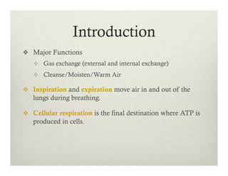
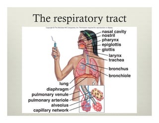
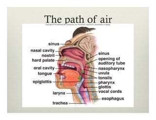
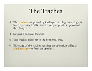
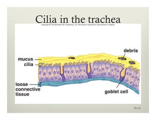
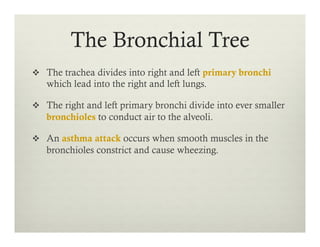
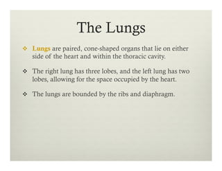
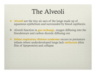
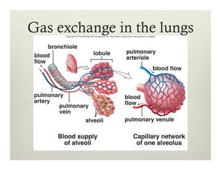
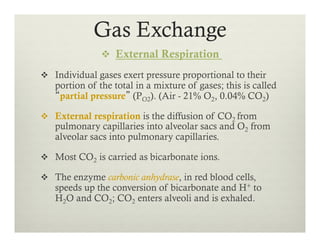
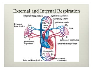
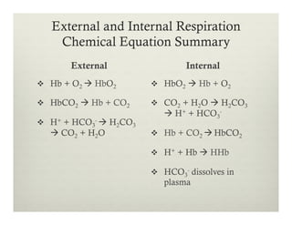
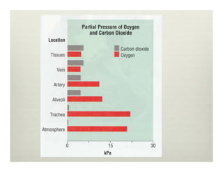
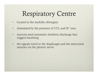
Respiratory
System
v  Major Functions
v  Gas exchange (external and internal exchange)
v  Cleanse/Moisten/Warm Air
v  Inspiration and expiration move air in and out of the
lungs during breathing.
v  Cellular respiration is the final destination where ATP is
produced in cells.
Introduction
The respiratory tract
The Nose
v  The two nasal cavities are divided by a septum.
v  They contain olfactory cells, receive tear ducts from
eyes, and communicate with sinuses.
v  The nasal cavities empty into the nasopharynx.
v  Auditory tubes lead from the middle ears to the
nasopharynx.
The path of air
The Pharynx
v  The pharynx (throat) is a passageway from the nasal
cavities to oral cavities and to the larynx.
v  The pharynx contains the tonsils; the respiratory tract
assists the immune system in maintaining homeostasis.
v  The pharynx takes air from the nose to the larynx and
takes food from the oral cavity to the esophagus.
The Larynx
v  The larynx is a cartilaginous structure lying between the
pharynx and the trachea.
v  The larynx houses the vocal cords.
v  A flap of tissue called the epiglottis covers the glottis, an
opening to the larynx.
v  In young men, rapid growth of the larynx and vocal cords
changes the voice.
Placement of the vocal cords
15-8
The Trachea
v  The trachea, supported by C-shaped cartilaginous rings, is
lined by ciliated cells, which sweep impurities up toward
the pharynx.
v  Smoking destroys the cilia.
v  The trachea takes air to the bronchial tree.
v  Blockage of the trachea requires an operation called a
tracheostomy to form an opening.
Cilia in the trachea
15-10
The Bronchial Tree
v  The trachea divides into right and left primary bronchi
which lead into the right and left lungs.
v  The right and left primary bronchi divide into ever smaller
bronchioles to conduct air to the alveoli.
v  An asthma attack occurs when smooth muscles in the
bronchioles constrict and cause wheezing.
The Lungs
v  Lungs are paired, cone-shaped organs that lie on either
side of the heart and within the thoracic cavity.
v  The right lung has three lobes, and the left lung has two
lobes, allowing for the space occupied by the heart.
v  The lungs are bounded by the ribs and diaphragm.
The Alveoli
v  Alveoli are the tiny air sacs of the lungs made up of
squamous epithelium and surrounded by blood capillaries.
v  Alveoli function in gas exchange, oxygen diffusing into the
bloodstream and carbon dioxide diffusing out.
v  Infant respiratory distress syndrome occurs in premature
infants where underdeveloped lungs lack surfactant (thin
film of lipoprotein) and collapse.
Gas exchange in the lungs
v  During breathing, air moves into the lungs during
inspiration (inhalation) from the nose or mouth, then
moves out again during expiration (exhalation).
v  A spirometer allows measurement of the components
of air during breathing.
Mechanism of Breathing
Respiratory Volumes
v  Tidal volume (VT) the normal amount of air
moved in and out of the lungs when relaxed, is
usually 500 ml.
v  Inspiratory reserve volume is the maximum
amount of forcibly inspired air.
v  Expiratory reserve volume is the maximum
amount of forcibly expired air.
v  Vital capacity is the maximum amount of air
moved in and out on deep breathing, and is the
sum of tidal, inspiratory reserve, and expiratory
reserve volumes.
v  Air that remains in the lungs is residual volume.
Vital capacity
Inspiration and Expiration
v  There is a continuous column of air from the pharynx to
the alveoli, and the lungs lie within the sealed-off thoracic
cavity.
v  The thoracic cavity is bounded by the rib cage and
diaphragm.
v  Pleural membranes line the thoracic cavity and lungs and
the intrapleural pressure is lower than atmospheric
pressure, keeping the lobules of the lungs from collapsing.
v  Minute Ventilation – The volume of inhaled and exhaled
air in one minute.
Inspiration
v  When we inhale (inspiration) impulses from the
respiratory center in the medulla oblongata cause the rib
cage to rise and the diaphragm to lower, causing the
thoracic cavity to expand.
v  The negative pressure or partial vacuum in the alveoli
causes the air to come in.
v  Changing amounts of blood of H+ and CO2 detected by
chemoreceptors in the carotid arteries and aorta increase
breathing rate.
O2 UPTAKE DURING
EXERCISE
v  VO2 - measured in
ml/Kg/min
v  VO2max - maximal
oxygen uptake
v  Metabolic Cart
v  Predicted VO2max
testing
v  Astrand 6 min. test
v  Balke Test
Nervous control of
breathing
Inspiration
Expiration
v  When we exhale (expiration), lack of impulses from the
respiratory center allow the rib cage to lower and the
diaphragm to resume a dome shape.
v  Expiration is passive, while inspiration is active.
v  The elastic recoil of the lungs causes expiration (gravity).
v  A deep breath causes alveoli to stretch; stretch receptors
then inhibit the respiratory center via the vagus nerve.
Expiration
Expiration: active
v  Under extreme conditions, exhalation becomes
dynamic.
v  Internal intercostal muscles contract forcing the ribs
down and inward.
v  Tightening of the abdominal walls also pushes on the
viscera which pushes the diaphragm and forces out
even more air.
v  FEV1 – the amount of air forcibly expired in one
second. Reduced in patients with COPD
Spirometry
v  External Respiration
v  Individual gases exert pressure proportional to their
portion of the total in a mixture of gases; this is called
“partial pressure” (PO2). (Air - 21% O2, 0.04% CO2)
v  External respiration is the diffusion of CO2 from
pulmonary capillaries into alveolar sacs and O2 from
alveolar sacs into pulmonary capillaries.
v  Most CO2 is carried as bicarbonate ions.
v  The enzyme carbonic anhydrase, in red blood cells,
speeds up the conversion of bicarbonate and H+ to
H2O and CO2; CO2 enters alveoli and is exhaled.
Gas Exchange
Partial
Pressures
Hemoglobin Structure
v Hemoglobin (Hb)
takes up oxygen
from alveoli and
becomes
oxyhemoglobin
(HbO2).
v 4 globular proteins
(globin) & 4 heme
units.
v (64 000 daltons)
Internal Respiration
v  Internal respiration is the diffusion of O2 from systemic
capillaries into tissues and CO2 from tissue fluid into
systemic capillaries.
v  Oxyhemoglobin gives up O2, which diffuses out of the
blood and into the tissues because the partial pressure of
O2 of tissues fluid is lower than that of the blood.
v  After CO2 diffuses from tissue cells into the blood, it enters
red blood cells where a small amount is taken up by
hemoglobin, forming carbaminohemoglobin.
Internal Respiration Cont.
v  Most of the CO2 combines with water to form
carbonic acid (H2CO3), which dissociates to release
hydrogen ions (H+) and bicarbonate ions (HCO3
-);
the enzyme carbonic anhydrase speeds this reaction.
v  The globin portion of hemoglobin combines with
excess hydrogen ions to become reduced hemoglobin
or HHb; this helps maintain a normal blood pH.
v  Blood leaving capillaries is a dark maroon color
because red blood cells contain reduced hemoglobin.
External and Internal Respiration
External
v  Hb + O2 à HbO2
v  HbCO2 à Hb + CO2
v  H+ + HCO3
- à H2CO3
à CO2 + H2O
Internal
v  HbO2 à Hb + O2
v  CO2 + H2O à H2CO3
à H+ + HCO3
-
v  Hb + CO2 à HbCO2
v  H+ + Hb à HHb
v  HCO3
- dissolves in
plasma
Chemical Equation Summary
Respiratory Centre
•  located in the medulla oblongata
•  stimulated by the presence of CO2 and H+ ions.
•  neurons send automatic rhythmic discharge that
triggers breathing
•  the signals travel to the diaphragm and the intercostal
muscles via the phrenic nerve.
v  Low O2 in blood
registered by carotid
and aortic bodies
v  Communication with
respiratory centre
v  Increase in minute
ventilation occurs
v  Hemoglobin has 200X
affinity for CO than O2
Respiratory Centre Cont.
Hemoglobin Saturation (SO2)
v  Lungs: 98-100% saturated
v  Tissues: 60-70% saturated
v  Conditions affecting Saturation:
v  Partial Pressure of O2 – incr. é SO2
v  pH – incr. pH é SO2
v  Temperature – incr. temp ê SO2
v  Exercise – increases temperature and decreases pH
causing a decrease in arterial oxygen saturation (SaO2)
Oxyhaemoglobin Dissociation Curve
v  Common Respiratory System Diseases:
v  Asthma
v  COPD
v  Chronic Bronchitis
v  Emphysema
v  Pleurisy
v  Cancer
v  Influenza
v  Sinusitis
Respiration and Health
Sites of upper respiratory infections
Sinusitis
v  Sinusitis is infection of the cranial sinuses within the
facial skeleton that drain into nasal cavities.
v  It occurs when nasal congestion blocks the sinus
openings and is relieved when drainage is restored.
v  Pain and tenderness over the lower forehead and
cheeks, and toothache, accompany this condition.
Lower respiratory tract disorders
COPD
Chronic Bronchitis
v  Long-term cough with
mucus
Emphysema
v  A breakdown of the
alveoli walls.
Causes, Risk Factors, Symptoms
v  Smoking
v  Exposure to gases and fumes, cooking fire
v  Cough, Fatigue, many R.T.I, Wheezing
Asthma
v  Muscle lining small airways becomes irritated
v  Causes a narrowing of airway passages
v  Steroids or bronchodilators can be used to prevent
constriction
Lung Cancer
v  Lung cancer follows this sequence of events: thickening of
airway cells, loss of cilia on the lining, cells with atypical
nuclei, tumor development, and finally metastasis.
v  Removal of a lobe or lung, called pneumonectomy, may
remove the cancer.
v  Smoking, whether active or passive, is a major cause of
lung cancer.
Normal lung versus cancerous
lung
15-47
Chapter Summary
v  Air passes through a series of tubes before gas exchange
takes place across an extensive moist surface in the alveoli
of the lungs.
v  Respiration comprises breathing, external and internal
respiration, and cellular respiration.
v  During inspiration, the pressure in the lungs decreases and
air comes rushing in; during expiration, increased pressure
in the thoracic cavity causes air to leave the lungs.
Chapter Summary Cont.
v  External respiration occurs in the lungs where oxygen
diffuses into the blood and carbon dioxide diffuses out
of the blood.
v  Internal respiration occurs in the tissues where oxygen
diffuses out of the blood into tissue cells and carbon
dioxide diffuses into the blood.
v  Spirometry is the measurement of lung capacities and
can be used to diagnose certain respiratory conditions
such as COPD.
Chapter Summary Cont.
v  The respiratory pigment hemoglobin transports
oxygen from the lungs to the tissues and aids in the
transport of carbon dioxide from the tissues to the
lungs.
v  Hemoglobin’s oxygen affinity is affected by pH, temp
and PO2
v  The respiratory tract is especially subject to disease
because it is exposed to infectious agents; also,
cigarette smoking contributes to two major lung
disorders—emphysema and cancer.

Bio12 Respiratory System Presentation

  • 1.
  • 2.
    v  Major Functions v Gas exchange (external and internal exchange) v  Cleanse/Moisten/Warm Air v  Inspiration and expiration move air in and out of the lungs during breathing. v  Cellular respiration is the final destination where ATP is produced in cells. Introduction
  • 3.
  • 4.
    The Nose v  Thetwo nasal cavities are divided by a septum. v  They contain olfactory cells, receive tear ducts from eyes, and communicate with sinuses. v  The nasal cavities empty into the nasopharynx. v  Auditory tubes lead from the middle ears to the nasopharynx.
  • 5.
  • 6.
    The Pharynx v  Thepharynx (throat) is a passageway from the nasal cavities to oral cavities and to the larynx. v  The pharynx contains the tonsils; the respiratory tract assists the immune system in maintaining homeostasis. v  The pharynx takes air from the nose to the larynx and takes food from the oral cavity to the esophagus.
  • 7.
    The Larynx v  Thelarynx is a cartilaginous structure lying between the pharynx and the trachea. v  The larynx houses the vocal cords. v  A flap of tissue called the epiglottis covers the glottis, an opening to the larynx. v  In young men, rapid growth of the larynx and vocal cords changes the voice.
  • 8.
    Placement of thevocal cords 15-8
  • 9.
    The Trachea v  Thetrachea, supported by C-shaped cartilaginous rings, is lined by ciliated cells, which sweep impurities up toward the pharynx. v  Smoking destroys the cilia. v  The trachea takes air to the bronchial tree. v  Blockage of the trachea requires an operation called a tracheostomy to form an opening.
  • 10.
    Cilia in thetrachea 15-10
  • 12.
    The Bronchial Tree v The trachea divides into right and left primary bronchi which lead into the right and left lungs. v  The right and left primary bronchi divide into ever smaller bronchioles to conduct air to the alveoli. v  An asthma attack occurs when smooth muscles in the bronchioles constrict and cause wheezing.
  • 13.
    The Lungs v  Lungsare paired, cone-shaped organs that lie on either side of the heart and within the thoracic cavity. v  The right lung has three lobes, and the left lung has two lobes, allowing for the space occupied by the heart. v  The lungs are bounded by the ribs and diaphragm.
  • 14.
    The Alveoli v  Alveoliare the tiny air sacs of the lungs made up of squamous epithelium and surrounded by blood capillaries. v  Alveoli function in gas exchange, oxygen diffusing into the bloodstream and carbon dioxide diffusing out. v  Infant respiratory distress syndrome occurs in premature infants where underdeveloped lungs lack surfactant (thin film of lipoprotein) and collapse.
  • 15.
    Gas exchange inthe lungs
  • 16.
    v  During breathing,air moves into the lungs during inspiration (inhalation) from the nose or mouth, then moves out again during expiration (exhalation). v  A spirometer allows measurement of the components of air during breathing. Mechanism of Breathing
  • 17.
    Respiratory Volumes v  Tidalvolume (VT) the normal amount of air moved in and out of the lungs when relaxed, is usually 500 ml. v  Inspiratory reserve volume is the maximum amount of forcibly inspired air. v  Expiratory reserve volume is the maximum amount of forcibly expired air. v  Vital capacity is the maximum amount of air moved in and out on deep breathing, and is the sum of tidal, inspiratory reserve, and expiratory reserve volumes. v  Air that remains in the lungs is residual volume.
  • 18.
  • 19.
    Inspiration and Expiration v There is a continuous column of air from the pharynx to the alveoli, and the lungs lie within the sealed-off thoracic cavity. v  The thoracic cavity is bounded by the rib cage and diaphragm. v  Pleural membranes line the thoracic cavity and lungs and the intrapleural pressure is lower than atmospheric pressure, keeping the lobules of the lungs from collapsing. v  Minute Ventilation – The volume of inhaled and exhaled air in one minute.
  • 20.
    Inspiration v  When weinhale (inspiration) impulses from the respiratory center in the medulla oblongata cause the rib cage to rise and the diaphragm to lower, causing the thoracic cavity to expand. v  The negative pressure or partial vacuum in the alveoli causes the air to come in. v  Changing amounts of blood of H+ and CO2 detected by chemoreceptors in the carotid arteries and aorta increase breathing rate.
  • 21.
    O2 UPTAKE DURING EXERCISE v VO2 - measured in ml/Kg/min v  VO2max - maximal oxygen uptake v  Metabolic Cart v  Predicted VO2max testing v  Astrand 6 min. test v  Balke Test
  • 22.
  • 23.
  • 24.
    Expiration v  When weexhale (expiration), lack of impulses from the respiratory center allow the rib cage to lower and the diaphragm to resume a dome shape. v  Expiration is passive, while inspiration is active. v  The elastic recoil of the lungs causes expiration (gravity). v  A deep breath causes alveoli to stretch; stretch receptors then inhibit the respiratory center via the vagus nerve.
  • 25.
  • 26.
    Expiration: active v  Underextreme conditions, exhalation becomes dynamic. v  Internal intercostal muscles contract forcing the ribs down and inward. v  Tightening of the abdominal walls also pushes on the viscera which pushes the diaphragm and forces out even more air. v  FEV1 – the amount of air forcibly expired in one second. Reduced in patients with COPD
  • 27.
  • 28.
    v  External Respiration v Individual gases exert pressure proportional to their portion of the total in a mixture of gases; this is called “partial pressure” (PO2). (Air - 21% O2, 0.04% CO2) v  External respiration is the diffusion of CO2 from pulmonary capillaries into alveolar sacs and O2 from alveolar sacs into pulmonary capillaries. v  Most CO2 is carried as bicarbonate ions. v  The enzyme carbonic anhydrase, in red blood cells, speeds up the conversion of bicarbonate and H+ to H2O and CO2; CO2 enters alveoli and is exhaled. Gas Exchange
  • 29.
  • 30.
    Hemoglobin Structure v Hemoglobin (Hb) takesup oxygen from alveoli and becomes oxyhemoglobin (HbO2). v 4 globular proteins (globin) & 4 heme units. v (64 000 daltons)
  • 31.
    Internal Respiration v  Internalrespiration is the diffusion of O2 from systemic capillaries into tissues and CO2 from tissue fluid into systemic capillaries. v  Oxyhemoglobin gives up O2, which diffuses out of the blood and into the tissues because the partial pressure of O2 of tissues fluid is lower than that of the blood. v  After CO2 diffuses from tissue cells into the blood, it enters red blood cells where a small amount is taken up by hemoglobin, forming carbaminohemoglobin.
  • 32.
    Internal Respiration Cont. v Most of the CO2 combines with water to form carbonic acid (H2CO3), which dissociates to release hydrogen ions (H+) and bicarbonate ions (HCO3 -); the enzyme carbonic anhydrase speeds this reaction. v  The globin portion of hemoglobin combines with excess hydrogen ions to become reduced hemoglobin or HHb; this helps maintain a normal blood pH. v  Blood leaving capillaries is a dark maroon color because red blood cells contain reduced hemoglobin.
  • 33.
  • 34.
    External and InternalRespiration External v  Hb + O2 à HbO2 v  HbCO2 à Hb + CO2 v  H+ + HCO3 - à H2CO3 à CO2 + H2O Internal v  HbO2 à Hb + O2 v  CO2 + H2O à H2CO3 à H+ + HCO3 - v  Hb + CO2 à HbCO2 v  H+ + Hb à HHb v  HCO3 - dissolves in plasma Chemical Equation Summary
  • 36.
    Respiratory Centre •  locatedin the medulla oblongata •  stimulated by the presence of CO2 and H+ ions. •  neurons send automatic rhythmic discharge that triggers breathing •  the signals travel to the diaphragm and the intercostal muscles via the phrenic nerve.
  • 37.
    v  Low O2in blood registered by carotid and aortic bodies v  Communication with respiratory centre v  Increase in minute ventilation occurs v  Hemoglobin has 200X affinity for CO than O2 Respiratory Centre Cont.
  • 38.
    Hemoglobin Saturation (SO2) v Lungs: 98-100% saturated v  Tissues: 60-70% saturated v  Conditions affecting Saturation: v  Partial Pressure of O2 – incr. é SO2 v  pH – incr. pH é SO2 v  Temperature – incr. temp ê SO2 v  Exercise – increases temperature and decreases pH causing a decrease in arterial oxygen saturation (SaO2)
  • 39.
  • 40.
    v  Common RespiratorySystem Diseases: v  Asthma v  COPD v  Chronic Bronchitis v  Emphysema v  Pleurisy v  Cancer v  Influenza v  Sinusitis Respiration and Health
  • 41.
    Sites of upperrespiratory infections
  • 42.
    Sinusitis v  Sinusitis isinfection of the cranial sinuses within the facial skeleton that drain into nasal cavities. v  It occurs when nasal congestion blocks the sinus openings and is relieved when drainage is restored. v  Pain and tenderness over the lower forehead and cheeks, and toothache, accompany this condition.
  • 43.
  • 44.
    COPD Chronic Bronchitis v  Long-termcough with mucus Emphysema v  A breakdown of the alveoli walls. Causes, Risk Factors, Symptoms v  Smoking v  Exposure to gases and fumes, cooking fire v  Cough, Fatigue, many R.T.I, Wheezing
  • 45.
    Asthma v  Muscle liningsmall airways becomes irritated v  Causes a narrowing of airway passages v  Steroids or bronchodilators can be used to prevent constriction
  • 46.
    Lung Cancer v  Lungcancer follows this sequence of events: thickening of airway cells, loss of cilia on the lining, cells with atypical nuclei, tumor development, and finally metastasis. v  Removal of a lobe or lung, called pneumonectomy, may remove the cancer. v  Smoking, whether active or passive, is a major cause of lung cancer.
  • 47.
    Normal lung versuscancerous lung 15-47
  • 48.
    Chapter Summary v  Airpasses through a series of tubes before gas exchange takes place across an extensive moist surface in the alveoli of the lungs. v  Respiration comprises breathing, external and internal respiration, and cellular respiration. v  During inspiration, the pressure in the lungs decreases and air comes rushing in; during expiration, increased pressure in the thoracic cavity causes air to leave the lungs.
  • 49.
    Chapter Summary Cont. v External respiration occurs in the lungs where oxygen diffuses into the blood and carbon dioxide diffuses out of the blood. v  Internal respiration occurs in the tissues where oxygen diffuses out of the blood into tissue cells and carbon dioxide diffuses into the blood. v  Spirometry is the measurement of lung capacities and can be used to diagnose certain respiratory conditions such as COPD.
  • 50.
    Chapter Summary Cont. v The respiratory pigment hemoglobin transports oxygen from the lungs to the tissues and aids in the transport of carbon dioxide from the tissues to the lungs. v  Hemoglobin’s oxygen affinity is affected by pH, temp and PO2 v  The respiratory tract is especially subject to disease because it is exposed to infectious agents; also, cigarette smoking contributes to two major lung disorders—emphysema and cancer.