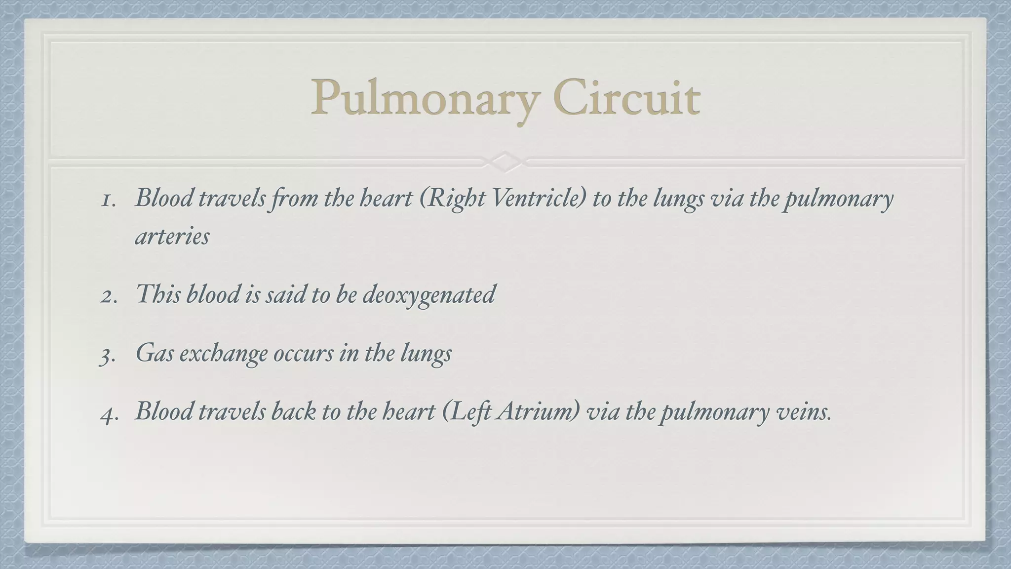
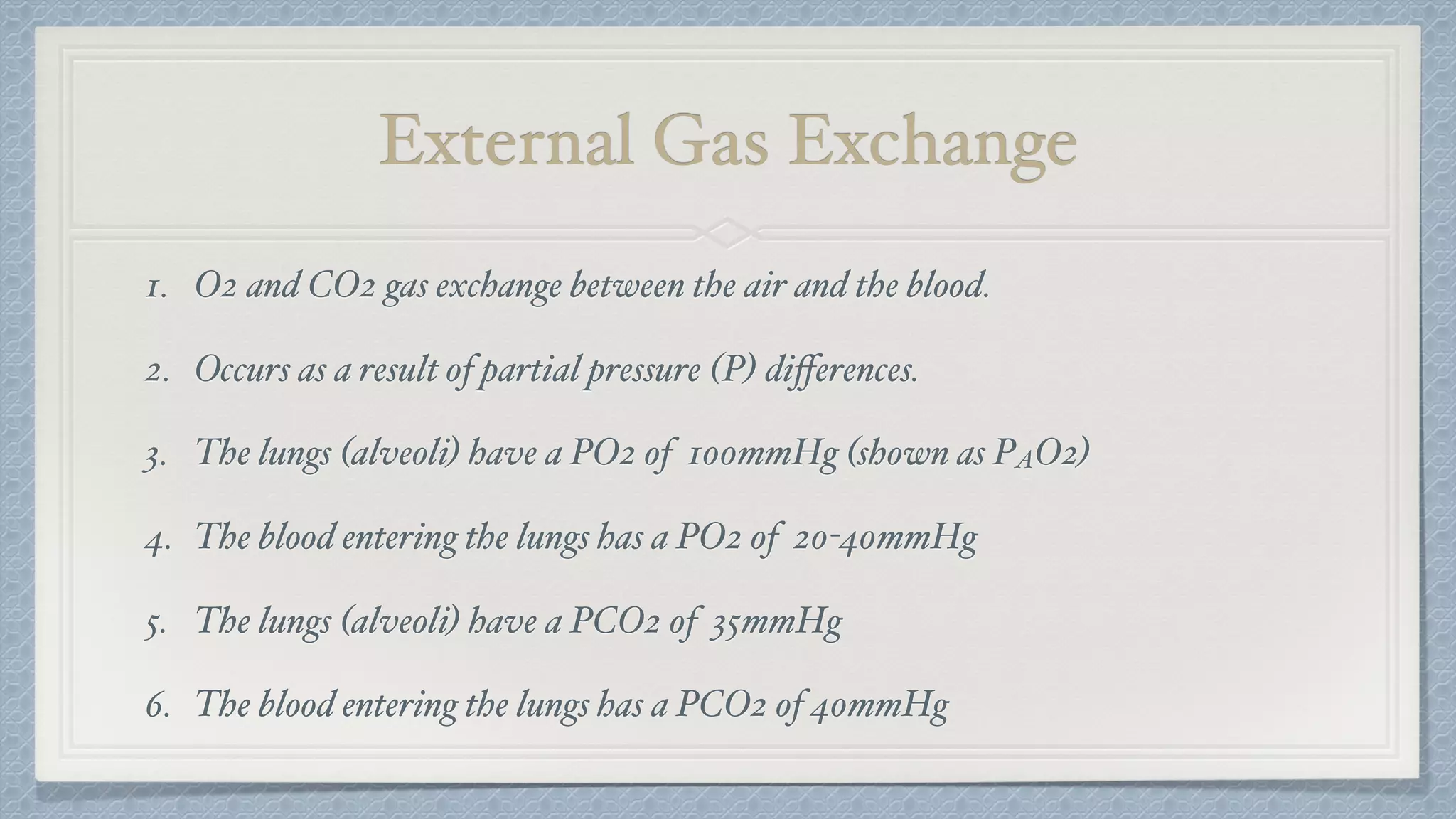
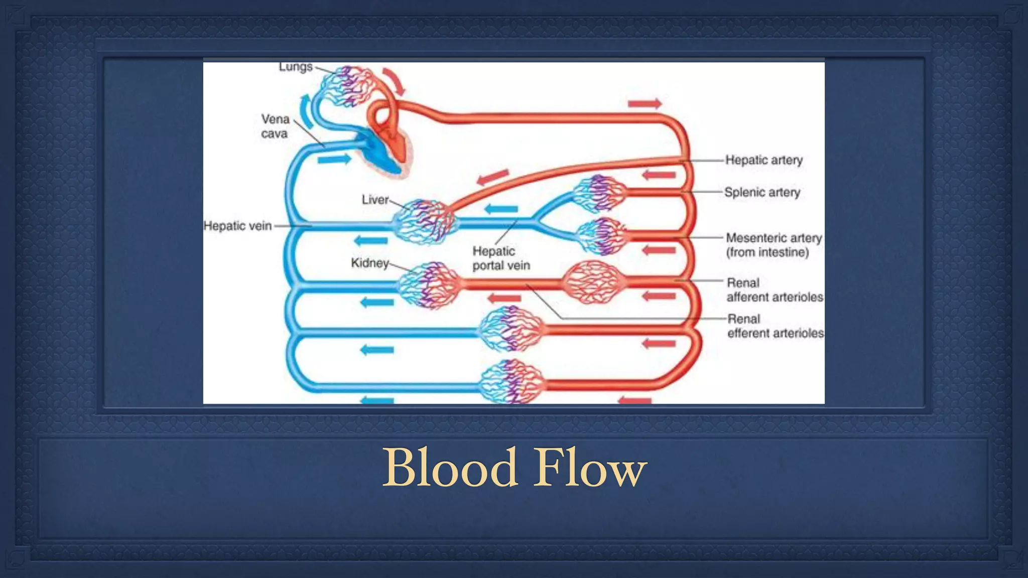
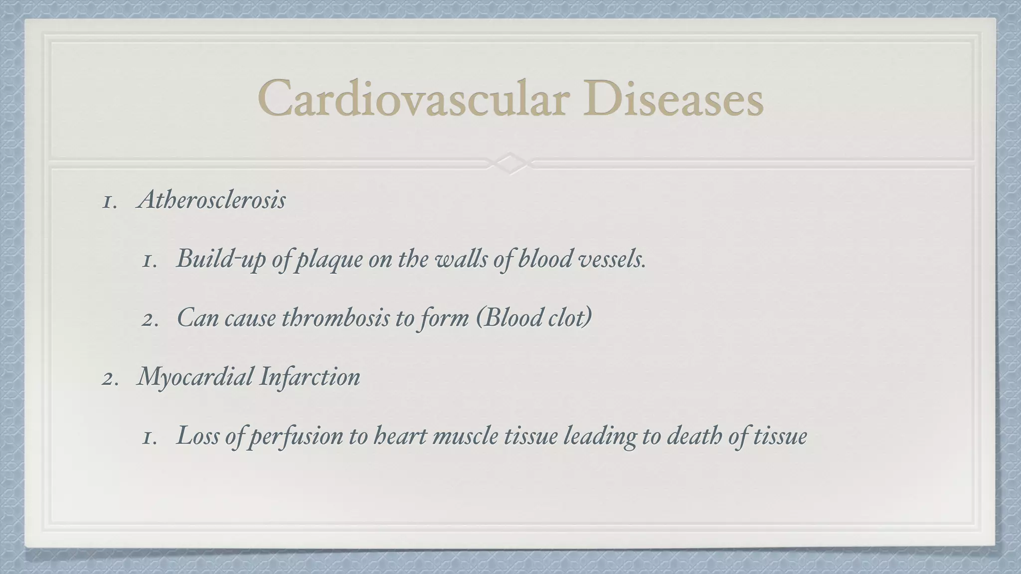
Cardiovascular and
Respiratory Systems
What are the parts of your circulatory system?
What are the functions of the parts?
What are the parts of your respiratory system?
What are the functions of the parts?
Did we miss anything?
The Brain!
These systems are all linked.
Circulation Functions
1. Transport nutrients to working tissue.
2. Deliver waste products from metabolic processes to urinary
and respiratory systems.
3. Transport components of the endocrine system to target tissue.
(Hormones)
Respiratory Functions
1. Oxygenation of blood.
Heart
1. Small upper chambers (Left and RightAtrium)
2. Large lower chambers
A. Right Ventricle - blood to lungs
B. Left Ventricle - blood to body
3. Valves control fluid travel between chambers and between vessels.
Heartbeat
1. Initiation - impulse from the SA node (RightAtrium) causes atria to contract
2. Electrical impulse is conveyed by theAV node (Central Heart) to the ventricles
3. Atrioventricular bundle relays signal through Purkinje fibres to the rest of the
ventricles
4. Delay in signal at theAV node allows for ventricular filling.
Electrocardiogram (ECG)
1. Measures electrical activity in the heart.
2. Normal sinus rhythm vs atrial fibrillation
3. AED - automated electrical defibrillator
Electrocardiogram
Pulmonary Circuit
1. Blood travels from the heart (Right Ventricle) to the lungs via the pulmonary
arteries
2. This blood is said to be deoxygenated
3. Gas exchange occurs in the lungs
4. Blood travels back to the heart (LeftAtrium) via the pulmonary veins.
Inspiration and Expiration
1. Air is drawn into the lungs as a result of decreased pressure in the thoracic
cavity.
2. We expand our thoracic cavity to decrease pressure by swinging our rib cage up
and out and flattening our diaphragm.
3. Inspiration is an active process - muscles contract to perform this task
4. Expiration is the opposite activity and is passive - muscles relax
External Gas Exchange
1. O2 and CO2 gas exchange between the air and the blood.
2. Occurs as a result of partial pressure (P) differences.
3. The lungs (alveoli) have a PO2 of 100mmHg (shown as PAO2)
4. The blood entering the lungs has a PO2 of 20-40mmHg
5. The lungs (alveoli) have a PCO2 of 35mmHg
6. The blood entering the lungs has a PCO2 of 40mmHg
Internal Gas Exchange
1. O2 and CO2 gas exchange between the blood and tissue.
2. The arterioles (blood) have a PO2 of 95mmHg
3. The interstitial fluid has a PO2 of 40mmHg
4. The arterioles (blood) have a PCO2 of 40mmHg
5. The interstitial fluid has a PCO2 of 45mmHg
Gas Transport in Blood
1. Oxygen is transported in the blood attached to RBC Hemoglobin
2. Carbon Dioxide is transported in three ways:
1. Attached to Hemoblogin as Carbaminohemoglobin (small amount)
2. Dissolved in the blood (small amount)
3. As bicarbonate ions (H+ and HCO3- Ions) - Majority
Hemoglobin Saturation
1. Definition - The percentage of hemoglobin molecules that are bound to oxygen,
written as %SO2.
2. Normal range from 95-100%
3. <90% is said to be hypoxic.
4. We measure this using a pulse oximeter.
5. pH and temperature can influence hemoglobin saturation.
Oxyhemoglobin
Dissociation
Curve
Regulation of HR and RR
Control of Heart Rate
1. Rate is fixed at 85 bpm without external controls (SA node)
2. Cardiac control centre in the Medulla Oblongata controls rate.
1. Sympathetic NS - increases HR
2. Parasympathetic NS - decreases HR
3. Hormonal Control - Epinephrine and Norepinephrine release increase HR
Control of Respiratory Rate
1. Resting rate is typically between 12-20 breaths per minute
2. Respiratory control centre in the Medulla Oblongata controls rate.
3. Nerve bundles (carotid and aortic bodies) stimulate the brain when CO2 levels
in the blood are elevated.
Blood Flow
Systemic Blood Circuit
1. Blood travels from the heart (Left Ventricle) to the working tissue of the body.
2. This blood brings nutrients to cells.
3. Major arteries lead to minor arteries, arterioles and capillaries.
4. Venules, minor veins, then major veins return blood to the heart.
Blood Pressure
1. Created by the heart.
2. Systolic blood pressure - pressure on blood vessels while ventricles contract
3. Diastolic BP - pressure while the heart is relaxed
4. Blood flow (velocity) and pressure decrease as you move farther away from the
heart.
Mean Arterial Pressure (MAP)
1. Organs require at least 60mmHg MAP to sustain function.
2. Determining MAP in post-operative patients is important as blood pressure
may drop causing organ damage.
3. The body can regulate BP a number of ways to compensate:
1. Decrease urine production to maintain blood volume.
2. Constriction of blood vessels to decrease vessel volume.
Spirometry
Spirometry Values
1. Tidal Volume (VT) - normal flow at rest (500mL)
2. Inspiratory Reserve Volume - volume of forced inhalation
3. Expiratory Reserve Volume - volume of forced exhalation
4. Vital Capacity -Addition of 1, 2, and 3
5. Residual Volume - volume left in lungs after forced exhalation.
Exercise
Respiratory Values
1. VT
2. Respiratory Rate (RR) - number of breaths in one minute
3. Minute Ventilation (VE) - total volume of gas entering the lungs in one minute.
Calculated as the RR x VT
Adaptation to Exercise
1. The metabolic requirements during exercise affects the respiratory and circulatory system.
2. These adaptations can be acute (short term) as well as long term adaptations.
Respiratory Circulatory
Short Term Inc. RR, small inc. VT Inc. HR
Long Term
Vascularization around
Alveoli
Inc. Stroke Volume
Increase RBC
VO2
1. Definition: The amount of oxygen consumed by the body.
2. VO2max is a measurement of the maximum amount of oxygen consumption.
3. Measurement is done using a metabolic cart and sampling of inspired and expired air.
4. Tests are done at maximal exertion.
5. Submaximal estimated tests can be performed, but are less accurate.
Cardiorespiratory Diseases
Respiratory Disease - COPD
1. Chronic Obstructive Pulmonary Disease
2. Diagnosed using Spirometry
3. FEV1 - Forced expiratory volume in one second
4. FVC - forced vital capacity
5. FEV1/FVC ration and FEV1 values provide information regarding
respiratory health
Cardiovascular Diseases
1. Atherosclerosis
1. Build-up of plaque on the walls of blood vessels.
2. Can cause thrombosis to form (Blood clot)
2. Myocardial Infarction
1. Loss of perfusion to heart muscle tissue leading to death of tissue
Cardiorespiratory presentation

Cardiorespiratory presentation

  • 1.
  • 2.
    What are theparts of your circulatory system?
  • 3.
    What are thefunctions of the parts?
  • 4.
    What are theparts of your respiratory system?
  • 5.
    What are thefunctions of the parts?
  • 6.
    Did we missanything?
  • 7.
  • 8.
    These systems areall linked.
  • 9.
    Circulation Functions 1. Transportnutrients to working tissue. 2. Deliver waste products from metabolic processes to urinary and respiratory systems. 3. Transport components of the endocrine system to target tissue. (Hormones)
  • 10.
  • 11.
    Heart 1. Small upperchambers (Left and RightAtrium) 2. Large lower chambers A. Right Ventricle - blood to lungs B. Left Ventricle - blood to body 3. Valves control fluid travel between chambers and between vessels.
  • 12.
    Heartbeat 1. Initiation -impulse from the SA node (RightAtrium) causes atria to contract 2. Electrical impulse is conveyed by theAV node (Central Heart) to the ventricles 3. Atrioventricular bundle relays signal through Purkinje fibres to the rest of the ventricles 4. Delay in signal at theAV node allows for ventricular filling.
  • 15.
    Electrocardiogram (ECG) 1. Measureselectrical activity in the heart. 2. Normal sinus rhythm vs atrial fibrillation 3. AED - automated electrical defibrillator
  • 16.
  • 17.
    Pulmonary Circuit 1. Bloodtravels from the heart (Right Ventricle) to the lungs via the pulmonary arteries 2. This blood is said to be deoxygenated 3. Gas exchange occurs in the lungs 4. Blood travels back to the heart (LeftAtrium) via the pulmonary veins.
  • 18.
    Inspiration and Expiration 1.Air is drawn into the lungs as a result of decreased pressure in the thoracic cavity. 2. We expand our thoracic cavity to decrease pressure by swinging our rib cage up and out and flattening our diaphragm. 3. Inspiration is an active process - muscles contract to perform this task 4. Expiration is the opposite activity and is passive - muscles relax
  • 20.
    External Gas Exchange 1.O2 and CO2 gas exchange between the air and the blood. 2. Occurs as a result of partial pressure (P) differences. 3. The lungs (alveoli) have a PO2 of 100mmHg (shown as PAO2) 4. The blood entering the lungs has a PO2 of 20-40mmHg 5. The lungs (alveoli) have a PCO2 of 35mmHg 6. The blood entering the lungs has a PCO2 of 40mmHg
  • 21.
    Internal Gas Exchange 1.O2 and CO2 gas exchange between the blood and tissue. 2. The arterioles (blood) have a PO2 of 95mmHg 3. The interstitial fluid has a PO2 of 40mmHg 4. The arterioles (blood) have a PCO2 of 40mmHg 5. The interstitial fluid has a PCO2 of 45mmHg
  • 22.
    Gas Transport inBlood 1. Oxygen is transported in the blood attached to RBC Hemoglobin 2. Carbon Dioxide is transported in three ways: 1. Attached to Hemoblogin as Carbaminohemoglobin (small amount) 2. Dissolved in the blood (small amount) 3. As bicarbonate ions (H+ and HCO3- Ions) - Majority
  • 23.
    Hemoglobin Saturation 1. Definition- The percentage of hemoglobin molecules that are bound to oxygen, written as %SO2. 2. Normal range from 95-100% 3. <90% is said to be hypoxic. 4. We measure this using a pulse oximeter. 5. pH and temperature can influence hemoglobin saturation.
  • 24.
  • 25.
  • 26.
    Control of HeartRate 1. Rate is fixed at 85 bpm without external controls (SA node) 2. Cardiac control centre in the Medulla Oblongata controls rate. 1. Sympathetic NS - increases HR 2. Parasympathetic NS - decreases HR 3. Hormonal Control - Epinephrine and Norepinephrine release increase HR
  • 27.
    Control of RespiratoryRate 1. Resting rate is typically between 12-20 breaths per minute 2. Respiratory control centre in the Medulla Oblongata controls rate. 3. Nerve bundles (carotid and aortic bodies) stimulate the brain when CO2 levels in the blood are elevated.
  • 28.
  • 29.
    Systemic Blood Circuit 1.Blood travels from the heart (Left Ventricle) to the working tissue of the body. 2. This blood brings nutrients to cells. 3. Major arteries lead to minor arteries, arterioles and capillaries. 4. Venules, minor veins, then major veins return blood to the heart.
  • 30.
    Blood Pressure 1. Createdby the heart. 2. Systolic blood pressure - pressure on blood vessels while ventricles contract 3. Diastolic BP - pressure while the heart is relaxed 4. Blood flow (velocity) and pressure decrease as you move farther away from the heart.
  • 33.
    Mean Arterial Pressure(MAP) 1. Organs require at least 60mmHg MAP to sustain function. 2. Determining MAP in post-operative patients is important as blood pressure may drop causing organ damage. 3. The body can regulate BP a number of ways to compensate: 1. Decrease urine production to maintain blood volume. 2. Constriction of blood vessels to decrease vessel volume.
  • 34.
  • 35.
    Spirometry Values 1. TidalVolume (VT) - normal flow at rest (500mL) 2. Inspiratory Reserve Volume - volume of forced inhalation 3. Expiratory Reserve Volume - volume of forced exhalation 4. Vital Capacity -Addition of 1, 2, and 3 5. Residual Volume - volume left in lungs after forced exhalation.
  • 37.
  • 38.
    Respiratory Values 1. VT 2.Respiratory Rate (RR) - number of breaths in one minute 3. Minute Ventilation (VE) - total volume of gas entering the lungs in one minute. Calculated as the RR x VT
  • 39.
    Adaptation to Exercise 1.The metabolic requirements during exercise affects the respiratory and circulatory system. 2. These adaptations can be acute (short term) as well as long term adaptations. Respiratory Circulatory Short Term Inc. RR, small inc. VT Inc. HR Long Term Vascularization around Alveoli Inc. Stroke Volume Increase RBC
  • 40.
    VO2 1. Definition: Theamount of oxygen consumed by the body. 2. VO2max is a measurement of the maximum amount of oxygen consumption. 3. Measurement is done using a metabolic cart and sampling of inspired and expired air. 4. Tests are done at maximal exertion. 5. Submaximal estimated tests can be performed, but are less accurate.
  • 42.
  • 43.
    Respiratory Disease -COPD 1. Chronic Obstructive Pulmonary Disease 2. Diagnosed using Spirometry 3. FEV1 - Forced expiratory volume in one second 4. FVC - forced vital capacity 5. FEV1/FVC ration and FEV1 values provide information regarding respiratory health
  • 44.
    Cardiovascular Diseases 1. Atherosclerosis 1.Build-up of plaque on the walls of blood vessels. 2. Can cause thrombosis to form (Blood clot) 2. Myocardial Infarction 1. Loss of perfusion to heart muscle tissue leading to death of tissue