The nervous system has four main functions:
1. Gathering sensory input from receptors
2. Integrating information in the central nervous system
3. Initiating motor responses via muscles or glands
4. Maintaining homeostasis through detection and response to internal and external changes.
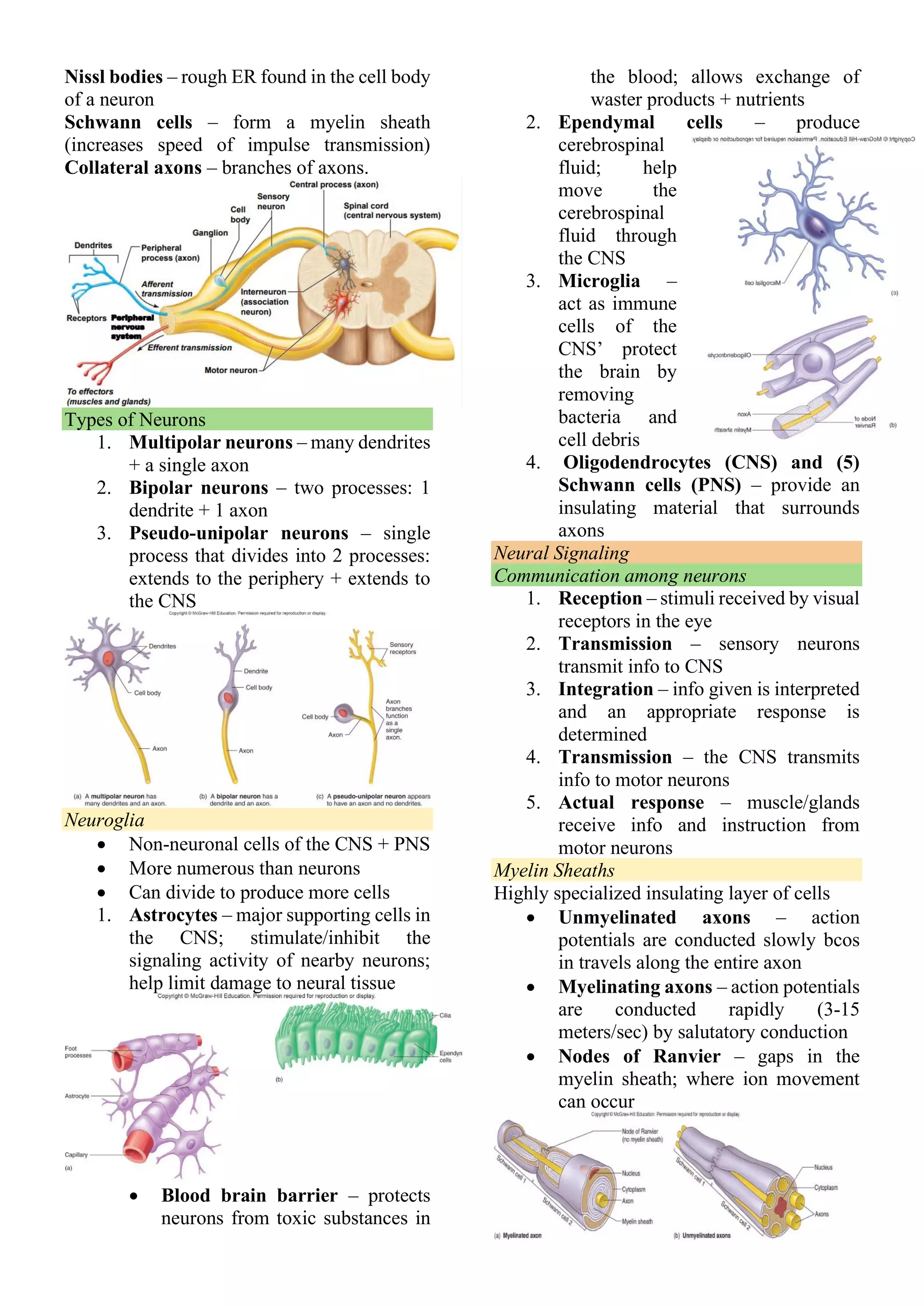
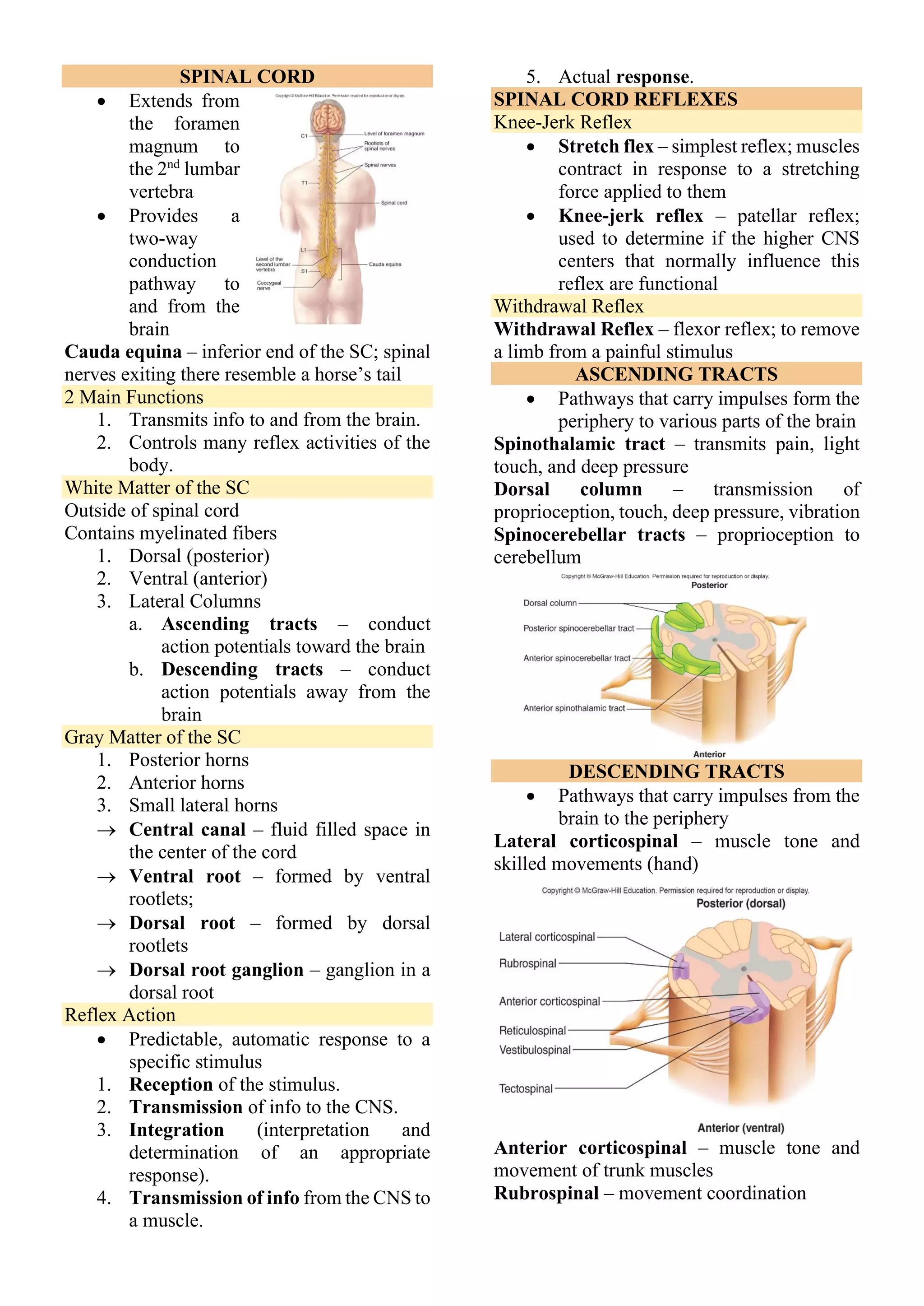
The nervous system is divided into the central nervous system (brain and spinal cord) and peripheral nervous system (nerves and ganglia). The central nervous system processes information while the peripheral nervous system connects to sensory receptors and muscles/glands. Neurons are the basic functional units that receive stimuli, conduct signals, and transmit to other neurons or tissues.