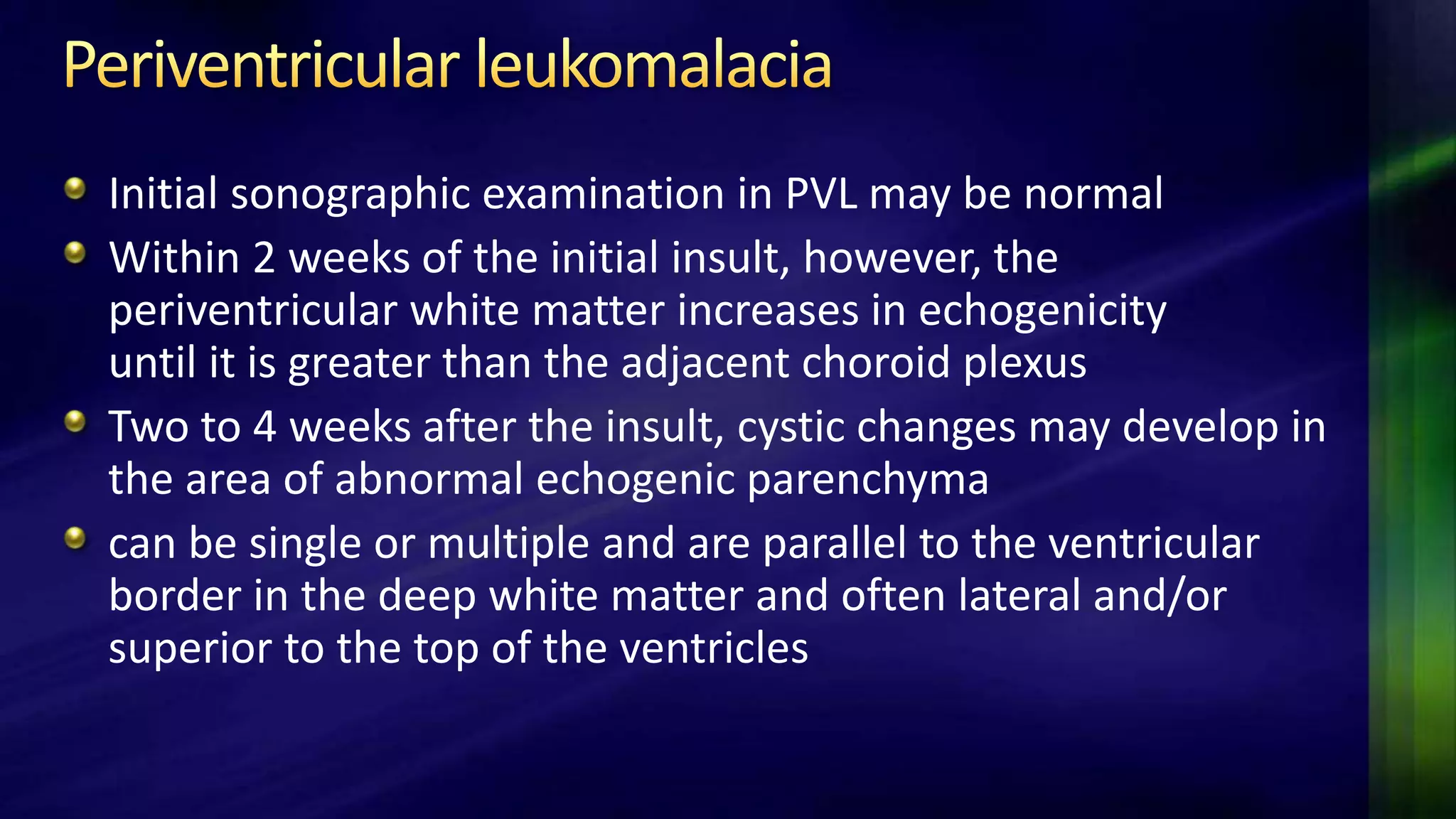
Neonatal sonography of the brain is an essential part of newborn care, particularly for preterm and unstable infants. It allows for portable, low-cost, and radiation-free evaluation of the brain for hemorrhages, abnormalities, and other issues like hydrocephalus. Standard imaging planes include coronal and sagittal views of the brain and ventricles. Key indications for neurosonography in newborns include detection of intraventricular hemorrhage in preterm infants and evaluation of periventricular leukomalacia, a common ischemic injury. Neurosonography is also used to identify other issues like cystic lesions, tumors, and hydrocephalus.
This lecture prides 8 easy steps of scanning the neonatal brain through anterior fontanelle ,followed by examples of most commonly encountered abnormalities.
Doppler ultrasound of visceral arteriesSamir Haffar
Doppler ultrasound of different diseases of visceral arteries including arterial stenosis and occlusion, arterial aneurysm, artrial pseudoaneurysm, arterio-venous fistula, artrial dissection, and abdominal vascular compression syndromes
In this presentation we will discuss role of high resolution in characterizing normal variant and pathologies of spinal pathologies.
This is a pictoral review.
By dr Rabab Hashem, MRCPCH, pediatrician at El Nasr hospital Port said.
Cranial sonography is the most widely used neuroimaging procedure in premature infants. US helps in assessing the neurologic status of the child, since clinical examination and symptoms are often nonspecific. It gives information about immediate and long term prognosis.
Hydrocephalous is a serious disease of the central nervous system which has both congenital and aquired subtypes. the congenital variety affects the children and is a considerable burden especially is the developing countries. I tleads to long term morbidity and high rates of mortality
Title: Sense of Smell
Presenter: Dr. Faiza, Assistant Professor of Physiology
Qualifications:
MBBS (Best Graduate, AIMC Lahore)
FCPS Physiology
ICMT, CHPE, DHPE (STMU)
MPH (GC University, Faisalabad)
MBA (Virtual University of Pakistan)
Learning Objectives:
Describe the primary categories of smells and the concept of odor blindness.
Explain the structure and location of the olfactory membrane and mucosa, including the types and roles of cells involved in olfaction.
Describe the pathway and mechanisms of olfactory signal transmission from the olfactory receptors to the brain.
Illustrate the biochemical cascade triggered by odorant binding to olfactory receptors, including the role of G-proteins and second messengers in generating an action potential.
Identify different types of olfactory disorders such as anosmia, hyposmia, hyperosmia, and dysosmia, including their potential causes.
Key Topics:
Olfactory Genes:
3% of the human genome accounts for olfactory genes.
400 genes for odorant receptors.
Olfactory Membrane:
Located in the superior part of the nasal cavity.
Medially: Folds downward along the superior septum.
Laterally: Folds over the superior turbinate and upper surface of the middle turbinate.
Total surface area: 5-10 square centimeters.
Olfactory Mucosa:
Olfactory Cells: Bipolar nerve cells derived from the CNS (100 million), with 4-25 olfactory cilia per cell.
Sustentacular Cells: Produce mucus and maintain ionic and molecular environment.
Basal Cells: Replace worn-out olfactory cells with an average lifespan of 1-2 months.
Bowman’s Gland: Secretes mucus.
Stimulation of Olfactory Cells:
Odorant dissolves in mucus and attaches to receptors on olfactory cilia.
Involves a cascade effect through G-proteins and second messengers, leading to depolarization and action potential generation in the olfactory nerve.
Quality of a Good Odorant:
Small (3-20 Carbon atoms), volatile, water-soluble, and lipid-soluble.
Facilitated by odorant-binding proteins in mucus.
Membrane Potential and Action Potential:
Resting membrane potential: -55mV.
Action potential frequency in the olfactory nerve increases with odorant strength.
Adaptation Towards the Sense of Smell:
Rapid adaptation within the first second, with further slow adaptation.
Psychological adaptation greater than receptor adaptation, involving feedback inhibition from the central nervous system.
Primary Sensations of Smell:
Camphoraceous, Musky, Floral, Pepperminty, Ethereal, Pungent, Putrid.
Odor Detection Threshold:
Examples: Hydrogen sulfide (0.0005 ppm), Methyl-mercaptan (0.002 ppm).
Some toxic substances are odorless at lethal concentrations.
Characteristics of Smell:
Odor blindness for single substances due to lack of appropriate receptor protein.
Behavioral and emotional influences of smell.
Transmission of Olfactory Signals:
From olfactory cells to glomeruli in the olfactory bulb, involving lateral inhibition.
Primitive, less old, and new olfactory systems with different path
Tom Selleck Health: A Comprehensive Look at the Iconic Actor’s Wellness Journeygreendigital
Tom Selleck, an enduring figure in Hollywood. has captivated audiences for decades with his rugged charm, iconic moustache. and memorable roles in television and film. From his breakout role as Thomas Magnum in Magnum P.I. to his current portrayal of Frank Reagan in Blue Bloods. Selleck's career has spanned over 50 years. But beyond his professional achievements. fans have often been curious about Tom Selleck Health. especially as he has aged in the public eye.
Follow us on: Pinterest
Introduction
Many have been interested in Tom Selleck health. not only because of his enduring presence on screen but also because of the challenges. and lifestyle choices he has faced and made over the years. This article delves into the various aspects of Tom Selleck health. exploring his fitness regimen, diet, mental health. and the challenges he has encountered as he ages. We'll look at how he maintains his well-being. the health issues he has faced, and his approach to ageing .
Early Life and Career
Childhood and Athletic Beginnings
Tom Selleck was born on January 29, 1945, in Detroit, Michigan, and grew up in Sherman Oaks, California. From an early age, he was involved in sports, particularly basketball. which played a significant role in his physical development. His athletic pursuits continued into college. where he attended the University of Southern California (USC) on a basketball scholarship. This early involvement in sports laid a strong foundation for his physical health and disciplined lifestyle.
Transition to Acting
Selleck's transition from an athlete to an actor came with its physical demands. His first significant role in "Magnum P.I." required him to perform various stunts and maintain a fit appearance. This role, which he played from 1980 to 1988. necessitated a rigorous fitness routine to meet the show's demands. setting the stage for his long-term commitment to health and wellness.
Fitness Regimen
Workout Routine
Tom Selleck health and fitness regimen has evolved. adapting to his changing roles and age. During his "Magnum, P.I." days. Selleck's workouts were intense and focused on building and maintaining muscle mass. His routine included weightlifting, cardiovascular exercises. and specific training for the stunts he performed on the show.
Selleck adjusted his fitness routine as he aged to suit his body's needs. Today, his workouts focus on maintaining flexibility, strength, and cardiovascular health. He incorporates low-impact exercises such as swimming, walking, and light weightlifting. This balanced approach helps him stay fit without putting undue strain on his joints and muscles.
Importance of Flexibility and Mobility
In recent years, Selleck has emphasized the importance of flexibility and mobility in his fitness regimen. Understanding the natural decline in muscle mass and joint flexibility with age. he includes stretching and yoga in his routine. These practices help prevent injuries, improve posture, and maintain mobilit
Knee anatomy and clinical tests 2024.pdfvimalpl1234
This includes all relevant anatomy and clinical tests compiled from standard textbooks, Campbell,netter etc..It is comprehensive and best suited for orthopaedicians and orthopaedic residents.
Couples presenting to the infertility clinic- Do they really have infertility...Sujoy Dasgupta
Dr Sujoy Dasgupta presented the study on "Couples presenting to the infertility clinic- Do they really have infertility? – The unexplored stories of non-consummation" in the 13th Congress of the Asia Pacific Initiative on Reproduction (ASPIRE 2024) at Manila on 24 May, 2024.
Ozempic: Preoperative Management of Patients on GLP-1 Receptor Agonists Saeid Safari
Preoperative Management of Patients on GLP-1 Receptor Agonists like Ozempic and Semiglutide
ASA GUIDELINE
NYSORA Guideline
2 Case Reports of Gastric Ultrasound
New Directions in Targeted Therapeutic Approaches for Older Adults With Mantl...i3 Health
i3 Health is pleased to make the speaker slides from this activity available for use as a non-accredited self-study or teaching resource.
This slide deck presented by Dr. Kami Maddocks, Professor-Clinical in the Division of Hematology and
Associate Division Director for Ambulatory Operations
The Ohio State University Comprehensive Cancer Center, will provide insight into new directions in targeted therapeutic approaches for older adults with mantle cell lymphoma.
STATEMENT OF NEED
Mantle cell lymphoma (MCL) is a rare, aggressive B-cell non-Hodgkin lymphoma (NHL) accounting for 5% to 7% of all lymphomas. Its prognosis ranges from indolent disease that does not require treatment for years to very aggressive disease, which is associated with poor survival (Silkenstedt et al, 2021). Typically, MCL is diagnosed at advanced stage and in older patients who cannot tolerate intensive therapy (NCCN, 2022). Although recent advances have slightly increased remission rates, recurrence and relapse remain very common, leading to a median overall survival between 3 and 6 years (LLS, 2021). Though there are several effective options, progress is still needed towards establishing an accepted frontline approach for MCL (Castellino et al, 2022). Treatment selection and management of MCL are complicated by the heterogeneity of prognosis, advanced age and comorbidities of patients, and lack of an established standard approach for treatment, making it vital that clinicians be familiar with the latest research and advances in this area. In this activity chaired by Michael Wang, MD, Professor in the Department of Lymphoma & Myeloma at MD Anderson Cancer Center, expert faculty will discuss prognostic factors informing treatment, the promising results of recent trials in new therapeutic approaches, and the implications of treatment resistance in therapeutic selection for MCL.
Target Audience
Hematology/oncology fellows, attending faculty, and other health care professionals involved in the treatment of patients with mantle cell lymphoma (MCL).
Learning Objectives
1.) Identify clinical and biological prognostic factors that can guide treatment decision making for older adults with MCL
2.) Evaluate emerging data on targeted therapeutic approaches for treatment-naive and relapsed/refractory MCL and their applicability to older adults
3.) Assess mechanisms of resistance to targeted therapies for MCL and their implications for treatment selection
NVBDCP.pptx Nation vector borne disease control programSapna Thakur
NVBDCP was launched in 2003-2004 . Vector-Borne Disease: Disease that results from an infection transmitted to humans and other animals by blood-feeding arthropods, such as mosquitoes, ticks, and fleas. Examples of vector-borne diseases include Dengue fever, West Nile Virus, Lyme disease, and malaria.
The prostate is an exocrine gland of the male mammalian reproductive system
It is a walnut-sized gland that forms part of the male reproductive system and is located in front of the rectum and just below the urinary bladder
Function is to store and secrete a clear, slightly alkaline fluid that constitutes 10-30% of the volume of the seminal fluid that along with the spermatozoa, constitutes semen
A healthy human prostate measures (4cm-vertical, by 3cm-horizontal, 2cm ant-post ).
It surrounds the urethra just below the urinary bladder. It has anterior, median, posterior and two lateral lobes
It’s work is regulated by androgens which are responsible for male sex characteristics
Generalised disease of the prostate due to hormonal derangement which leads to non malignant enlargement of the gland (increase in the number of epithelial cells and stromal tissue)to cause compression of the urethra leading to symptoms (LUTS
Explore natural remedies for syphilis treatment in Singapore. Discover alternative therapies, herbal remedies, and lifestyle changes that may complement conventional treatments. Learn about holistic approaches to managing syphilis symptoms and supporting overall health.
Prix Galien International 2024 Forum ProgramLevi Shapiro
June 20, 2024, Prix Galien International and Jerusalem Ethics Forum in ROME. Detailed agenda including panels:
- ADVANCES IN CARDIOLOGY: A NEW PARADIGM IS COMING
- WOMEN’S HEALTH: FERTILITY PRESERVATION
- WHAT’S NEW IN THE TREATMENT OF INFECTIOUS,
ONCOLOGICAL AND INFLAMMATORY SKIN DISEASES?
- ARTIFICIAL INTELLIGENCE AND ETHICS
- GENE THERAPY
- BEYOND BORDERS: GLOBAL INITIATIVES FOR DEMOCRATIZING LIFE SCIENCE TECHNOLOGIES AND PROMOTING ACCESS TO HEALTHCARE
- ETHICAL CHALLENGES IN LIFE SCIENCES
- Prix Galien International Awards Ceremony
Flu Vaccine Alert in Bangalore Karnatakaaddon Scans
As flu season approaches, health officials in Bangalore, Karnataka, are urging residents to get their flu vaccinations. The seasonal flu, while common, can lead to severe health complications, particularly for vulnerable populations such as young children, the elderly, and those with underlying health conditions.
Dr. Vidisha Kumari, a leading epidemiologist in Bangalore, emphasizes the importance of getting vaccinated. "The flu vaccine is our best defense against the influenza virus. It not only protects individuals but also helps prevent the spread of the virus in our communities," he says.
This year, the flu season is expected to coincide with a potential increase in other respiratory illnesses. The Karnataka Health Department has launched an awareness campaign highlighting the significance of flu vaccinations. They have set up multiple vaccination centers across Bangalore, making it convenient for residents to receive their shots.
To encourage widespread vaccination, the government is also collaborating with local schools, workplaces, and community centers to facilitate vaccination drives. Special attention is being given to ensuring that the vaccine is accessible to all, including marginalized communities who may have limited access to healthcare.
Residents are reminded that the flu vaccine is safe and effective. Common side effects are mild and may include soreness at the injection site, mild fever, or muscle aches. These side effects are generally short-lived and far less severe than the flu itself.
Healthcare providers are also stressing the importance of continuing COVID-19 precautions. Wearing masks, practicing good hand hygiene, and maintaining social distancing are still crucial, especially in crowded places.
Protect yourself and your loved ones by getting vaccinated. Together, we can help keep Bangalore healthy and safe this flu season. For more information on vaccination centers and schedules, residents can visit the Karnataka Health Department’s official website or follow their social media pages.
Stay informed, stay safe, and get your flu shot today!
5. Evaluation for hemorrhage or parenchymal abnormalities in
preterm and term infants.
Evaluation for hydrocephalus.
Evaluation for the presence of vascular abnormalities
Evaluation for the presence of congenital malformations.
Evaluation of signs and/or symptoms of central nervous system
disorders.
Evaluation of congenital or acquired brain infections
Evaluation of trauma
Follow-up or surveillance of previously documented
abnormalities, including prenatal abnormalities
6. a 7.5-MHz or higher transducer is recommended to obtain the
highest resolution possible. (phased array transducer)
5-MHz transducer may be necessary to allow for
adequate sound penetration of a larger infant head.
11. Very useful view to evaluate the occipitals horns for the
diagnosis of intraventricular hemorrhage
The choroid glomus will be seen with extensions into the
ventricular body and temporal horn.
The occipital horn does not contain choroid plexus and should
be completely anechoic.
12.
13. The caudothalamic
groove at the junction
of these two structures
is an important area to
recognize, because this
is the most common
site of germinal matrix
hemorrhage in the
subependymal region
of the ventricle.
14. Brain perfusion patterns
Vascular supply changes with maturation
and affect the pattern of brain injury in
HIE
Premature neonatal brain:
Has a ventriculopetal vascular pattern
and hypo perfusion results in a
periventricular border zone of white
matter injury
15. In term neonatal brain
Ventriculofugal vascular pattern
develops as the brain matures
and the border zone during hypo
perfusion is more peripheral with
subcortical white matter and
parasagittal cortical injury
16. Documentation of Doppler
imaging of the circle of Willis
and the region of vein of Galen
is an essential part of the
assessment.
Spectral tracing with peak
systolic velocity (PSV),
end-diastolic velocity (EDV),
and resistive index (RI) need to
be recorded for evaluation of
ischemia
17. There is a large range of ventricular size both antenatally and in
the newborn. Reliable measurements are needed to:
● Define enlargement of the ventricles
● Monitor hydrocephalus
● Minimize interobserver variations
● Provide proper and adequate documentation.
18. It is important to have a standard technique and measurement
within a department so that it is consistently reproducible
between sonographers undertaking the examinations and so
that the clinicians clearly understand the figures produced
Still no consensus as to which is best, but the most widely
accepted measure is the ventricular index as described by
Levene
19. Chart is for preterm infants.
Index measures the distance
from the falx to the lateral
border of the lateral ventricle.
This is measured coronally in
the plane of the third ventricle
posterior to the foramen of
Monro
The only problem with
Levene’s ventricular index
occurs when there is midline
shift.
20.
21.
22.
23.
24. One of the most important indications of neurosonography is
the demonstration of intracranial hemorrhage in a premature
infant.
Routine screening cranial USG should be performed in all
infants of under 30 weeks gestation, once between 7 and 14
days of age
Should be optimally repeated between 36 and 40 weeks
postmenstrual age
25.
26.
27.
28.
29.
30.
31.
32.
33. Periventricular Leukomalacia:
the principal ischemic lesion of the premature infant, is infarction
and necrosis of the periventricular white matter.
PVL the white matter most affected is in the arterial border zones
at the level of the optic radiations adjacent to the trigones of the
lateral ventricles and the frontal cerebral white matter near the
foramina of Monro.
34. Initial sonographic examination in PVL may be normal
Within 2 weeks of the initial insult, however, the
periventricular white matter increases in echogenicity
until it is greater than the adjacent choroid plexus
Two to 4 weeks after the insult, cystic changes may develop in
the area of abnormal echogenic parenchyma
can be single or multiple and are parallel to the ventricular
border in the deep white matter and often lateral and/or
superior to the top of the ventricles
35.
36.
37.
38.
39. is a common result of hypoxic-ischemic events in full-term
infants
Initially, the brain edema will cause slitlike ventricles in a
diffusely echogenic brain with poorly defned sulci.
This echogenicity may cause silhouetting of the sulci so that the
sulci seem to disappear.
brain parenchyma appears echogenic in the distribution of the
injury, and the sulci are difficult to appreciate because of
surrounding echogenic edematous brain
40.
41. Doppler examination of the intracranial vessels and circle of
Willis is helpful in evaluating the severity of intracranial ischemia.
Diastolic flow, reflected in Resistive Index (RI) is a measure that
will indicate the hemodynamic status of intracranial flow.
42.
43.
44.
45.
46.
47. Cystic intracranial lesions are
quite common, and
ultrasound is the best method
for evaluating such lesions
most cystic masses of the
brain are quite benign, so it is
important to recognize them
for what they are
48.
49.
50. 11% of children with brain neoplasms present before 2 years of
age.
Tumors that do present before 2 years are usually congenital.
Brain tumors can be difficult to diagnosis in the neonate
MRI or CT is generally the imaging modality of choice in these
infants.
51.
52. Neonatal hydrocephalus is easily
recognized by routine coronal and
sagittal imaging.
Ventricular size is slightly larger in
newborns than in older children
53. The entire ventricular system should be evaluated to identify the
level
at which a transition occurs from a large to a small ventricle.
Dilation of the lateral and third ventricles indicates an aqueduct
of Sylvius obstruction, most often
Most common causes of intraventricular obstructive
hydrocephalus (IVOH) include infection or hemorrhage (causing
obstruction to the exiting foramina of the third or fourth
ventricle), congenital anomalies (e.g., aqueductal stenosis
54. The most common causes of extraventricular obstructive
hydrocephalus (EVOH) are hemorrhage and infection with
fibrosis at the basal cisterns, incisura, convexity cisterns, or
parasagittal region
Ventricular enlargement does not always mean obstruction.
Severe cases of hypoxic-ischemic injury result in large ventricles
due to brain atrophy 2 to 4 weeks after the insult rather than
obstructive hydrocephalus
Editor's Notes
P, Putamen; C, caudate nucleus; f, frontal hornsof lateral ventricles; TL, temporal lobe; arrowhead, corpus callosum; closed arrow, sylvian fssure; open arrow, bifurcation of internal carotidartery. (On images A and B black arrow represents interhemispheric fssure.) C, B, Brainstem; 3, location of third ventricle (third andfourth ventricles are diffcult to see in normal patients on coronal cuts). D, S, Centrum semiovale; b, body of lateral ventricle; c, choroidplexus; T, thalamus; V, vermis of cerebellum; curved arrow, tentorium cerebelli; straight white arrow, cingulate sulcus. E, PL, Parietal lobe;G, glomus of choroid plexus; CB, cerebellum. F, OL, Occipital lobe.
Normal midline sagittal ultrasound brain scan. FL, Frontal lobe; P, parietal lobe; OL, occipitallobe; short arrow, corpus callosum; csp, cavum septi pellucidi; 3, third ventricle; 4, fourth ventricle; CB, cerebellar vermis; long arrow,cingulate sulcus. opf, occipitoparietal fssure