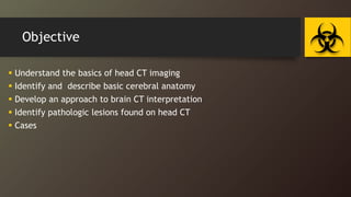
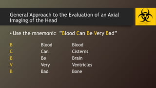
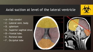
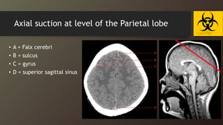
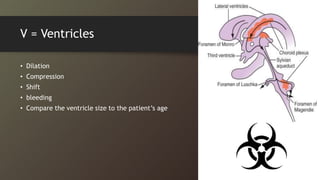
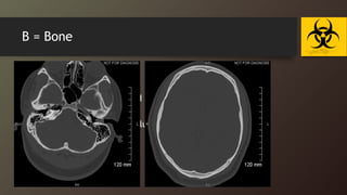
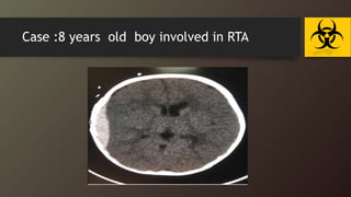
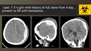
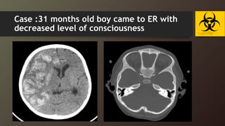
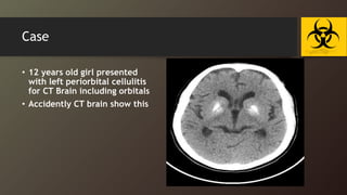
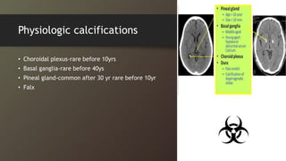
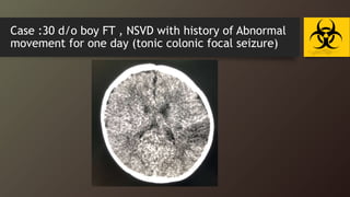
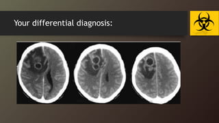
Head and Brain CT
Presented by Dr. shaikhah A.O.Bawazir
Pediatric resident R3
Saudi pediatric program – MCH – Buridah
Objective
 Understand the basics of head CT imaging
 Identify and describe basic cerebral anatomy
 Develop an approach to brain CT interpretation
 Identify pathologic lesions found on head CT
 Cases
Introduction
Introduction
• CT Fast tool
• high radiation risk.
• Best for bone and calcification.
• radiation exposure during childhood causes larger additional
cancer risk 10 % than a corresponding exposure in adulthood.
Indications
• Acute head trauma
• Acute neurologic deficits
• Increased intracranial pressure;
• Suspected acute intracranial hemorrhage;
• Suspected intra cranial lesion ( mass , tumor or
abscess)
• Suspected shunt malfunctions, or shunt revisions
• Suspected acute hydrocephalus, Brain herniation
• Non-febrile seizures
• Skull bone lesions (Langerhans cell histiocytosis,
neuroblastoma, etc)
• Craniosynostosis/ plagiocephaly
• Detection of calcification
• Immediate postoperative evaluation following brain
surgery (evacuation of hematoma, abscess drainage,
etc)
• When magnetic resonance imaging (MRI) imaging is
unavailable,
Basics of CT Imaging
Section and density
• Sagittal
• Coronal
• Cross section
• Transverse
• axial
• Bright (hyper density)
• Isodense ( gray)
• Dark (hypo density)
Bone
Contrast
Acute blood
Soft tissue
Water
Fat
Air
Hyperdense things on CT
acute blood
ocular lens
calcifications
contrast (dye)bone
metal (bullets w/
streak artifact)
Isodense things on CT
• Note that white matter is less dense than gray
matter and therefore:
(white matter is darker than gray matter)
Gray matter (cerebral
cortex)
Gray matter (basal
ganglia)
White matter
Hypodense things on CT
fat
air
CSF (water)
Area of infarction
Three window
Approach
General Approach to the Evaluation of an Axial
Imaging of the Head
• Use the mnemonic ”Blood Can Be Very Bad”
B Blood Blood
C Can Cisterns
B Be Brain
V Very Ventricles
B Bad Bone
B = blood
• Look for any evidence of
bleeding in:
 epidural, subdural,
 intraparenchymal,
intraventricular,
 subarachnoid
 Hyperdense (bright): Acute
blood
 Isodense: Subacute (1
week)
 Hypodense: Subacute to
chronic (2 weeks)
C = Cisterns
• Look for blood in the cisterns and to see if they are open:
• Suprasellar : contains the optic chiasm and pituitary stalk
• Quadrigeminal : extends laterally around the midbrain, from the great cerebral vein to the
third ventricle
• Sylvian : contains several arteries, including the middle cerebral artery.
• cisterna magna (cerebellomedullary cistern): is the largest of the subarachnoid cisterns
• Cerebellopontine angle cistern : Schwannomas (nearly 80% of all CP angle tumors) ,
Medulloblastoma, arachnoid cysts
B = Brain
Look COMPARTIVLY at both hemispheres of the brain:
• the grey-white mater differentiation-- > signs of atrophy ,ischemia
and earl stroke.
• the symmetry of the brain
• midline shift -- > indicates an intracerebral mass, edema, or a herniation.
• the parenchyma
-- > anatomical distortion or altered attenuation -- >
indicative of a mass, bleed, or vascular malformation
Axial suction at orbital level
• A = orbit
• B = sphenoid sinus
• C = temporal lobe
• D = external auditory canal
• E = mastoid air cell
• F = cerebellar hemispheres
• J = medulla oblongata J
Axial suction at level of Pons
• A = frontal lobe
• B = superior surface of
orbital part
• C = dorsum sellae
• D = basilar artery
• E = temporal lobe
• F = mastoid air cells
• G = Cerebellar hemisphere FM
Pons
cisterna magna
Axial suction at level of the 4th ventricle
• A . Frontal lobe
• B. sylvian Fissure
• C . Temporal lobe
• D . Suprasellar cistern
• E . Midbrain
• F . 4th ventricle
• G . Cerebellar hemisphere
Axial suction at level of the Basal ganglia
• A . Genu of corpus call.
• B. Ant.horn of Lat.Vent.
• C . Internal capsule
• D . Thalamus
• E . Pineal gland
• F . Choroid plexus
• G . Straight siuns
3rd vent
Axial suction at level of the lateral ventricle
• A = Falx cerebri
• C . Lateral vent. body
• D . splenium
• G . Superior sagittal sinus
• B = frontal lobe.
• E . Parietal lobe
• F . Occipital lobe
3rd vent
Axial suction at level of the Parietal lobe
• A = Falx cerebri
• B = sulcus
• C = gyrus
• D = superior sagittal sinus
V = Ventricles
• Dilation
• Compression
• Shift
• bleeding
• Compare the ventricle size to the patient’s age
B = Bone
• Examine by bone window :
• fractures
• evaluate the sinuses for fluid or soft tissues accumulation
• Mastoid air cell
• Fluid in the sinus may be a clue to a facial injury !!
Additional Areas to Examine
• Major venous sinuses to detect hypo density which is indicative of
thrombosis
• Venous thrombosis is a difficult diagnosis to make
• Soft tissues and skin through the parenchymal window looking for
lesions or hematomas
• Soft tissue injuries noted on the scan should initiate a detailed evaluation of
the subjacent structures.
• Check the orbits
• Look at every image
• Review sagittal and coronal if available
C A S
Case :8 years old boy involved in RTA
case: 1 Y/o girl with history of full done from 4 day ,
present to ER with hematoma.
Case :31 months old boy came to ER with
decreased level of consciousness
Case :A Flat neonate delivered by emergency C/S
after abruption placenta , CPR done in OR room
Bilateral diffuse cerebral hypo-densities.
Mild dilatation of the ventricular system.
Case
• 12 years old girl presented
with left periorbital cellulitis
for CT Brain including orbitals
• Accidently CT brain show this
Physiologic calcifications
• Choroidal plexus-rare before 10yrs
• Basal ganglia-rare before 40ys
• Pineal gland-common after 30 yr rare before 10yr
• Falx
Case :30 d/o boy FT , NSVD with history of Abnormal
movement for one day (tonic colonic focal seizure)
Your differential diagnosis:
In the END Systemic Approach to Head CT
 Symmetry : Compare left and right side of the cranium
 Midline: Look for midline shift
 Cross-sectional anatomy (Review anatomical landmark for each section)
 Brain tissue : the gray matter, white matter and intracerebral lesions
 CSF space (ventricle (dilated or not) )
 Skull and soft tissue : scalp swelling, fractures, sinuses, orbit
 Subdural windows : Look for blood collection adjacent to the skull
 Bone windows : Skull, orbit and sinuses, intracranial air
Our use ”Blood Can Be Very Bad”
ANY QUESTION !

brian CT introdaction pediatric radiology

  • 1.
    Head and BrainCT Presented by Dr. shaikhah A.O.Bawazir Pediatric resident R3 Saudi pediatric program – MCH – Buridah
  • 2.
    Objective  Understand thebasics of head CT imaging  Identify and describe basic cerebral anatomy  Develop an approach to brain CT interpretation  Identify pathologic lesions found on head CT  Cases
  • 3.
  • 4.
    Introduction • CT Fasttool • high radiation risk. • Best for bone and calcification. • radiation exposure during childhood causes larger additional cancer risk 10 % than a corresponding exposure in adulthood.
  • 5.
    Indications • Acute headtrauma • Acute neurologic deficits • Increased intracranial pressure; • Suspected acute intracranial hemorrhage; • Suspected intra cranial lesion ( mass , tumor or abscess) • Suspected shunt malfunctions, or shunt revisions • Suspected acute hydrocephalus, Brain herniation
  • 6.
    • Non-febrile seizures •Skull bone lesions (Langerhans cell histiocytosis, neuroblastoma, etc) • Craniosynostosis/ plagiocephaly • Detection of calcification • Immediate postoperative evaluation following brain surgery (evacuation of hematoma, abscess drainage, etc) • When magnetic resonance imaging (MRI) imaging is unavailable,
  • 7.
    Basics of CTImaging
  • 8.
    Section and density •Sagittal • Coronal • Cross section • Transverse • axial • Bright (hyper density) • Isodense ( gray) • Dark (hypo density) Bone Contrast Acute blood Soft tissue Water Fat Air
  • 9.
    Hyperdense things onCT acute blood ocular lens calcifications contrast (dye)bone metal (bullets w/ streak artifact)
  • 10.
    Isodense things onCT • Note that white matter is less dense than gray matter and therefore: (white matter is darker than gray matter) Gray matter (cerebral cortex) Gray matter (basal ganglia) White matter
  • 11.
    Hypodense things onCT fat air CSF (water) Area of infarction
  • 12.
  • 13.
  • 14.
    General Approach tothe Evaluation of an Axial Imaging of the Head • Use the mnemonic ”Blood Can Be Very Bad” B Blood Blood C Can Cisterns B Be Brain V Very Ventricles B Bad Bone
  • 15.
    B = blood •Look for any evidence of bleeding in:  epidural, subdural,  intraparenchymal, intraventricular,  subarachnoid  Hyperdense (bright): Acute blood  Isodense: Subacute (1 week)  Hypodense: Subacute to chronic (2 weeks)
  • 16.
    C = Cisterns •Look for blood in the cisterns and to see if they are open: • Suprasellar : contains the optic chiasm and pituitary stalk • Quadrigeminal : extends laterally around the midbrain, from the great cerebral vein to the third ventricle • Sylvian : contains several arteries, including the middle cerebral artery. • cisterna magna (cerebellomedullary cistern): is the largest of the subarachnoid cisterns • Cerebellopontine angle cistern : Schwannomas (nearly 80% of all CP angle tumors) , Medulloblastoma, arachnoid cysts
  • 18.
    B = Brain LookCOMPARTIVLY at both hemispheres of the brain: • the grey-white mater differentiation-- > signs of atrophy ,ischemia and earl stroke. • the symmetry of the brain • midline shift -- > indicates an intracerebral mass, edema, or a herniation. • the parenchyma -- > anatomical distortion or altered attenuation -- > indicative of a mass, bleed, or vascular malformation
  • 19.
    Axial suction atorbital level • A = orbit • B = sphenoid sinus • C = temporal lobe • D = external auditory canal • E = mastoid air cell • F = cerebellar hemispheres • J = medulla oblongata J
  • 20.
    Axial suction atlevel of Pons • A = frontal lobe • B = superior surface of orbital part • C = dorsum sellae • D = basilar artery • E = temporal lobe • F = mastoid air cells • G = Cerebellar hemisphere FM Pons cisterna magna
  • 21.
    Axial suction atlevel of the 4th ventricle • A . Frontal lobe • B. sylvian Fissure • C . Temporal lobe • D . Suprasellar cistern • E . Midbrain • F . 4th ventricle • G . Cerebellar hemisphere
  • 22.
    Axial suction atlevel of the Basal ganglia • A . Genu of corpus call. • B. Ant.horn of Lat.Vent. • C . Internal capsule • D . Thalamus • E . Pineal gland • F . Choroid plexus • G . Straight siuns 3rd vent
  • 23.
    Axial suction atlevel of the lateral ventricle • A = Falx cerebri • C . Lateral vent. body • D . splenium • G . Superior sagittal sinus • B = frontal lobe. • E . Parietal lobe • F . Occipital lobe 3rd vent
  • 24.
    Axial suction atlevel of the Parietal lobe • A = Falx cerebri • B = sulcus • C = gyrus • D = superior sagittal sinus
  • 25.
    V = Ventricles •Dilation • Compression • Shift • bleeding • Compare the ventricle size to the patient’s age
  • 26.
    B = Bone •Examine by bone window : • fractures • evaluate the sinuses for fluid or soft tissues accumulation • Mastoid air cell • Fluid in the sinus may be a clue to a facial injury !!
  • 27.
    Additional Areas toExamine • Major venous sinuses to detect hypo density which is indicative of thrombosis • Venous thrombosis is a difficult diagnosis to make • Soft tissues and skin through the parenchymal window looking for lesions or hematomas • Soft tissue injuries noted on the scan should initiate a detailed evaluation of the subjacent structures. • Check the orbits • Look at every image • Review sagittal and coronal if available
  • 28.
  • 29.
    Case :8 yearsold boy involved in RTA
  • 30.
    case: 1 Y/ogirl with history of full done from 4 day , present to ER with hematoma.
  • 31.
    Case :31 monthsold boy came to ER with decreased level of consciousness
  • 32.
    Case :A Flatneonate delivered by emergency C/S after abruption placenta , CPR done in OR room Bilateral diffuse cerebral hypo-densities. Mild dilatation of the ventricular system.
  • 33.
    Case • 12 yearsold girl presented with left periorbital cellulitis for CT Brain including orbitals • Accidently CT brain show this
  • 34.
    Physiologic calcifications • Choroidalplexus-rare before 10yrs • Basal ganglia-rare before 40ys • Pineal gland-common after 30 yr rare before 10yr • Falx
  • 35.
    Case :30 d/oboy FT , NSVD with history of Abnormal movement for one day (tonic colonic focal seizure)
  • 36.
  • 37.
    In the ENDSystemic Approach to Head CT  Symmetry : Compare left and right side of the cranium  Midline: Look for midline shift  Cross-sectional anatomy (Review anatomical landmark for each section)  Brain tissue : the gray matter, white matter and intracerebral lesions  CSF space (ventricle (dilated or not) )  Skull and soft tissue : scalp swelling, fractures, sinuses, orbit  Subdural windows : Look for blood collection adjacent to the skull  Bone windows : Skull, orbit and sinuses, intracranial air Our use ”Blood Can Be Very Bad”
  • 38.

Editor's Notes

  • #17 magna: vertebral arteries, glossopharyngeal nerve  (cranial nerve 9) vagus nerve (cranial nerve 10), accessory nerve (cranial nerve 11),chordal plexus Quadrigeminal : the pineal gland, trochlear nerves (cranial nerve 4) ,the great cerebral vein, posterior cerebral arteries, superior cerebellar arteries The infratentorial portion contains the posterior cerebral artery and the 4th cranial nerve (the trochlear nerve ). This nerve innervates the superior oblique muscle, which moves the eye down and out. It is vulnerable to damage in raised intracranial pressure due to its long winding course from the back of the brainstem to the superior orbital fissure, as well as its relatively thin caliber. children between ages of three and eight
  • #28 High clinical suspicion helps make diagnosis (risk factors) Look for a non-arterial distribution of ischemic change
  • #32 PRIGED VEIN
  • #33 MIDDEL MENINGIAL ARTERY INJURY
  • #34 Acute bleeding is seen in the cortical gyrai on the right Fronto- parietal area (haemorrhagic infarction). · Mixed density subdural hematoma is seen in the right front-parietal region (sub-acute sub-dural hematoma). · Diffuse brain oedema is seen on the right fronto-parietal and temporal parenchyma with loss of gray-white matter differentiation and effacement of the right lateral ventricle and minimal midline shift to the other side. · Fracture of the occipital bone seen.
  • #39 Initially unilateral --> "sequential bilaterality" is highly suggestive of HSE1
  • #40 Infection ( toxo , absence Neoplasm primary or seconder Demylenaton