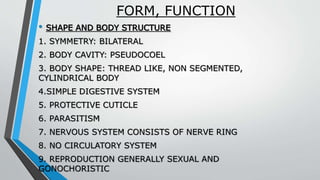
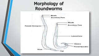
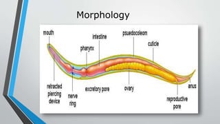
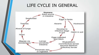
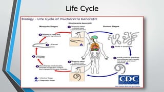
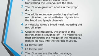
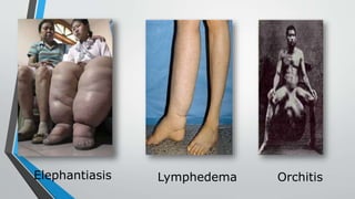
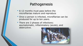
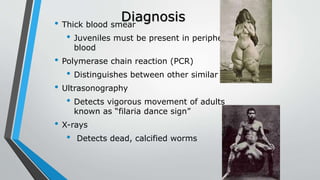
NEMATODA
•VIVEK DEV
FORM, FUNCTION
• SHAPE AND BODY STRUCTURE
1. SYMMETRY: BILATERAL
2. BODY CAVITY: PSEUDOCOEL
3. BODY SHAPE: THREAD LIKE, NON SEGMENTED,
CYLINDRICAL BODY
4.SIMPLE DIGESTIVE SYSTEM
5. PROTECTIVE CUTICLE
6. PARASITISM
7. NERVOUS SYSTEM CONSISTS OF NERVE RING
8. NO CIRCULATORY SYSTEM
9. REPRODUCTION GENERALLY SEXUAL AND
GONOCHORISTIC
Morphology of
Roundworms
Morphology
LIFE CYCLE IN GENERAL
CLASSIFICATIONThey are divided into two classes
distinguished by differences in sensory
and excretory system.
1. Class Enoplea
-Subclass Dorylaimia
Amphids are well developed .Excretory
system are found in
single,ventral,glandular cells or entirely
absent. Mostly free-living and some are
parasiitic on plants and animals.
Orders –
Trichurida,Dioctophymatida,Muspiceida,Me
Trichuris trichiura
-”Whipworm”
-Most common gastrointestinal parasites
of human
-Eggs posses plugs
-Have stichosome esophagus
-Male have coiled posterior ends.
-Pathogenesis- Trichuriasis
•Kingdom-Animalia
Phylum- Nematoda
Class- Enoplea
Order- Trichurida
Family- Trichuridae
Genus- Trichuris
Species- trichiura
Class Rhabditea
-Subclass Rhabditia
Amphids are generally poorly
developed. Phasmids are present .
Excretory system with one or two
lateral canals,with or without
associated glandular cells .Both free
living and parasitic.
Orders-
Rhabditida,Strongylida,Ascaridida,Ox
yurida,Spirurida.
Kingdom
Phylum
Class
Order
Family
Genus
Species
Animalia
Nematoda
Rhabditea
Ascaridida
Ascarididae
Ascaris
lumbricoides
Ascaris
lumbricoides
MORPHOLOGY
•Because of their great size, abundance, and
cosmopolitan distribution, these nematodes
may well have been the first parasites known
to humans.
•These species are characterized by, in addition
to their great size, having three prominent
lips, each with a dentigerous ridge, and no
interlabia or alae.
•Male ( 15 cms to 31 cms) and females (20
cms to 49 cms) are of different size.
Fertilized egg of Ascaris
Biology of Ascaris
•Ascaris lumbricoides, a roundworm, infects
humans when an ingested fertilised egg
becomes a larval worm (called rhabditiform
larva) that penetrates the wall of
the duodenum and enters the blood stream.
From there, it is carried to the liver and heart,
and enters pulmonary circulation to break free
in the alveoli, where it grows and molts. In
three weeks, the larva passes from
the respiratory system to be coughed up,
swallowed, and thus returned to the small
intestine, where it matures to an adult male
Pathogenesis
•Little damage is caused by penetration of
intestinal mucosa by newly hatched worms.
•Infection with Ascaris lumbricoides often
causes no symptoms.
• Infections with a large number of worms
may cause abdominal pain or intestinal
obstruction. Adults feed on the contents of
the small intestine and in heavy infections this
may compound problems in malnourished
individuals (especially children).
Life cycle of Ascaris
lumbricoides
Diagnosis and Treatment
DIAGNOSIS — The diagnosis of ascariasis is
usually made via stool microscopy. Other forms
of diagnosis are
through eosinophilia, imaging, ultrasound,
or serology examination.
TREATMENT — Treatment consists of choosing
the right drugs, therapy, follow-up,
and supportive care for each patient.
PREVENTION — Prevention of reinfection poses a
substantial problem since Ascaris parasites are
abundant in soil. Good sanitation to prevent fecal
contamination of soil is required. An education
program advising against the use of human feces
as a fertilizer is also needed in some areas. Soil
treatments have been attempted but are
HOOKWORM
FAMILY ANCYLOSTOMIDAE
Kingdom Animalia
Phylum Nematoda
Class Secernentea
Order Strongylida
Family Ancylostomatidae
Genus Ancylostoma
Species duodenale
C. Name Hookworm
MORPHOLOGY
 MOST SPECIES ARE RATHER STOUT, AND THE
ANTERIOR END IS CURVED DORSALLY, GIVING THE
WORM A HOOKLIKE APPEARANCE
 THE BUCCAL CAPSULE IS LARGE AND HEAVILY
SCLEROTIZED AND USUALLY IS ARMED WITH
CUTTING PLATES, TEETH, LANCETS, OR A DORSAL
CONE. LIPS ARE REDUCED OR ABSENT.
THE ESOPHAGUS IS STOUT, WITH A SWOLLEN
POSTERIOR END, GIVING IT A CLUB SHAPE
MORPHOLOGY
MALES HAVE A CONSPICUOUS COPULATORY
BURSA, CONSISTING OF TWO BROAD LATERAL
LOBES AND A SMALLER DORSAL LOBE, ALL
SUPPORTED BY FLESHY RAYS
FEMALES HAVE A SIMPLE, CONICAL TAIL. THE
VULVA IS POSTEQUATORIAL, AND TWO OVARIES
ARE PRESENT. ABOUT 5% OF THE DAILY OUTPUT OF
EGGS IS FOUND IN THE UTERI AT ANY ONE TIME;
THE TOTAL PRODUCTION IS SEVERAL THOUSAND
PER DAY FOR AS LONG AS NINE YEARS.
BIOLOGY OF HOOKWORM
 HOOKWORMS MATURE AND MATE IN THE SMALL
INTESTINE OF THEIR HOST
SPECIES INFECTING HUMANS CANNOT BE
DIAGNOSED RELIABLY BY THEIR EGGS ALONE
 EGGS REQUIRE WARMTH, SHADE, AND
MOISTURE FOR CONTINUED DEVELOPMENT
JUVENILES LIVE IN THE FECES, FEEDING ON
FECAL MATTER, AND MOLT THEIR CUTICLE IN
TWO TO THREE DAYS
BIOLOGY OF HOOKWORM
 WHEN THE GROUND SURFACE IS DRY, THEY
MIGRATE A SHORT DISTANCE INTO THE SOIL,
FOLLOWING THE RETREATING WATER. UNDER
IDEAL CONDITIONS, THEY CAN LIVE FOR SEVERAL
WEEKS.INFECTION OCCURS WHEN WORM CONTACT A
HOST’S SKIN AND BURROW INTO IT, AND THEY
 SECOND STAGE JUVENILES, WHICH ALSO HAVE A
RHABDITIFORM ESOPHAGUS, CONTINUE TO FEED
AND GROW AND, AFTER ABOUT FIVE DAYS, MOLT
TO THE THIRD STAGE, WHICH IS INFECTIVE TO A
HOST. SECOND-STAGE CUTICLE MAY BE RETAINED
AS A LOOSEFITTING SHEATH UNTIL PENETRATION
OF A NEW HOST, OR IT MAY BE LOST EARLIER
Diagnosis, Symptoms and
Treatment
Symptoms
•abdominal pain
•colic, or cramping
and excessive
crying in infants
•intestinal cramps
•nausea
• fever
•blood in stool
•a loss of appetite
Treatment & Prevention
•Albendazole,
Mebendazole
•wearing shoes
when you walk
outdoors, especially
in areas that might
have feces in the
soil
•drinking safe water
•properly cleaning
Diagnosis:
The standard method for diagnosing the
presence of hookworm is by identifying
hookworm eggs in a stool sample using a
microscope
Wuchereria
bancrofti
Scientific Classification
Kingdom: Animalia
Phylum: Nematoda
Class: Secernentea
Order: Spirurida
Family: Filariidae
Genus: Wuchereria
Species: bancrofti
Wuchereria bancrofti
• Definitive Host: Humans
• Intermediate Host: Mosquitoes (especially night-feeding
mosquitoes)
• Species: Anopheles, Aedes, Culex, Mansonia
• Morphology:
• Males:
• 40 mm long & 100 μm wide
• Fingerlike tail
• Females:
• 6-10 cm long & 300 μm wide
• They are viviparous
• Their vulva is near the level of the middle of
Geographic
Distribution• Its infection is mainly found in the tropical
region, pacific islands and sometimes in the
Caribbean.
• West and south parts of Africa
• Egypt
• Southern Asia
• Western Pacific islands
• Northeastern coast of Brazil
• Guyana
• Caribbean island of Hispaniola
Life Cycle
1. The mosquito takes a blood meal,
transferring the L3 larva into the skin.
2. The L3 larva grow into adults in the lymph
ducts.
3. The adults reproduce, producing sheathed
microfilariae; the microfilariae migrate into
the blood and lymph channels
4. A mosquito takes a blood meal, ingesting the
microfilariae.
5. Once in the mosquito, the sheath of the
microfilariae is sloughed off. The microfilariae
then penetrates the midgut of the mosquito,
making its way into the thoracic muscles.
6. L1 larvae form
7. L3 larvae form
8. The L3 larvae are the infective stage,
• High levels of microfilaria in the blood
• No symptoms present
• Inflammatory (Acute) Phase:
• Inflammatory responses happen in response to
antigens from adult worms
• Lymphedema—swelling due to blockage of lymph
vessels
• Orchitis—inflammation of the testes
• Epididymitis—inflammation of the spermatic cord
• Obstructive Phase:
• Lymph varices—enlarged lymph vessels
(synonymous with varicose veins)
• Lymph scrotum— enlarged lymph vessels in the
scrotum
• Chyluria—lymph in urine (milky and sometimes
bloody urine)
• Elephantiasis—enlargement of limbs and thickening
• Elephantiasis
Elephantiasis Lymphedema Orchitis
Pathogenesis
• 6-12 months must pass before the
microfilariae mature and reproduce
• Once a person is infected, microfilariae can be
produced for up to ten years.
• There are 3 phases of infections:
asymptomatic, inflammatory (acute), and
obstructive.
Diagnosis• Thick blood smear
• Juveniles must be present in peripheral
blood
• Polymerase chain reaction (PCR)
• Distinguishes between other similar species
• Ultrasonography
• Detects vigorous movement of adults
known as “filaria dance sign”
• X-rays
• Detects dead, calcified worms
Treatment
• Primary drug of choice: diethylcarbamazine
(DEC)
• Eliminates microfilaria from the blood and
(if administered correctly) kills adult worms
• Ivermectin
• Albendazole & Mectizan
• Application of pressure bandages
Control
• Administration of Albendazole & Mectizan to
control the spread of the disease.
• There are currently no vaccines
• Elimination of common mosquito breeding
grounds
• Fallen coconuts
• containers filled with stagnant water
• Marshy and swampy areas.
• They hope to eradicate it by year 2020; so far,
the diseased population has declined
significantly
Nematoda
Nematoda

Nematoda

  • 1.
  • 2.
    FORM, FUNCTION • SHAPEAND BODY STRUCTURE 1. SYMMETRY: BILATERAL 2. BODY CAVITY: PSEUDOCOEL 3. BODY SHAPE: THREAD LIKE, NON SEGMENTED, CYLINDRICAL BODY 4.SIMPLE DIGESTIVE SYSTEM 5. PROTECTIVE CUTICLE 6. PARASITISM 7. NERVOUS SYSTEM CONSISTS OF NERVE RING 8. NO CIRCULATORY SYSTEM 9. REPRODUCTION GENERALLY SEXUAL AND GONOCHORISTIC
  • 3.
  • 4.
  • 5.
  • 6.
    CLASSIFICATIONThey are dividedinto two classes distinguished by differences in sensory and excretory system. 1. Class Enoplea -Subclass Dorylaimia Amphids are well developed .Excretory system are found in single,ventral,glandular cells or entirely absent. Mostly free-living and some are parasiitic on plants and animals. Orders – Trichurida,Dioctophymatida,Muspiceida,Me
  • 7.
    Trichuris trichiura -”Whipworm” -Most commongastrointestinal parasites of human -Eggs posses plugs -Have stichosome esophagus -Male have coiled posterior ends. -Pathogenesis- Trichuriasis
  • 9.
    •Kingdom-Animalia Phylum- Nematoda Class- Enoplea Order-Trichurida Family- Trichuridae Genus- Trichuris Species- trichiura
  • 10.
    Class Rhabditea -Subclass Rhabditia Amphidsare generally poorly developed. Phasmids are present . Excretory system with one or two lateral canals,with or without associated glandular cells .Both free living and parasitic. Orders- Rhabditida,Strongylida,Ascaridida,Ox yurida,Spirurida.
  • 11.
  • 12.
    MORPHOLOGY •Because of theirgreat size, abundance, and cosmopolitan distribution, these nematodes may well have been the first parasites known to humans. •These species are characterized by, in addition to their great size, having three prominent lips, each with a dentigerous ridge, and no interlabia or alae. •Male ( 15 cms to 31 cms) and females (20 cms to 49 cms) are of different size.
  • 13.
  • 14.
    Biology of Ascaris •Ascarislumbricoides, a roundworm, infects humans when an ingested fertilised egg becomes a larval worm (called rhabditiform larva) that penetrates the wall of the duodenum and enters the blood stream. From there, it is carried to the liver and heart, and enters pulmonary circulation to break free in the alveoli, where it grows and molts. In three weeks, the larva passes from the respiratory system to be coughed up, swallowed, and thus returned to the small intestine, where it matures to an adult male
  • 15.
    Pathogenesis •Little damage iscaused by penetration of intestinal mucosa by newly hatched worms. •Infection with Ascaris lumbricoides often causes no symptoms. • Infections with a large number of worms may cause abdominal pain or intestinal obstruction. Adults feed on the contents of the small intestine and in heavy infections this may compound problems in malnourished individuals (especially children).
  • 16.
    Life cycle ofAscaris lumbricoides
  • 17.
    Diagnosis and Treatment DIAGNOSIS— The diagnosis of ascariasis is usually made via stool microscopy. Other forms of diagnosis are through eosinophilia, imaging, ultrasound, or serology examination. TREATMENT — Treatment consists of choosing the right drugs, therapy, follow-up, and supportive care for each patient. PREVENTION — Prevention of reinfection poses a substantial problem since Ascaris parasites are abundant in soil. Good sanitation to prevent fecal contamination of soil is required. An education program advising against the use of human feces as a fertilizer is also needed in some areas. Soil treatments have been attempted but are
  • 18.
    HOOKWORM FAMILY ANCYLOSTOMIDAE Kingdom Animalia PhylumNematoda Class Secernentea Order Strongylida Family Ancylostomatidae Genus Ancylostoma Species duodenale C. Name Hookworm
  • 19.
    MORPHOLOGY  MOST SPECIESARE RATHER STOUT, AND THE ANTERIOR END IS CURVED DORSALLY, GIVING THE WORM A HOOKLIKE APPEARANCE  THE BUCCAL CAPSULE IS LARGE AND HEAVILY SCLEROTIZED AND USUALLY IS ARMED WITH CUTTING PLATES, TEETH, LANCETS, OR A DORSAL CONE. LIPS ARE REDUCED OR ABSENT. THE ESOPHAGUS IS STOUT, WITH A SWOLLEN POSTERIOR END, GIVING IT A CLUB SHAPE
  • 20.
    MORPHOLOGY MALES HAVE ACONSPICUOUS COPULATORY BURSA, CONSISTING OF TWO BROAD LATERAL LOBES AND A SMALLER DORSAL LOBE, ALL SUPPORTED BY FLESHY RAYS FEMALES HAVE A SIMPLE, CONICAL TAIL. THE VULVA IS POSTEQUATORIAL, AND TWO OVARIES ARE PRESENT. ABOUT 5% OF THE DAILY OUTPUT OF EGGS IS FOUND IN THE UTERI AT ANY ONE TIME; THE TOTAL PRODUCTION IS SEVERAL THOUSAND PER DAY FOR AS LONG AS NINE YEARS.
  • 21.
    BIOLOGY OF HOOKWORM HOOKWORMS MATURE AND MATE IN THE SMALL INTESTINE OF THEIR HOST SPECIES INFECTING HUMANS CANNOT BE DIAGNOSED RELIABLY BY THEIR EGGS ALONE  EGGS REQUIRE WARMTH, SHADE, AND MOISTURE FOR CONTINUED DEVELOPMENT JUVENILES LIVE IN THE FECES, FEEDING ON FECAL MATTER, AND MOLT THEIR CUTICLE IN TWO TO THREE DAYS
  • 22.
    BIOLOGY OF HOOKWORM WHEN THE GROUND SURFACE IS DRY, THEY MIGRATE A SHORT DISTANCE INTO THE SOIL, FOLLOWING THE RETREATING WATER. UNDER IDEAL CONDITIONS, THEY CAN LIVE FOR SEVERAL WEEKS.INFECTION OCCURS WHEN WORM CONTACT A HOST’S SKIN AND BURROW INTO IT, AND THEY  SECOND STAGE JUVENILES, WHICH ALSO HAVE A RHABDITIFORM ESOPHAGUS, CONTINUE TO FEED AND GROW AND, AFTER ABOUT FIVE DAYS, MOLT TO THE THIRD STAGE, WHICH IS INFECTIVE TO A HOST. SECOND-STAGE CUTICLE MAY BE RETAINED AS A LOOSEFITTING SHEATH UNTIL PENETRATION OF A NEW HOST, OR IT MAY BE LOST EARLIER
  • 26.
    Diagnosis, Symptoms and Treatment Symptoms •abdominalpain •colic, or cramping and excessive crying in infants •intestinal cramps •nausea • fever •blood in stool •a loss of appetite Treatment & Prevention •Albendazole, Mebendazole •wearing shoes when you walk outdoors, especially in areas that might have feces in the soil •drinking safe water •properly cleaning Diagnosis: The standard method for diagnosing the presence of hookworm is by identifying hookworm eggs in a stool sample using a microscope
  • 27.
    Wuchereria bancrofti Scientific Classification Kingdom: Animalia Phylum:Nematoda Class: Secernentea Order: Spirurida Family: Filariidae Genus: Wuchereria Species: bancrofti
  • 28.
    Wuchereria bancrofti • DefinitiveHost: Humans • Intermediate Host: Mosquitoes (especially night-feeding mosquitoes) • Species: Anopheles, Aedes, Culex, Mansonia • Morphology: • Males: • 40 mm long & 100 μm wide • Fingerlike tail • Females: • 6-10 cm long & 300 μm wide • They are viviparous • Their vulva is near the level of the middle of
  • 30.
    Geographic Distribution• Its infectionis mainly found in the tropical region, pacific islands and sometimes in the Caribbean. • West and south parts of Africa • Egypt • Southern Asia • Western Pacific islands • Northeastern coast of Brazil • Guyana • Caribbean island of Hispaniola
  • 31.
  • 32.
    1. The mosquitotakes a blood meal, transferring the L3 larva into the skin. 2. The L3 larva grow into adults in the lymph ducts. 3. The adults reproduce, producing sheathed microfilariae; the microfilariae migrate into the blood and lymph channels 4. A mosquito takes a blood meal, ingesting the microfilariae. 5. Once in the mosquito, the sheath of the microfilariae is sloughed off. The microfilariae then penetrates the midgut of the mosquito, making its way into the thoracic muscles. 6. L1 larvae form 7. L3 larvae form 8. The L3 larvae are the infective stage,
  • 33.
    • High levelsof microfilaria in the blood • No symptoms present • Inflammatory (Acute) Phase: • Inflammatory responses happen in response to antigens from adult worms • Lymphedema—swelling due to blockage of lymph vessels • Orchitis—inflammation of the testes • Epididymitis—inflammation of the spermatic cord • Obstructive Phase: • Lymph varices—enlarged lymph vessels (synonymous with varicose veins) • Lymph scrotum— enlarged lymph vessels in the scrotum • Chyluria—lymph in urine (milky and sometimes bloody urine) • Elephantiasis—enlargement of limbs and thickening
  • 34.
  • 35.
    Pathogenesis • 6-12 monthsmust pass before the microfilariae mature and reproduce • Once a person is infected, microfilariae can be produced for up to ten years. • There are 3 phases of infections: asymptomatic, inflammatory (acute), and obstructive.
  • 36.
    Diagnosis• Thick bloodsmear • Juveniles must be present in peripheral blood • Polymerase chain reaction (PCR) • Distinguishes between other similar species • Ultrasonography • Detects vigorous movement of adults known as “filaria dance sign” • X-rays • Detects dead, calcified worms
  • 37.
    Treatment • Primary drugof choice: diethylcarbamazine (DEC) • Eliminates microfilaria from the blood and (if administered correctly) kills adult worms • Ivermectin • Albendazole & Mectizan • Application of pressure bandages
  • 38.
    Control • Administration ofAlbendazole & Mectizan to control the spread of the disease. • There are currently no vaccines • Elimination of common mosquito breeding grounds • Fallen coconuts • containers filled with stagnant water • Marshy and swampy areas. • They hope to eradicate it by year 2020; so far, the diseased population has declined significantly

Editor's Notes

  • #21 Bursa = a bodily pouch or sac