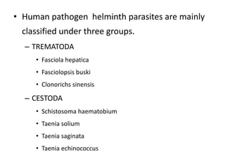
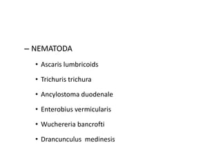
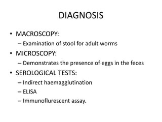
HUMAN PATHOGEN
HELMINTH PARASITES
RATHEESH R.L
• Human pathogen helminth parasites are mainly
classified under three groups.
– TREMATODA
• Fasciola hepatica
• Fasciolopsis buski
• Clonorichs sinensis
– CESTODA
• Schistosoma haematobium
• Taenia solium
• Taenia saginata
• Taenia echinococcus
– NEMATODA
• Ascaris lumbricoids
• Trichuris trichura
• Ancylostoma duodenale
• Enterobius vermicularis
• Wuchereria bancrofti
• Drancunculus medinesis
Fasciola hepatica ( sheep liver fluke)
• HABITAT: It lives in the bile duct of sheep, ox,
goat and man
LIFE CYCLE
• The life cycle is completing in two hosts: man
and snails.
• Fasciola hepatica is a parasite living in the liver
and bile duct of man and sheep.
• The adult worm discharges immature eggs in
the bile duct and in stool.
• Eggs become embryonated in water and
releases larvae which invade a suitable snail.
• A human eats the raw freshwater plant
containing the cyst will get the infection.
• After ingestion it will spreads to the
duodenum, intestinal wall, peritoneal cavity
and liver.
CLINICAL FEATURES
• The disease caused by the fasciola hepatica is
known as fascioliasis.
• In the chronic phase of fascioliasis it cause
liver inflammation and obstruction of the
biliary fluid. other symptoms include:
– diarrhea
– eosinophilia (high number of white blood cells)
– fever
– nausea
– stomach ache
– vomiting.
LABORATORY DIAGNOSIS
• MICROSCOPY:
– Eggs can be identify in the stool of infected
persons under a microscope.
• ANTIBODY DETECTION TEST:
TREATMENT
• Triclabendazole is the drug of choice
PREVENTION
• Avoid eating raw fresh water plants
• Avoid drinking contaminated water
• Drink only purified and boiled water
Taenia solium (pork tape worm)
Taenia saginata (beef tape worm)
HABITAT:
Taenia solium: Lives in the intestine
Taenia saginata: Lives in the mucosal wall
of ileum.
LIFE CYCLE
• Taeniasis is the infection of humans with the
adult tapeworm of Taenia saginata or Taenia
solium.
• Eggs are passed with feces ; the eggs can
survive for days to months in the environment.
• Cattle (T. saginata) and pigs (T. solium) become
infected by ingesting vegetation contaminated
with eggs
• In the animal intestine the eggs rupture with
liberation of onchospheres.
• These onchospheres invade to the intestinal
wall and migrate to the striated muscles of the
animal and they develop into cysticerci.
• A cysticercus can survive for several years in
the animal. Humans become infected by
ingesting raw or undercooked infected meat
• In the human intestine, the cysticercus
develops over 2 months into an adult
tapeworm, which can survive for years.
• The adult tapeworms attach to the small
intestine.
• Then the adult female tapeworm starts to
produce eggs and the life cycle will be
repeated.
CLINICAL FEATURES
• The infection with the taenia species is known
as taeniasis.
• Most people that have taeniasis don’t have
any symptoms. If signs and symptoms are
present they may include:
– pain
– unexplained weight loss
– blockage of the intestine
– digestive problems
– Fiscomfort
– Hunger
– constipation
DIAGNOSIS
• MACROSCOPY:
– Examination of stool for adult worms
• MICROSCOPY:
– Demonstrates the presence of eggs in the feces
• SEROLOGICAL TESTS:
– Indirect haemagglutination
– ELISA
– Immunoflurescent assay.
TREATMENT
• The drug of choice are
– Niclosamide
– Albendazol
– mebendazole and
– praziquantal
TRANSMISSION
• By eating under cooked beef or pork
• By drinking contaminated water
PREVENTION
• Eat the food items only after proper cooking.
NEMATODA
(round worm)
• The family includes the parasites,
• Ascaris lumbricoids
• Trichuris trichura
• Ancylostoma duodenale
• Enterobius vermicularis
• Wuchereria bancrofti
• Drancunculus medinesis
Ascaris lumbricoids ( human round worm)
HABITAT:
lumen of the small intestine
LIFE CYCLE
• Life cycle is completed only in man.
• Adult worms live in the lumen of the small
intestine.
• A female may produce up to 240,000 eggs per
day, which are passed with the feces
• Fertile eggs embryonate and become infective
after 18 days to several weeks , depending on the
environmental conditions (optimum: moist,
warm, shaded soil).
• After infective eggs are swallowed , the larvae
invade the intestinal mucosa, and are carried
via the portal and systemic circulation to the
lungs.
• The larvae mature further in the lungs (10-14
days), penetrate the alveolar walls, ascend the
bronchial tree to the throat, and are
swallowed.
• Upon reaching the small intestine, they
develop into adult worms and the life cycle is
repeated.
CLINICAL PICTURE
• The infection caused by the Ascaris
lumbricoids is known as intestinal ascariasis.
• Infections with a large number of worms may
cause abdominal pain or intestinal
obstruction.
• Other symptoms are, abdominal pain,
intestinal obstruction, poor digestion,
diarrhea, appendicitis, cough, dyspnoea and
hemoptysis.
DIAGNOSIS
• MICROSCOPY:
– During pulmonary migration larvae can be
identified in sputum and gastric aspirate.
• MACROSCOPY
– Detection of adult worms in stool
– Scratch test
TREATMENT
• Albendazole
• Mebendazole
• Piperazine
• Decaris and
• pyrantel pamoate
TRANSMISSION
• Contaminated water, foods especially raw
vegetables which may contain eggs of the
parasite.
• Unhygienic sanitation
PREVENTION
• Drink clean and boiled water
• Wash hands thoroughly
• Use properly cooked vegetables.
ANCYLOSTOMA DUODENALE( HOOK
WORM)
HABITAT:
Small intestine particularly in the jejunum.
LIFE CYCLE
• Man is the only host. Adult worms live inside the
jejunum part of the small intestine of man.
• Females lay egg after fertilization which are
passed out with stool.
• Under favorable conditions the eggs will remain
in the soil.
• Then the larvaes are produced in the soil/feces
from the eggs known as rhabditiform larvae.
• These rhabditiform larvae will transform to
the form of filariform larvae that are infective
and it will remain in the grass and soil.
• When a person walking bare footed or coming
contact with the filariform larvae, it will
penetrate directly through the skin or hair
follicles to the subcutaneous tissue.
• From the subcutaneous tissue, it will finally
reaches the lymphatic system and small
venules.
• Through the blood stream it will reaches to
the heart and lungs.
• In lungs they penetrate pulmonary alveoli,
bronchioles, trachea, larynx and swallowed
back to stomach.
• Again the larvae reach to small intestine and
the cycle is repeated.
CLINICAL PICTURE
• The infection by the ancylostoma duodenale is known
as ancylostomiasis or hookworm disease.
• if the patient experience symptoms, they generally
start with itchiness and a small rash caused by an
allergic reaction as the larvae enters to the skin.
• Commonly the symptoms of anemia will also occur.
• Other symptoms include:
– abdominal pain
– intestinal cramps
– nausea
– fever
– blood in your stool
– appetite loss
– itchy rash
DIAGNOSIS
• MICROSCOPY:
done to demonstrate eggs in stool.
• BLOOD STUDIES
To reveal anemia
• STOOL EXAMINATION
To find out the presence of occult blood in
stool
TREATMENT
• Albendazole
• Mebandazole
• Pyrantel pamoate.
TRANSMISSION
• Transmission occurs by walking bare footed or
coming in direct contacted with the
contaminated soil.
• Rarely transmission can be occur through oral
route by the accidental drinking of
contaminated water.
PREVENTION
• Avoid walking bare foot
• Hygienic sanitary conditions
Wuchereria bancrofti
HABITAT:
Adult worms lives in the lymphatic
vessels and lymph nodes of man while as
microfilaria are found in the blood.
LIFE CYCLE
• It is a digenetic parasite as it completes its life
cycle in two hosts.
• The primary or definitive host is man, while the
secondary or intermediate host is several species
of mosquitoes belonging to genus Сulex, Aedes
and Anopheles.
• The adult worm lives inside the lymphatic vessels
and lymph nodes of human beings.
• After fertilization the female produces larvae.
During day time it remains in deeper blood
vessels and at night in the capillaries of the
skin.
• These larvae does not develop until it is
sucked by the mosquitos.
• During bite the mosquito the larvae reaches to
the mosquito gut.
• Then the larvae penetrate the gut and reaches
to the thoracic muscles.
• In the thoracic muscles it changes to the
infective form. ( filariform larvae)
• After 2 moultings these larvae moves to the
mouth of the mosquito and when such
mosquito bites an individual these larvae
enters to the human body especially on the
lymphatics of the patient.
• From the lymphatic system it reaches to
various parts of the body and the cycle is
repeated.
CLINICAL PICTURE
• The disease caused by the Wuchereria bancrofti
is known as lymphatic filariasis.
• Other symptoms are,
– Fever
– Headache
– Myalgia
– Lymphadenitis
– Lymphadenopathy
– Inflammation of lymphatics
– Lymphatic dysfunction
– Progressive swelling of limbs
– Progressive swelling of genitalia
– Elephantiasis
DIAGNOSIS
• MICROSCOPY:
• Doing to demonstrate the larvae form.
• DEMONSTRATION OF ADULT WORM
• By lymph node biopsy
• SEROLOGICAL STUDIES
• ELISA, RIA, CFT, IFA & PCR
TREATMENT
• Diethyl carbamazine is the drug of choice.
PREVENTION
• Using mesh doors and nets.
• Use mosquito repellents
• Eliminate the breeding grounds of mosquito
• Chemical sprays.
Drancunculus medinesis (guinea worm)
HABITAT:
the adult female guinea worm lives in the
sub cutaneous tissue of legs , arms and back.
LIFE CYCLE
• Humans become infected by drinking unfiltered
water containing with the of Drancunculus
medinesis.
• Following ingestion, the eggs will die and release
the larvae, which penetrate the host stomach and
intestinal wall and enter the abdominal cavity and
retroperitoneal space.
• After maturation into adults, the male worms die
and the females migrate in the subcutaneous
tissues towards the skin surface.
• Approximately one year after infection, the
female worm induces a blister on the skin,
generally on the distal lower extremity, which
ruptures.
• When this lesion comes into contact with
water the patients develop local discomfort
and the larvae will spreads to the water.
• Thus is the cycle is repeated.
CLINICAL PICTURE
• The infection in man is called as dracunculosis
and is commonly known as guinea worm disease.
• The other symptoms are,
• Fever
• Local swelling
• Local pain
• Chronic skin ulcers
• Visible worms in skin ulcers
• Painful skin bumps
• Nausea
• Vomiting
• Diarrhea
DIAGNOSIS
• Detection of adult worm
• Detection of embryo
• Intra dermal test
TREATMENT
• Local cleansing of lesion and local application
of antibiotics.
• Mechanical extraction of worms.
• Ambithus is the drug of choice
PREVENTION
• DRINK CLEAN AND BOILED WATER.
• USE CLEAN WATER FOR BATHING ALSO.
30. human pathogen  helminthic parasites

30. human pathogen helminthic parasites

  • 1.
  • 2.
    • Human pathogenhelminth parasites are mainly classified under three groups. – TREMATODA • Fasciola hepatica • Fasciolopsis buski • Clonorichs sinensis – CESTODA • Schistosoma haematobium • Taenia solium • Taenia saginata • Taenia echinococcus
  • 3.
    – NEMATODA • Ascarislumbricoids • Trichuris trichura • Ancylostoma duodenale • Enterobius vermicularis • Wuchereria bancrofti • Drancunculus medinesis
  • 5.
    Fasciola hepatica (sheep liver fluke) • HABITAT: It lives in the bile duct of sheep, ox, goat and man
  • 6.
    LIFE CYCLE • Thelife cycle is completing in two hosts: man and snails. • Fasciola hepatica is a parasite living in the liver and bile duct of man and sheep. • The adult worm discharges immature eggs in the bile duct and in stool. • Eggs become embryonated in water and releases larvae which invade a suitable snail.
  • 7.
    • A humaneats the raw freshwater plant containing the cyst will get the infection. • After ingestion it will spreads to the duodenum, intestinal wall, peritoneal cavity and liver.
  • 8.
    CLINICAL FEATURES • Thedisease caused by the fasciola hepatica is known as fascioliasis. • In the chronic phase of fascioliasis it cause liver inflammation and obstruction of the biliary fluid. other symptoms include: – diarrhea – eosinophilia (high number of white blood cells) – fever – nausea – stomach ache – vomiting.
  • 9.
    LABORATORY DIAGNOSIS • MICROSCOPY: –Eggs can be identify in the stool of infected persons under a microscope. • ANTIBODY DETECTION TEST:
  • 10.
  • 11.
    PREVENTION • Avoid eatingraw fresh water plants • Avoid drinking contaminated water • Drink only purified and boiled water
  • 13.
    Taenia solium (porktape worm) Taenia saginata (beef tape worm) HABITAT: Taenia solium: Lives in the intestine Taenia saginata: Lives in the mucosal wall of ileum.
  • 14.
    LIFE CYCLE • Taeniasisis the infection of humans with the adult tapeworm of Taenia saginata or Taenia solium. • Eggs are passed with feces ; the eggs can survive for days to months in the environment. • Cattle (T. saginata) and pigs (T. solium) become infected by ingesting vegetation contaminated with eggs
  • 15.
    • In theanimal intestine the eggs rupture with liberation of onchospheres. • These onchospheres invade to the intestinal wall and migrate to the striated muscles of the animal and they develop into cysticerci. • A cysticercus can survive for several years in the animal. Humans become infected by ingesting raw or undercooked infected meat
  • 16.
    • In thehuman intestine, the cysticercus develops over 2 months into an adult tapeworm, which can survive for years. • The adult tapeworms attach to the small intestine. • Then the adult female tapeworm starts to produce eggs and the life cycle will be repeated.
  • 17.
    CLINICAL FEATURES • Theinfection with the taenia species is known as taeniasis. • Most people that have taeniasis don’t have any symptoms. If signs and symptoms are present they may include: – pain – unexplained weight loss – blockage of the intestine – digestive problems – Fiscomfort – Hunger – constipation
  • 18.
    DIAGNOSIS • MACROSCOPY: – Examinationof stool for adult worms • MICROSCOPY: – Demonstrates the presence of eggs in the feces • SEROLOGICAL TESTS: – Indirect haemagglutination – ELISA – Immunoflurescent assay.
  • 19.
    TREATMENT • The drugof choice are – Niclosamide – Albendazol – mebendazole and – praziquantal
  • 20.
    TRANSMISSION • By eatingunder cooked beef or pork • By drinking contaminated water
  • 21.
    PREVENTION • Eat thefood items only after proper cooking.
  • 22.
  • 23.
    • The familyincludes the parasites, • Ascaris lumbricoids • Trichuris trichura • Ancylostoma duodenale • Enterobius vermicularis • Wuchereria bancrofti • Drancunculus medinesis
  • 24.
    Ascaris lumbricoids (human round worm) HABITAT: lumen of the small intestine
  • 25.
    LIFE CYCLE • Lifecycle is completed only in man. • Adult worms live in the lumen of the small intestine. • A female may produce up to 240,000 eggs per day, which are passed with the feces • Fertile eggs embryonate and become infective after 18 days to several weeks , depending on the environmental conditions (optimum: moist, warm, shaded soil).
  • 26.
    • After infectiveeggs are swallowed , the larvae invade the intestinal mucosa, and are carried via the portal and systemic circulation to the lungs. • The larvae mature further in the lungs (10-14 days), penetrate the alveolar walls, ascend the bronchial tree to the throat, and are swallowed.
  • 27.
    • Upon reachingthe small intestine, they develop into adult worms and the life cycle is repeated.
  • 28.
    CLINICAL PICTURE • Theinfection caused by the Ascaris lumbricoids is known as intestinal ascariasis. • Infections with a large number of worms may cause abdominal pain or intestinal obstruction. • Other symptoms are, abdominal pain, intestinal obstruction, poor digestion, diarrhea, appendicitis, cough, dyspnoea and hemoptysis.
  • 29.
    DIAGNOSIS • MICROSCOPY: – Duringpulmonary migration larvae can be identified in sputum and gastric aspirate. • MACROSCOPY – Detection of adult worms in stool – Scratch test
  • 30.
    TREATMENT • Albendazole • Mebendazole •Piperazine • Decaris and • pyrantel pamoate
  • 31.
    TRANSMISSION • Contaminated water,foods especially raw vegetables which may contain eggs of the parasite. • Unhygienic sanitation
  • 32.
    PREVENTION • Drink cleanand boiled water • Wash hands thoroughly • Use properly cooked vegetables.
  • 33.
    ANCYLOSTOMA DUODENALE( HOOK WORM) HABITAT: Smallintestine particularly in the jejunum.
  • 34.
    LIFE CYCLE • Manis the only host. Adult worms live inside the jejunum part of the small intestine of man. • Females lay egg after fertilization which are passed out with stool. • Under favorable conditions the eggs will remain in the soil. • Then the larvaes are produced in the soil/feces from the eggs known as rhabditiform larvae.
  • 35.
    • These rhabditiformlarvae will transform to the form of filariform larvae that are infective and it will remain in the grass and soil. • When a person walking bare footed or coming contact with the filariform larvae, it will penetrate directly through the skin or hair follicles to the subcutaneous tissue. • From the subcutaneous tissue, it will finally reaches the lymphatic system and small venules.
  • 36.
    • Through theblood stream it will reaches to the heart and lungs. • In lungs they penetrate pulmonary alveoli, bronchioles, trachea, larynx and swallowed back to stomach. • Again the larvae reach to small intestine and the cycle is repeated.
  • 37.
    CLINICAL PICTURE • Theinfection by the ancylostoma duodenale is known as ancylostomiasis or hookworm disease. • if the patient experience symptoms, they generally start with itchiness and a small rash caused by an allergic reaction as the larvae enters to the skin. • Commonly the symptoms of anemia will also occur.
  • 38.
    • Other symptomsinclude: – abdominal pain – intestinal cramps – nausea – fever – blood in your stool – appetite loss – itchy rash
  • 39.
    DIAGNOSIS • MICROSCOPY: done todemonstrate eggs in stool. • BLOOD STUDIES To reveal anemia • STOOL EXAMINATION To find out the presence of occult blood in stool
  • 40.
  • 41.
    TRANSMISSION • Transmission occursby walking bare footed or coming in direct contacted with the contaminated soil. • Rarely transmission can be occur through oral route by the accidental drinking of contaminated water.
  • 42.
    PREVENTION • Avoid walkingbare foot • Hygienic sanitary conditions
  • 43.
    Wuchereria bancrofti HABITAT: Adult wormslives in the lymphatic vessels and lymph nodes of man while as microfilaria are found in the blood.
  • 44.
    LIFE CYCLE • Itis a digenetic parasite as it completes its life cycle in two hosts. • The primary or definitive host is man, while the secondary or intermediate host is several species of mosquitoes belonging to genus Сulex, Aedes and Anopheles. • The adult worm lives inside the lymphatic vessels and lymph nodes of human beings.
  • 45.
    • After fertilizationthe female produces larvae. During day time it remains in deeper blood vessels and at night in the capillaries of the skin. • These larvae does not develop until it is sucked by the mosquitos. • During bite the mosquito the larvae reaches to the mosquito gut.
  • 46.
    • Then thelarvae penetrate the gut and reaches to the thoracic muscles. • In the thoracic muscles it changes to the infective form. ( filariform larvae) • After 2 moultings these larvae moves to the mouth of the mosquito and when such mosquito bites an individual these larvae enters to the human body especially on the lymphatics of the patient.
  • 47.
    • From thelymphatic system it reaches to various parts of the body and the cycle is repeated.
  • 48.
    CLINICAL PICTURE • Thedisease caused by the Wuchereria bancrofti is known as lymphatic filariasis. • Other symptoms are, – Fever – Headache – Myalgia – Lymphadenitis – Lymphadenopathy – Inflammation of lymphatics – Lymphatic dysfunction – Progressive swelling of limbs – Progressive swelling of genitalia – Elephantiasis
  • 49.
    DIAGNOSIS • MICROSCOPY: • Doingto demonstrate the larvae form. • DEMONSTRATION OF ADULT WORM • By lymph node biopsy • SEROLOGICAL STUDIES • ELISA, RIA, CFT, IFA & PCR
  • 50.
    TREATMENT • Diethyl carbamazineis the drug of choice.
  • 51.
    PREVENTION • Using meshdoors and nets. • Use mosquito repellents • Eliminate the breeding grounds of mosquito • Chemical sprays.
  • 52.
    Drancunculus medinesis (guineaworm) HABITAT: the adult female guinea worm lives in the sub cutaneous tissue of legs , arms and back.
  • 53.
    LIFE CYCLE • Humansbecome infected by drinking unfiltered water containing with the of Drancunculus medinesis. • Following ingestion, the eggs will die and release the larvae, which penetrate the host stomach and intestinal wall and enter the abdominal cavity and retroperitoneal space.
  • 54.
    • After maturationinto adults, the male worms die and the females migrate in the subcutaneous tissues towards the skin surface. • Approximately one year after infection, the female worm induces a blister on the skin, generally on the distal lower extremity, which ruptures.
  • 55.
    • When thislesion comes into contact with water the patients develop local discomfort and the larvae will spreads to the water. • Thus is the cycle is repeated.
  • 56.
    CLINICAL PICTURE • Theinfection in man is called as dracunculosis and is commonly known as guinea worm disease. • The other symptoms are, • Fever • Local swelling • Local pain • Chronic skin ulcers • Visible worms in skin ulcers • Painful skin bumps • Nausea • Vomiting • Diarrhea
  • 57.
    DIAGNOSIS • Detection ofadult worm • Detection of embryo • Intra dermal test
  • 58.
    TREATMENT • Local cleansingof lesion and local application of antibiotics. • Mechanical extraction of worms. • Ambithus is the drug of choice
  • 59.
    PREVENTION • DRINK CLEANAND BOILED WATER. • USE CLEAN WATER FOR BATHING ALSO.