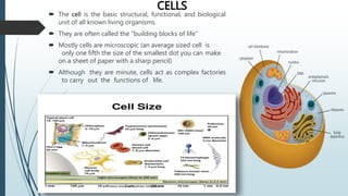
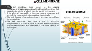
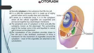
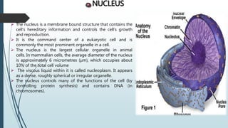
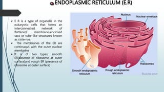
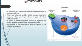
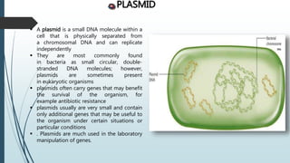
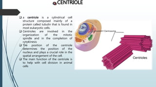
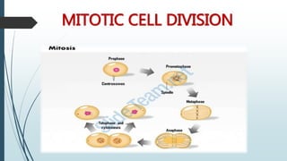
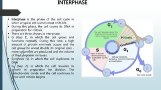
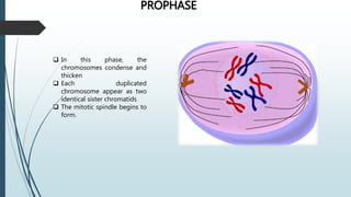
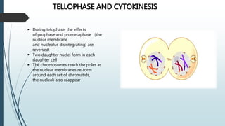
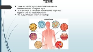
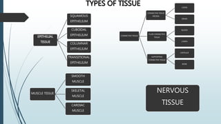
The document provides information on cells and their structures. It defines the cell as the basic unit of life and describes key cellular components including the cell membrane, cytoplasm, nucleus, organelles like mitochondria and lysosomes, and other structures. It also explains the process of mitosis and how cells divide to form two daughter cells. Additionally, it discusses the four main types of tissues - epithelial, connective, muscle and nervous tissue - and provides details on their characteristics and functions.
The cell is the basic structural, functional, and biological unit of all known organisms. A cell is the smallest unit of life. Cells are often called the "building blocks of life". The study of cells is called cell biology, cellular biology, or cytology.
. Definition of Tissues Biological tissue is a collection of interconnected cells that perform a similar function within an organism. In other words, it is a group of cells working together mainly inside an organ.
3. Classification of Tissues Human body is composed of 4 basic types of tissue: •Epithelial tissue •Connective tissue •Muscular tissue •Nervous tissue
4. Origin of Tissue A fertilized egg divides to produce 3 primary germ cell layers. These layers differentiate to form the tissues of the body.
5. Epithelial Tissue Epithelial cells cover or line all body surfaces, cavities and tubes. So, These are called covering epithelia. Epithelial cells form the functional units of secretory glands. So, These are called glandular epithelia.
Skeletal system. anatomy and physiology of skeletal system. appendicular skel...mamtabisht10
SKELETAL SYSTEM
bones, cartilage and ligaments are tightly joined to form a strong, flexible framework called skeletal system
anatomy and physiology of axial and appendicular skeletal system
Axial Skeleton: The axial skeleton includes the skull, spine, ribs and sternum.
Appendicular Skeleton:
The appendicular skeleton includes the appendages of the body, which are the shoulders, arms, hips, and legs.
Glands, Types, classification and functions(Anatomy Topic)Swatilekha Das
Anatomy Topic for B.Sc & GNM nursing students- easy explanation.
what is gland? what are the types of glands? classification of glands according to structure,and according to function. functions of exocrine and endocrine glands.
lymphatic system, a subsystem of the circulatory system in the vertebrate body that consists of a complex network of vessels, tissues, and organs. The lymphatic system helps maintain fluid balance in the body by collecting excess fluid and particulate matter from tissues and depositing them in the bloodstream
Tissues- types and functions, glands and membranes bhartisharma175
it consist of tissues their types, glands and membranes. it also consist of colorful images which will help the students to identify and differentiate the different types of tissues from one another. different types of glands and membranes are also explained with the help of images.
This power point helpful for diploma students. this presentation include classification of tissue- epithelial tissue, muscular tissue,skeletal muscle, cardiac muscle, nervous tissue ,difference between smooth muscle and skeletal muscle
The cellular components of blood are erythrocytes (red blood cells, or RBCs), leukocytes (white blood cells, or WBCs), and thrombocytes (platelets). By volume, the RBCs constitute about 45% of whole blood, the plasma about 54.3%, and white blood cells about 0.7%. Platelets make up less than 1%.
discussion about nematode,
their form, general characteristics, life cycle, discussion about their host.
diseases caused by nematodes
and discussion about different class of nematodes.
Respond to all of the initial postings from your peers in your dismickietanger
Respond to all of the initial postings from your peers in your discussion group on different days of the week.
Peer 1
Initial Post: Identify all the components of a cell. Describe the function of each of these components.
All living organisms are composed of a cell which is the smallest structural unit of living matter. A cell acts as a powerhouse to sustain the functions of living organisms for them to be alive, and it is made up of different components performing different functions. The organelles that make up a cell include the nucleus, mitochondria, plasma membrane, cytoplasm, vacuoles, ribosomes, endoplasmic reticulum, Golgi apparatus, Cytoskeleton, lysosomes, and peroxisomes, each of them acting independently to sustain the cell's life.
The nucleus is the main headquarter of a cell and contains most of the cell's DNA, genetic code that contains hereditary information. It's bounded in the eukaryotic cell membrane, and it controls and regulates cell growth and metabolism. Within the nucleus, a nucleolus is responsible for producing and assembling the cell's ribosomes. Ribosomes as cell components consist of RNA and associated proteins that synthesize proteins (McCance & Huether, 2018). This protein is required in many cellular functions, such as directing chemical processes. The lysosome is another cell organelle that contains digestive enzymes. These lysosomes break down worn-out parts of a cell and may destroy invading viruses and bacteria. Peroxisomes are another bag of enzymes that destroy toxic materials.
Mitochondria is the cell's powerhouse located in the cell's cytoplasm, which plays an imperative role in cell respiration. It generates most of the chemical energy required to power the biochemical reactions of a cell (McCance & Huether, 2018). This chemical energy produced by the mitochondrion is stored in adenosine triphosphate (ATP). The cytoplasm is another cell organelle that holds components of a cell and protects them from damage. It also stores molecules needed for cellular processes and gives the cell shape.
As another cell organelle, Cytoskeleton is responsible for maintaining the cell's shape and provides mechanical support that enhances cell division and movement.
The plasma membrane, also known as the cell membrane, transports nutrients into the cell and toxic substances out of the cell. It also provides additional support to the cell. Within a cell, we also have the Golgi apparatus, also known as Golgi bodies, which process and package proteins destined to be exported from the cell and lipid molecules (Wayne, 2019). Another organelle is the smooth and rough endoplasmic reticulum responsible for producing proteins for the rest of the cell to function.
The vacuole is another cell component that stores molecules, including water, food product, and enzyme. It also maintains the proper cell hydrostatic pressure as well as maintaining an acidic internal ...
These lecture slides, by Dr Sidra Arshad, offer a quick overview of physiological basis of a normal electrocardiogram.
Learning objectives:
1. Define an electrocardiogram (ECG) and electrocardiography
2. Describe how dipoles generated by the heart produce the waveforms of the ECG
3. Describe the components of a normal electrocardiogram of a typical bipolar leads (limb II)
4. Differentiate between intervals and segments
5. Enlist some common indications for obtaining an ECG
Study Resources:
1. Chapter 11, Guyton and Hall Textbook of Medical Physiology, 14th edition
2. Chapter 9, Human Physiology - From Cells to Systems, Lauralee Sherwood, 9th edition
3. Chapter 29, Ganong’s Review of Medical Physiology, 26th edition
4. Electrocardiogram, StatPearls - https://www.ncbi.nlm.nih.gov/books/NBK549803/
5. ECG in Medical Practice by ABM Abdullah, 4th edition
6. ECG Basics, http://www.nataliescasebook.com/tag/e-c-g-basics
Pulmonary Thromboembolism - etilogy, types, medical- Surgical and nursing man...VarunMahajani
Disruption of blood supply to lung alveoli due to blockage of one or more pulmonary blood vessels is called as Pulmonary thromboembolism. In this presentation we will discuss its causes, types and its management in depth.
Tom Selleck Health: A Comprehensive Look at the Iconic Actor’s Wellness Journeygreendigital
Tom Selleck, an enduring figure in Hollywood. has captivated audiences for decades with his rugged charm, iconic moustache. and memorable roles in television and film. From his breakout role as Thomas Magnum in Magnum P.I. to his current portrayal of Frank Reagan in Blue Bloods. Selleck's career has spanned over 50 years. But beyond his professional achievements. fans have often been curious about Tom Selleck Health. especially as he has aged in the public eye.
Follow us on: Pinterest
Introduction
Many have been interested in Tom Selleck health. not only because of his enduring presence on screen but also because of the challenges. and lifestyle choices he has faced and made over the years. This article delves into the various aspects of Tom Selleck health. exploring his fitness regimen, diet, mental health. and the challenges he has encountered as he ages. We'll look at how he maintains his well-being. the health issues he has faced, and his approach to ageing .
Early Life and Career
Childhood and Athletic Beginnings
Tom Selleck was born on January 29, 1945, in Detroit, Michigan, and grew up in Sherman Oaks, California. From an early age, he was involved in sports, particularly basketball. which played a significant role in his physical development. His athletic pursuits continued into college. where he attended the University of Southern California (USC) on a basketball scholarship. This early involvement in sports laid a strong foundation for his physical health and disciplined lifestyle.
Transition to Acting
Selleck's transition from an athlete to an actor came with its physical demands. His first significant role in "Magnum P.I." required him to perform various stunts and maintain a fit appearance. This role, which he played from 1980 to 1988. necessitated a rigorous fitness routine to meet the show's demands. setting the stage for his long-term commitment to health and wellness.
Fitness Regimen
Workout Routine
Tom Selleck health and fitness regimen has evolved. adapting to his changing roles and age. During his "Magnum, P.I." days. Selleck's workouts were intense and focused on building and maintaining muscle mass. His routine included weightlifting, cardiovascular exercises. and specific training for the stunts he performed on the show.
Selleck adjusted his fitness routine as he aged to suit his body's needs. Today, his workouts focus on maintaining flexibility, strength, and cardiovascular health. He incorporates low-impact exercises such as swimming, walking, and light weightlifting. This balanced approach helps him stay fit without putting undue strain on his joints and muscles.
Importance of Flexibility and Mobility
In recent years, Selleck has emphasized the importance of flexibility and mobility in his fitness regimen. Understanding the natural decline in muscle mass and joint flexibility with age. he includes stretching and yoga in his routine. These practices help prevent injuries, improve posture, and maintain mobilit
Recomendações da OMS sobre cuidados maternos e neonatais para uma experiência pós-natal positiva.
Em consonância com os ODS – Objetivos do Desenvolvimento Sustentável e a Estratégia Global para a Saúde das Mulheres, Crianças e Adolescentes, e aplicando uma abordagem baseada nos direitos humanos, os esforços de cuidados pós-natais devem expandir-se para além da cobertura e da simples sobrevivência, de modo a incluir cuidados de qualidade.
Estas diretrizes visam melhorar a qualidade dos cuidados pós-natais essenciais e de rotina prestados às mulheres e aos recém-nascidos, com o objetivo final de melhorar a saúde e o bem-estar materno e neonatal.
Uma “experiência pós-natal positiva” é um resultado importante para todas as mulheres que dão à luz e para os seus recém-nascidos, estabelecendo as bases para a melhoria da saúde e do bem-estar a curto e longo prazo. Uma experiência pós-natal positiva é definida como aquela em que as mulheres, pessoas que gestam, os recém-nascidos, os casais, os pais, os cuidadores e as famílias recebem informação consistente, garantia e apoio de profissionais de saúde motivados; e onde um sistema de saúde flexível e com recursos reconheça as necessidades das mulheres e dos bebês e respeite o seu contexto cultural.
Estas diretrizes consolidadas apresentam algumas recomendações novas e já bem fundamentadas sobre cuidados pós-natais de rotina para mulheres e neonatos que recebem cuidados no pós-parto em unidades de saúde ou na comunidade, independentemente dos recursos disponíveis.
É fornecido um conjunto abrangente de recomendações para cuidados durante o período puerperal, com ênfase nos cuidados essenciais que todas as mulheres e recém-nascidos devem receber, e com a devida atenção à qualidade dos cuidados; isto é, a entrega e a experiência do cuidado recebido. Estas diretrizes atualizam e ampliam as recomendações da OMS de 2014 sobre cuidados pós-natais da mãe e do recém-nascido e complementam as atuais diretrizes da OMS sobre a gestão de complicações pós-natais.
O estabelecimento da amamentação e o manejo das principais intercorrências é contemplada.
Recomendamos muito.
Vamos discutir essas recomendações no nosso curso de pós-graduação em Aleitamento no Instituto Ciclos.
Esta publicação só está disponível em inglês até o momento.
Prof. Marcus Renato de Carvalho
www.agostodourado.com
263778731218 Abortion Clinic /Pills In Harare ,sisternakatoto
263778731218 Abortion Clinic /Pills In Harare ,ABORTION WOMEN’S CLINIC +27730423979 IN women clinic we believe that every woman should be able to make choices in her pregnancy. Our job is to provide compassionate care, safety,affordable and confidential services. That’s why we have won the trust from all generations of women all over the world. we use non surgical method(Abortion pills) to terminate…Dr.LISA +27730423979women Clinic is committed to providing the highest quality of obstetrical and gynecological care to women of all ages. Our dedicated staff aim to treat each patient and her health concerns with compassion and respect.Our dedicated group ABORTION WOMEN’S CLINIC +27730423979 IN women clinic we believe that every woman should be able to make choices in her pregnancy. Our job is to provide compassionate care, safety,affordable and confidential services. That’s why we have won the trust from all generations of women all over the world. we use non surgical method(Abortion pills) to terminate…Dr.LISA +27730423979women Clinic is committed to providing the highest quality of obstetrical and gynecological care to women of all ages. Our dedicated staff aim to treat each patient and her health concerns with compassion and respect.Our dedicated group of receptionists, nurses, and physicians have worked together as a teamof receptionists, nurses, and physicians have worked together as a team wwww.lisywomensclinic.co.za/
Flu Vaccine Alert in Bangalore Karnatakaaddon Scans
As flu season approaches, health officials in Bangalore, Karnataka, are urging residents to get their flu vaccinations. The seasonal flu, while common, can lead to severe health complications, particularly for vulnerable populations such as young children, the elderly, and those with underlying health conditions.
Dr. Vidisha Kumari, a leading epidemiologist in Bangalore, emphasizes the importance of getting vaccinated. "The flu vaccine is our best defense against the influenza virus. It not only protects individuals but also helps prevent the spread of the virus in our communities," he says.
This year, the flu season is expected to coincide with a potential increase in other respiratory illnesses. The Karnataka Health Department has launched an awareness campaign highlighting the significance of flu vaccinations. They have set up multiple vaccination centers across Bangalore, making it convenient for residents to receive their shots.
To encourage widespread vaccination, the government is also collaborating with local schools, workplaces, and community centers to facilitate vaccination drives. Special attention is being given to ensuring that the vaccine is accessible to all, including marginalized communities who may have limited access to healthcare.
Residents are reminded that the flu vaccine is safe and effective. Common side effects are mild and may include soreness at the injection site, mild fever, or muscle aches. These side effects are generally short-lived and far less severe than the flu itself.
Healthcare providers are also stressing the importance of continuing COVID-19 precautions. Wearing masks, practicing good hand hygiene, and maintaining social distancing are still crucial, especially in crowded places.
Protect yourself and your loved ones by getting vaccinated. Together, we can help keep Bangalore healthy and safe this flu season. For more information on vaccination centers and schedules, residents can visit the Karnataka Health Department’s official website or follow their social media pages.
Stay informed, stay safe, and get your flu shot today!
NVBDCP.pptx Nation vector borne disease control programSapna Thakur
NVBDCP was launched in 2003-2004 . Vector-Borne Disease: Disease that results from an infection transmitted to humans and other animals by blood-feeding arthropods, such as mosquitoes, ticks, and fleas. Examples of vector-borne diseases include Dengue fever, West Nile Virus, Lyme disease, and malaria.
micro teaching on communication m.sc nursing.pdfAnurag Sharma
Microteaching is a unique model of practice teaching. It is a viable instrument for the. desired change in the teaching behavior or the behavior potential which, in specified types of real. classroom situations, tends to facilitate the achievement of specified types of objectives.
Title: Sense of Smell
Presenter: Dr. Faiza, Assistant Professor of Physiology
Qualifications:
MBBS (Best Graduate, AIMC Lahore)
FCPS Physiology
ICMT, CHPE, DHPE (STMU)
MPH (GC University, Faisalabad)
MBA (Virtual University of Pakistan)
Learning Objectives:
Describe the primary categories of smells and the concept of odor blindness.
Explain the structure and location of the olfactory membrane and mucosa, including the types and roles of cells involved in olfaction.
Describe the pathway and mechanisms of olfactory signal transmission from the olfactory receptors to the brain.
Illustrate the biochemical cascade triggered by odorant binding to olfactory receptors, including the role of G-proteins and second messengers in generating an action potential.
Identify different types of olfactory disorders such as anosmia, hyposmia, hyperosmia, and dysosmia, including their potential causes.
Key Topics:
Olfactory Genes:
3% of the human genome accounts for olfactory genes.
400 genes for odorant receptors.
Olfactory Membrane:
Located in the superior part of the nasal cavity.
Medially: Folds downward along the superior septum.
Laterally: Folds over the superior turbinate and upper surface of the middle turbinate.
Total surface area: 5-10 square centimeters.
Olfactory Mucosa:
Olfactory Cells: Bipolar nerve cells derived from the CNS (100 million), with 4-25 olfactory cilia per cell.
Sustentacular Cells: Produce mucus and maintain ionic and molecular environment.
Basal Cells: Replace worn-out olfactory cells with an average lifespan of 1-2 months.
Bowman’s Gland: Secretes mucus.
Stimulation of Olfactory Cells:
Odorant dissolves in mucus and attaches to receptors on olfactory cilia.
Involves a cascade effect through G-proteins and second messengers, leading to depolarization and action potential generation in the olfactory nerve.
Quality of a Good Odorant:
Small (3-20 Carbon atoms), volatile, water-soluble, and lipid-soluble.
Facilitated by odorant-binding proteins in mucus.
Membrane Potential and Action Potential:
Resting membrane potential: -55mV.
Action potential frequency in the olfactory nerve increases with odorant strength.
Adaptation Towards the Sense of Smell:
Rapid adaptation within the first second, with further slow adaptation.
Psychological adaptation greater than receptor adaptation, involving feedback inhibition from the central nervous system.
Primary Sensations of Smell:
Camphoraceous, Musky, Floral, Pepperminty, Ethereal, Pungent, Putrid.
Odor Detection Threshold:
Examples: Hydrogen sulfide (0.0005 ppm), Methyl-mercaptan (0.002 ppm).
Some toxic substances are odorless at lethal concentrations.
Characteristics of Smell:
Odor blindness for single substances due to lack of appropriate receptor protein.
Behavioral and emotional influences of smell.
Transmission of Olfactory Signals:
From olfactory cells to glomeruli in the olfactory bulb, involving lateral inhibition.
Primitive, less old, and new olfactory systems with different path
New Directions in Targeted Therapeutic Approaches for Older Adults With Mantl...i3 Health
i3 Health is pleased to make the speaker slides from this activity available for use as a non-accredited self-study or teaching resource.
This slide deck presented by Dr. Kami Maddocks, Professor-Clinical in the Division of Hematology and
Associate Division Director for Ambulatory Operations
The Ohio State University Comprehensive Cancer Center, will provide insight into new directions in targeted therapeutic approaches for older adults with mantle cell lymphoma.
STATEMENT OF NEED
Mantle cell lymphoma (MCL) is a rare, aggressive B-cell non-Hodgkin lymphoma (NHL) accounting for 5% to 7% of all lymphomas. Its prognosis ranges from indolent disease that does not require treatment for years to very aggressive disease, which is associated with poor survival (Silkenstedt et al, 2021). Typically, MCL is diagnosed at advanced stage and in older patients who cannot tolerate intensive therapy (NCCN, 2022). Although recent advances have slightly increased remission rates, recurrence and relapse remain very common, leading to a median overall survival between 3 and 6 years (LLS, 2021). Though there are several effective options, progress is still needed towards establishing an accepted frontline approach for MCL (Castellino et al, 2022). Treatment selection and management of MCL are complicated by the heterogeneity of prognosis, advanced age and comorbidities of patients, and lack of an established standard approach for treatment, making it vital that clinicians be familiar with the latest research and advances in this area. In this activity chaired by Michael Wang, MD, Professor in the Department of Lymphoma & Myeloma at MD Anderson Cancer Center, expert faculty will discuss prognostic factors informing treatment, the promising results of recent trials in new therapeutic approaches, and the implications of treatment resistance in therapeutic selection for MCL.
Target Audience
Hematology/oncology fellows, attending faculty, and other health care professionals involved in the treatment of patients with mantle cell lymphoma (MCL).
Learning Objectives
1.) Identify clinical and biological prognostic factors that can guide treatment decision making for older adults with MCL
2.) Evaluate emerging data on targeted therapeutic approaches for treatment-naive and relapsed/refractory MCL and their applicability to older adults
3.) Assess mechanisms of resistance to targeted therapies for MCL and their implications for treatment selection
Evaluation of antidepressant activity of clitoris ternatea in animals
Cell and tissue
1.
2.
3. CELLS
The cell is the basic structural, functional, and biological
unit of all known living organisms.
They are often called the "building blocks of life”
Mostly cells are microscopic (an average sized cell is
only one fifth the size of the smallest dot you can make
on a sheet of paper with a sharp pencil)
Although they are minute, cells act as complex factories
to carry out the functions of life.
5. CELL MEMBRANE
The cell membrane (also known as the plasma
membrane or cytoplasmic membrane) is a biological membrane that
separates the interior of all cells from the outside environment.
It is selectively permeable to ions and organic molecules and
controls the movement of substances in and out of cells.
The basic function of the cell membrane is to protect the cell from
its surroundings.
The cell membrane also plays a role in anchoring
the cytoskeleton to provide shape to the cell, and in attaching to
the extracellular matrix and other cells to hold them together to
form tissues.
6. CYTOPLASM
Basically cytoplasm is the substance that fills the cell.
It is a jelly-like substance and it is made up of eighty
percent water and is usually clear and colourless.
It serves as a molecular soup, it is in the cytoplasm
where all the cellular organelles are suspended and
are bound together by a lipid bilayer membrane.
The peripheral zone of cytoplasm is thick and jelly-like
substance, known as the plasmogel. The surrounding
area of the nuclear zone is thin and liquefied in nature
and is known as the plasmosol
The cytoskeleton of the cytoplasm provides shape to
the cell and it also facilitates movement. It forms a
framework for the movement of organelles around the
cytoplasm - most of the organelles are attached to the
cytoskeleton.
7. NUCLEUS
The nucleus is a membrane bound structure that contains the
cell's hereditary information and controls the cell's growth
and reproduction.
It is the command center of a eukaryotic cell and is
commonly the most prominent organelle in a cell.
The nucleus is the largest cellular organelle in animal
cells. In mammalian cells, the average diameter of the nucleus
is approximately 6 micrometres (µm), which occupies about
10% of the total cell volume
The viscous liquid within it is called nucleoplasm. It appears
as a dense, roughly spherical or irregular organelle.
The nucleus controls many of the functions of the cell (by
controlling protein synthesis) and contains DNA (in
chromosomes).
8. ENDOPLASMIC RETICULUM (E.R)
E R is a type of organelle in the
eukaryotic cells that forms an
interconnected network of
flattened, membrane-enclosed
sacs or tube-like structures known
as cisternae.
The membranes of the ER are
continuous with the outer nuclear
membrane
It is of two types; smooth
ER(absence of ribosome at outer
surface)and rough ER (presence of
ribosome at outer surface)
9. GOLGI APPARATUS
The Golgi apparatus, also known as the Golgi complex, Golgi
body, or simply the Golgi, is an organelle found in
most eukaryotic cells
The Golgi apparatus is a major collection and dispatch station
of protein products received from the endoplasmic reticulum
A major function is the modifying, sorting and packaging of
proteins for secretion
. It is also involved in the transport of lipids around the cell,
and the creation of lysosomes
. The sacs or folds of the Golgi apparatus are called cisternae
10. MITOCHONDRIA
The mitochondrion (plural mitochondria) is a
double membrane-bound organelle found in
all eukaryotic organisms, although some cells in some
organisms may lack them (e.g. Red blood cells)
The main job of mitochondria is to perform cellular
respiration. This means it takes in nutrients from the cell,
breaks it down, and turns it into energy
This energy is then in turn used by the cell to carry out
various functions
Also known as power house of cell because they generate
most of the cell's supply of adenosine triphosphate (ATP),
used as a source of chemical energy.
The number of mitochondria in a cell can vary widely
by organism, tissue, and cell type. For instance, red blood
cells have no mitochondria, whereas liver cells can have
more than 2000
11. LYSOSOMES
A lysosome is a membrane-bounded organelle found in
most animal cells.
They are spherical vesicles which contain hydrolytic
enzymes that can break down virtually all kinds
of biomolecules.
It is One of the key organelles involved in digestion and
waste removal as hey contain digestive enzymes.
the interior of the lysosomes is acidic compared to the
slightly basic cytosol
12. PLASMID
A plasmid is a small DNA molecule within a
cell that is physically separated from
a chromosomal DNA and can replicate
independently
They are most commonly found
in bacteria as small circular, double-
stranded DNA molecules; however,
plasmids are sometimes present
in eukaryotic organisms
plasmids often carry genes that may benefit
the survival of the organism, for
example antibiotic resistance
plasmids usually are very small and contain
only additional genes that may be useful to
the organism under certain situations or
particular conditions
. Plasmids are much used in the laboratory
manipulation of genes.
13. VACUOLE
A vacuole is a membrane-bound organelle which is present
in all plant and fungal cells and some protest,
animal and bacterial cells
Vacuoles are essentially enclosed compartments which are
filled with water containing inorganic and organic molecules
including enzymes in solution
Vacuoles are formed by the fusion of multiple
membrane vesicles and are effectively just larger forms of
these.
It has no basic shape or size; its structure varies according to
the needs of the cell.
It is surrounded by a membrane and functions to hold
materials and wastes
. It also functions to maintain the proper pressure within the
plant cells to provide structure and support for the growing
plant
14. RIBOSOME
The ribosome is a complex molecular machine,
found within all living cells, that serves as the
site of biological protein synthesis
Ribosomes link amino acids together in the
order specified by messenger RNA(mRNA)
molecules.
Ribosomes can be found floating within the
cytoplasm or attached to the endoplasmic
reticulum
15. CENTRIOLE
a centriole is a cylindrical cell
structure composed mainly of a
protein called tubulin that is found in
most eukaryotic cells.
Centrioles are involved in the
organization of the mitotic
spindle and in the completion of
cytokinesis
The position of the centriole
determines the position of the
nucleus and plays a crucial role in the
spatial arrangement of the cell.
The main function of the centriole is
to help with cell division in animal
cells
17. INTERPHASE
Interphase is the phase of the cell cycle in
which a typical cell spends most of its life.
During this phase, the cell copies its DNA in
preparation for mitosis
There are three phases in interphase
G (Gap 1), in which the cell grows and
functions normally. During this time, a high
amount of protein synthesis occurs and the
cell grows (to about double its original size) -
more organelles are produced and the volume
of the cytoplasm increases.
Synthesis (S), in which the cell duplicates its
DNA
G2 (Gap 2), in which the cell resumes its
growth in preparation for division. The
mitochondria divide and the cell continues to
grow until mitosis begins
18. PROPHASE
In this phase, the
chromosomes condense and
thicken
Each duplicated
chromosome appear as two
identical sister chromatids
The mitotic spindle begins to
form.
19. METAPHASE
In metaphase, the centromeres of the
chromosomes convene themselves on
the metaphase plate
20. ANAPHASE
The spindle fibers begin to
contract
This starts to pull the sister
chromatids spart
At the end of anaphase a
complete set of daughter
chromosomes is found in each
pole
21. TELLOPHASE AND CYTOKINESIS
During telophase, the effects
of prophase and prometaphase (the
nuclear membrane
and nucleolus disintegrating) are
reversed.
Two daughter nuclei form in each
daughter cell
The chromosomes reach the poles as
the nuclear membranes re-form
around each set of chromatids,
the nucleoli also reappear
22.
23. TISSUE
tissue is a cellular organizational level intermediate
between cells and a complete organ.
is an ensemble of similar cells from the same origin that
together carry out a specific function.
The study of tissue is known as histology
25. EPITHELIAL TISSUE
Epithelial tissues line the cavities and surfaces of blood
vessels and organs throughout the body.
1.
26. EPITHELIAL TISSUE
SQUAMOUS EPITHELIUM
It has surface whose outermost (apical) layer
consists of thin, flat cells.
they may be composed of one layer of cells, in
which case it is referred to as simple squamous
epithelium, or it may possess multiple layers,
referred to then as stratified squamous epithelium.
squamous epithelial cells have
a polygonal appearance when viewed from above
CUBOIDAL EPITHELIUM
cuboidal epithelia are epithelial cells having a
cube-like shape; that is, their width is
approximately equal to their height.
They may exist in single layers (simple cuboidal
epithelium) or multiple layers (stratified cuboidal
epithelium) depending on their location (and thus
function) in the body.
27. EPITHELIAL TISSUE
COLUMNAR EPITHELIUM
Columnar epithelial cells are epithelial
cells whose height are at least four times their
width.
Columnar epithelia are divided into simple (or
unilayered), and stratified (or multi-layered)
They are found in the inner lining of the
intestine. They secrete digestive enzymes and
absorb digested food.
TRANSITIONAL EPITHELIUM
Transitional epithelium is a type of tissue consisting
of multiple layers of epithelial cells which can
contract and expand.
This tissue structure type is found in urothelium,
including that of the urinary bladder, the ureters,
and the superior urethra and gland ducts of
the prostate.
They also functions as barrier between the lumen,
or inside hollow space, of the tract that it lines and
the bloodstream.
28. CONNECTIVE TISSUE
The different types of connective tissue maintain the form of organs throughout the body
Connective tissues provide a matrix that supports and physically connects other tissues
and cells together in organs
29. CONNECTIVE TISSUE
CONNECTIVE TISSUE PROPER
it is categorised into two:
1) Loose:
includes areolar tissue, reticular tissue,
and adipose tissue
It holds organs in place and
attaches epithelial tissue to other underlying
tissues
It may be found in tissue sections from
almost every part of the body
2) DENSE:
also called dense fibrous tissue
is a type of connective tissue with fibers as its
main matrix element
forms strong, rope-like structures such
as tendons and ligament
FLUID CONNECTIVE TISSUE
It is categorised into two:
1) BLOOD
Blood is considered a connective tissue for two
basic reasons: (1) embryologically, it has the same
origin (mesodermal) as do the other connective
tissue types and (2) blood connects the body
systems
The cell found in greatest abundance in blood is
the erythrocyte, responsible for transporting
oxygen to body tissues
2) LYMPH
Lymph is a clear fluid that is similar to plasma but
contains less protein
It flows through lymphatic vessels throughout the
body and includes chemicals and cells whose
composition varies according to location within
the body.
30. SUPPORTING CONNECTIVE TISSUE
It is categorised into two:
CONNECTIVE TISSUE
1) CARTILAJE
Cartilage is a resilient and
smooth elastic tissue, covering and
protecting the ends of the
long bones at joints, and is a structural
component of the rib cage, the ear,
the nose, the bronchial tubes,
the intervertebral discs, and many more
other body components
It is not as hard and rigid as bone, but
it is stiffer and less flexible than muscle
2) BONE
Bone tissue is the major structural and
supportive connective tissue of the
body
Bone tissue forms the rigid part of
the bones that make up the skeleton.
Supports muscles, organs, and soft
tissues.
It is formed by cells, called osteoblasts
31. MUSCLE TISSUE
Muscle tissue is a soft tissue that composes muscles in animal bodies, and
gives rise to muscles' ability to contract
It is formed during embryonic development through a process known
as myogenesis
Muscle tissue varies with function and location in the body
32. MUSCLE TISSUE
SMOOTH MUSCLE
Smooth muscle is an involuntary
non-striated muscle
Smooth muscle is found within
the walls of blood vessels
Smooth muscle is also found in
lymphatic vessels, the urinary
bladder, uterus, male and
female reproductive
tracts, gastrointestinal tract,
respiratory tract, the ciliary
muscle, and iris of the eye
SKELETAL MUSCLE
It is a form of striated muscle tissue which
is under the voluntary control of
the somatic nervous system
Most skeletal muscles are attached
to bones by bundles of collagen fibers
known as tendons
Muscle fibres, or muscle cells, are formed
from the fusion of
developmental myoblasts in a process
known as myogenesis
33. MUSCLE TISSUE
CARDIAC MUSCLE
Cardiac muscle (heart muscle) is an involuntary, striated
muscle that is found in the walls and histological
foundation of the heart, specifically the myocardium
These three types of muscle all form in the process
of myogenesis
The myocardium is the muscle tissue of the heart, and
forms a thick middle layer between the
outer epicardium layer and the inner
endocardium layer.
Cardiac muscle cells, unlike most other tissues in the
body, rely on an available blood and electrical supply
to deliver oxygen and nutrients and remove waste
products such as carbon dioxide
34. NERVOUS TISSUE
Nervous tissue or nerve tissue is the
main tissue component of the two parts of the nervous
system; the brain and spinal cord of the central
nervous system (CNS), and the branching peripheral
nerves of the peripheral nervous system (PNS), which
regulates and controls bodily functions and activity
The function of nervous tissue is to form the
communication network of the nervous system by
conducting electric signals across tissue
It is composed of neurons, or nerve cells, which
receive and transmit impulses
Editor's Notes
Tendons attach skeletal muscles to bones; ligaments connect bones to bones at joints