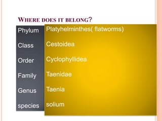
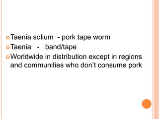
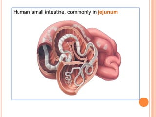
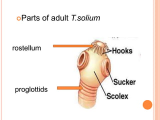
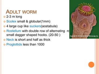
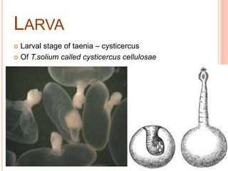
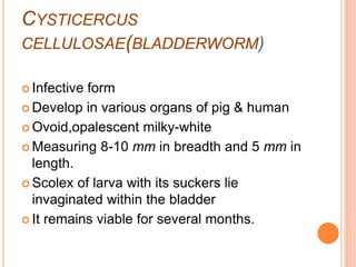
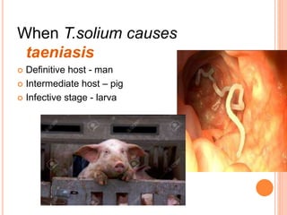
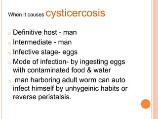
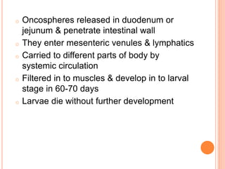
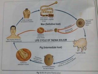
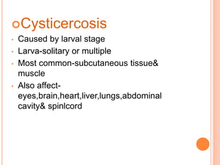
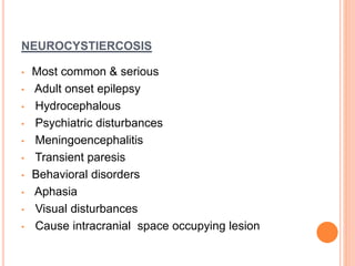
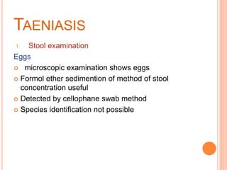
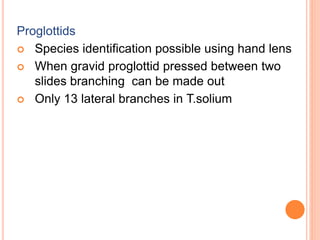
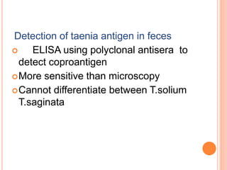
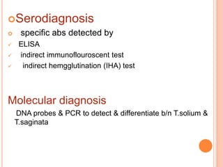
TAENIA SOLIUM
Shifa
58th batch
CMC
WHERE DOES IT BELONG?
order
order
Phylum
Class
Order
Family
Genus
species
Platyhelminthes( flatworms)
Cestoidea
Cyclophyllidea
Taenidae
Taenia
solium
Taenia solium - pork tape worm
Taenia - band/tape
Worldwide in distribution except in regions
and communities who don’t consume pork
Human small intestine, commonly in jejunum
Parts of adult T.solium
rostellum
proglottids
ADULT WORM
 2-3 m long
 Scolex small & globular(1mm)
 4 large cup like suckers(acetabula)
 Rostellum with double row of alternating round &
small dagger shaped hooks. (20-50 )
 Neck is short and half as thick
 Proglottids less than 1000
 Gravid segments are twice as long & broad.
 Testes composed of 150-200 follicles
 An accessory lobe for ovary
 Vaginal sphincter absent
 Uterus has 5 to 10 thick lateral branches
 A lateral thick lipped genital pore is present
alternating the right & left sides of adjacent
segments.
 Gravid segments pass passively out as short
chains. Eggs escape from ruptured uterus
EGGS
 Egg similar to T.saginata
 Spherical 30-40 micro.m
 Thin hyaline membrane around it which disappears
after release
 Inner embryophore radially striated & is yellow
brown due to bile staining.
 In the centre a fully developed embryo(oncosphere)
with 3 pairs of hooklets(hexacanth embryo)
 Eggs do not float in saturated salt solution
 Eggs are infective both pigs & humans.
LARVA
 Larval stage of taenia – cysticercus
 Of T.solium called cysticercus cellulosae

CYSTICERCUS
CELLULOSAE(BLADDERWORM)
 Infective form
 Develop in various organs of pig & human
 Ovoid,opalescent milky-white
 Measuring 8-10 mm in breadth and 5 mm in
length.
 Scolex of larva with its suckers lie
invaginated within the bladder
 It remains viable for several months.
When T.solium causes
taeniasis
 Definitive host - man
 Intermediate host – pig
 Infective stage - larva
When it causes cysticercosis
o Definitive host - man
o Intermediate - man
o Infective stage- eggs
o Mode of infection- by ingesting eggs
with contaminated food & water
o man harboring adult worm can auto
infect himself by unhygeinic habits or
reverse peristalsis.
o Oncospheres released in duodenum or
jejunum & penetrate intestinal wall
o They enter mesenteric venules & lymphatics
o Carried to different parts of body by
systemic circulation
o Filtered in to muscles & develop in to larval
stage in 60-70 days
o Larvae die without further development
Intestinal taeniasis
 caused by T.solium & T.saginata
 Sometimes asymptomatic
 When symptomatic causes abdominal
discomfort,indigestion,nausea,diarrhea&
weight loss
 Occasionaly acute appendicitis &
pancreatitis
Cysticercosis
• Caused by larval stage
• Larva-solitary or multiple
• Most common-subcutaneous tissue&
muscle
• Also affect-
eyes,brain,heart,liver,lungs,abdominal
cavity& spinlcord
• Larvae can evoke a cellular reaction starting
with infiltration of
neutrophils,eosinophils,lymphocytes,plasma
cells & giant cells
• Subcutaneous – asymptomatic
• Cysticercus surrounded by a fibrous capsule
except in eye & ventricles of brain
CLINICAL FEATURES-
Subcutaneous
cysticercosis
myocysticercosis
neurocysticercosis
oculocysticercosis
SUBCUTANEOUS CYSTICERCOSIS
 Mostly asymptomatic
 Subcutneous nodules
MYOCYSTICERCOSIS
 May cause acute myositis
NEUROCYSTIERCOSIS
• Most common & serious
• Adult onset epilepsy
• Hydrocephalous
• Psychiatric disturbances
• Meningoencephalitis
• Transient paresis
• Behavioral disorders
• Aphasia
• Visual disturbances
• Cause intracranial space occupying lesion
•
•
MRI of brain showing
neurocysticercosis
OCULAR CYSTICERCOSIS
Cysts found in vitreous humor,subretinal space & Conjunctiva
May present as blurred vision or loss of vision
Iritis ,uveitis & palpebral conjuctivitis
TAENIASIS
1. Stool examination
Eggs
 microscopic examination shows eggs
 Formol ether sedimention of method of stool
concentration useful
 Detected by cellophane swab method
 Species identification not possible
Proglottids
 Species identification possible using hand lens
 When gravid proglottid pressed between two
slides branching can be made out
 Only 13 lateral branches in T.solium
Detection of taenia antigen in feces
 ELISA using polyclonal antisera to
detect coproantigen
More sensitive than microscopy
Cannot differentiate between T.solium
T.saginata
Serodiagnosis
 specific abs detected by
 ELISA
 indirect immunoflouroscent test
 indirect hemgglutination (IHA) test
Molecular diagnosis
DNA probes & PCR to detect & differentiate b/n T.solium &
T.saginata
Others
Ocular cysticercosis-by opthalmoscopy
Eosinophilia in early stage
Intestinal taeniasis
 Single dose of praziquantel(10-20 mg)
 Nicolsamide (2 g) single dose
 Purgation is not necessary
Cysticercosis
 Excision best method
 Asymptomatic neurocysticercosis no treatment
 Symptomatic cerebral cysticercosis-praziquantel – 50
mg/kg in divided dose for 20-30 days
 Albentazole – 400 mg twice daily for 30 days
 Corticosteroids to reduce inflammatory reactions
 Antiepileptic drugs
 Operative intervention indicated for hydrocephalous
 Inspection for cysticerci in slaughter house
 Avoid undercooked pork
(temp 56 `c for 5 mts)
 maintain clean personal habits & general
Sanitary measures
 Prevention of fecal contamination
 Detection & treatment of hosts
Taenia solium

Taenia solium

  • 1.
  • 2.
    WHERE DOES ITBELONG? order order Phylum Class Order Family Genus species Platyhelminthes( flatworms) Cestoidea Cyclophyllidea Taenidae Taenia solium
  • 3.
    Taenia solium -pork tape worm Taenia - band/tape Worldwide in distribution except in regions and communities who don’t consume pork
  • 5.
    Human small intestine,commonly in jejunum
  • 7.
    Parts of adultT.solium rostellum proglottids
  • 8.
    ADULT WORM  2-3m long  Scolex small & globular(1mm)  4 large cup like suckers(acetabula)  Rostellum with double row of alternating round & small dagger shaped hooks. (20-50 )  Neck is short and half as thick  Proglottids less than 1000
  • 9.
     Gravid segmentsare twice as long & broad.  Testes composed of 150-200 follicles  An accessory lobe for ovary  Vaginal sphincter absent  Uterus has 5 to 10 thick lateral branches  A lateral thick lipped genital pore is present alternating the right & left sides of adjacent segments.  Gravid segments pass passively out as short chains. Eggs escape from ruptured uterus
  • 10.
    EGGS  Egg similarto T.saginata  Spherical 30-40 micro.m  Thin hyaline membrane around it which disappears after release  Inner embryophore radially striated & is yellow brown due to bile staining.  In the centre a fully developed embryo(oncosphere) with 3 pairs of hooklets(hexacanth embryo)  Eggs do not float in saturated salt solution  Eggs are infective both pigs & humans.
  • 11.
    LARVA  Larval stageof taenia – cysticercus  Of T.solium called cysticercus cellulosae 
  • 12.
    CYSTICERCUS CELLULOSAE(BLADDERWORM)  Infective form Develop in various organs of pig & human  Ovoid,opalescent milky-white  Measuring 8-10 mm in breadth and 5 mm in length.  Scolex of larva with its suckers lie invaginated within the bladder  It remains viable for several months.
  • 14.
    When T.solium causes taeniasis Definitive host - man  Intermediate host – pig  Infective stage - larva
  • 15.
    When it causescysticercosis o Definitive host - man o Intermediate - man o Infective stage- eggs o Mode of infection- by ingesting eggs with contaminated food & water o man harboring adult worm can auto infect himself by unhygeinic habits or reverse peristalsis.
  • 16.
    o Oncospheres releasedin duodenum or jejunum & penetrate intestinal wall o They enter mesenteric venules & lymphatics o Carried to different parts of body by systemic circulation o Filtered in to muscles & develop in to larval stage in 60-70 days o Larvae die without further development
  • 19.
    Intestinal taeniasis  causedby T.solium & T.saginata  Sometimes asymptomatic  When symptomatic causes abdominal discomfort,indigestion,nausea,diarrhea& weight loss  Occasionaly acute appendicitis & pancreatitis
  • 20.
    Cysticercosis • Caused bylarval stage • Larva-solitary or multiple • Most common-subcutaneous tissue& muscle • Also affect- eyes,brain,heart,liver,lungs,abdominal cavity& spinlcord
  • 21.
    • Larvae canevoke a cellular reaction starting with infiltration of neutrophils,eosinophils,lymphocytes,plasma cells & giant cells • Subcutaneous – asymptomatic • Cysticercus surrounded by a fibrous capsule except in eye & ventricles of brain
  • 22.
  • 23.
    SUBCUTANEOUS CYSTICERCOSIS  Mostlyasymptomatic  Subcutneous nodules
  • 24.
  • 25.
    NEUROCYSTIERCOSIS • Most common& serious • Adult onset epilepsy • Hydrocephalous • Psychiatric disturbances • Meningoencephalitis • Transient paresis • Behavioral disorders • Aphasia • Visual disturbances • Cause intracranial space occupying lesion
  • 26.
    • • MRI of brainshowing neurocysticercosis
  • 27.
    OCULAR CYSTICERCOSIS Cysts foundin vitreous humor,subretinal space & Conjunctiva May present as blurred vision or loss of vision Iritis ,uveitis & palpebral conjuctivitis
  • 29.
    TAENIASIS 1. Stool examination Eggs microscopic examination shows eggs  Formol ether sedimention of method of stool concentration useful  Detected by cellophane swab method  Species identification not possible
  • 30.
    Proglottids  Species identificationpossible using hand lens  When gravid proglottid pressed between two slides branching can be made out  Only 13 lateral branches in T.solium
  • 31.
    Detection of taeniaantigen in feces  ELISA using polyclonal antisera to detect coproantigen More sensitive than microscopy Cannot differentiate between T.solium T.saginata
  • 32.
    Serodiagnosis  specific absdetected by  ELISA  indirect immunoflouroscent test  indirect hemgglutination (IHA) test Molecular diagnosis DNA probes & PCR to detect & differentiate b/n T.solium & T.saginata
  • 33.
  • 35.
    Intestinal taeniasis  Singledose of praziquantel(10-20 mg)  Nicolsamide (2 g) single dose  Purgation is not necessary
  • 36.
    Cysticercosis  Excision bestmethod  Asymptomatic neurocysticercosis no treatment  Symptomatic cerebral cysticercosis-praziquantel – 50 mg/kg in divided dose for 20-30 days  Albentazole – 400 mg twice daily for 30 days  Corticosteroids to reduce inflammatory reactions  Antiepileptic drugs  Operative intervention indicated for hydrocephalous
  • 38.
     Inspection forcysticerci in slaughter house  Avoid undercooked pork (temp 56 `c for 5 mts)  maintain clean personal habits & general Sanitary measures  Prevention of fecal contamination  Detection & treatment of hosts