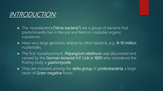
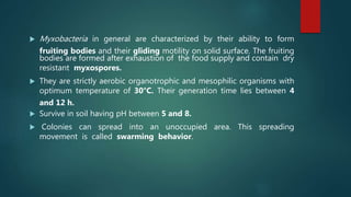
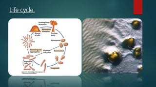
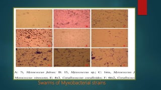
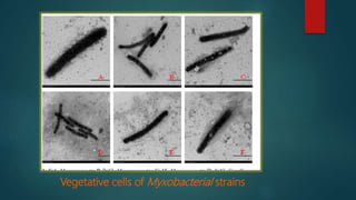
MYXOBACTERIA
ASIF NAWAZ
M.PHIL 3RD MICROBIOLOGY
INTRODUCTION:
 The myxobacteria ("slime bacteria") are a group of bacteria that
predominantly live in the soil and feed on insoluble organic
substances.
 Have very large genomes relative to other bacteria, e.g. 9–10 million
nucleotides.
 The first myxobacterium, Polyangium vitellinum, was discovered and
named by the German botanist H.F. Link in 1809 who considered the
fruiting body a gastromycete.
 They are included among the delta group of proteobacteria, a large
taxon of Gram-negative forms.
 Myxobacteria in general are characterized by their ability to form
fruiting bodies and their gliding motility on solid surface. The fruiting
bodies are formed after exhaustion of the food supply and contain dry
resistant myxospores.
 They are strictly aerobic organotrophic and mesophilic organisms with
optimum temperature of 30°C. Their generation time lies between 4
and 12 h.
 Survive in soil having pH between 5 and 8.
 Colonies can spread into an unoccupied area. This spreading
movement is called swarming behavior.
 The Myxobacteria belong to order Myxococcales and the most
common members are Myxococcus xanthus, Sorangium
cellulosum, etc.
 Myxobacteria are also capable of lysing living cells, for this reason,
they are called predators.
Life cycle:
Types of myxobacteria:
Based on the specialization of Myxobacteria in degrading bio
macro-molecules, they are divided into :
 Bacteriolytic myxobacteria that lyse whole cells of other
microorganisms.
 Cellulolytic myxobacteria that efficiently decompose cellulose
instead of living cells.
The shape, size, color, or arrangement of vegetative cells, swarms,
fruiting bodies, and myxospores are important in determining genus
of myxobacteria.
Isolation methods from soil:
Samples are taken from soil, air dried quickly as soon as possible and
store at room.
The following four different isolation methods are used:
 Baiting with dung pellets.
 Baiting with Escherichia coli streaks.
 Incubation of bark in moist chambers.
 Inoculation of filter paper with soil .
1. Baiting with dung pellets:
 Petri dishes are filled with soil moistened with distilled water
containing 100 μg/ml cycloheximide.
 In each plate autoclaved and dried rabbit dung pellets are
embedded leaving about half of the pellet protruding from the
soil.
 After 1 week of incubation at 32°C, fruiting bodies of
myxobacteria, which had migrated onto the dung from the soil,
are observed at the stereomicroscope.
2. Baiting with Escherichia coli streaks.
 E. coli cells are grown in Luria broth, centrifuged and resuspended
in distilled water containing 100 μg/ml cycloheximide till a thick
slurry is obtained, and cross streaked on the surface of water
cycloheximide (WCX) agar (CaCl2.2H2O 0.1%; agar 1.5%).
 The Centre of the cross is inoculated with a pea‐sized amount of
soil. The plates are incubated at 32°C and examined for swarms
and fruiting bodies after 1–4 weeks.
3. Incubation of bark in moist chambers.
 Pieces of bark are placed on water‐soaked filter paper on Petri
dishes and submerged in distilled water containing 100 μg/ml
cycloheximide.
 Bark checked for fruiting bodies after 1–3 weeks of incubation
at 32°C.
4. Inoculation of filter paper with soil.
 Filter paper is placed on the surface of mineral ST21CX agar (KNO3
0.1%, FeCl3·6 H2O 0.02%, K2HPO4 0.1%, MnSO4·7 H2O 0.01%,
CaCl2·2 H2O 0.1%, MgSO4·7 H2O 0.1%, yeast extract 0.002%, agar
1.5%) containing cellulose as the only carbon and energy source.
 Pea‐sized mass of soil is placed on the filter paper.
 Plates are incubated at 32°C and examined for fruiting bodies after
1–3 weeks.
 The fruiting bodies are picked daily and examined under a
dissecting microscope. They are transferred to VY/2 medium.
Purification of strains:
 For purification, the agar pieces from the outer edges of the
swarms are picked with needles and transfer to VY/2 (Barkers yeast
0.5%, CaCl2·2 H2O 0.1%, agar 1.5%).
 The procedure is repeated until the swarms are pure.
Morphological identification.
 All strains are incubated in VY/2 medium.
 Some myxobacterial fruiting bodies, swarms, and vegetative cells
appear.
 Growth and morphogenesis are observed with dissecting
microscope, optical microscope, and electronic microscope.
 The swarms are carefully scraped to glass slides for detection.
Fruiting bodies are crash to release myxospores for detection.
 The taxonomy of the isolates is determined by the morphologies
of vegetative cells, fruiting bodies, myxospores, and swarms.
 For example, in case of Myxococcus, the fruiting bodies are always
spherical, pale yellow, yellow, or orange red. Vegetative cells are
always rod-shaped with sharp ends, 2–10 µm in length.
 While for Corallcoccus, the fruiting bodies are always coralloid
branched in shape; brown or orange red. Vegetative cells are rod-
shaped with sharp ends, between 3–7 µm in length.
Morphological characteristics of Myxobacteria fruiting bodies.
Swarms of Myxobacterial strains
Vegetative cells of Myxobacterial strains
Applications of Myxobacterial secondary
metabolites :
 Myxobacteria are called as “Microbial factories” due to their ability
to produce distinct secondary metabolites.
 These Secondary metabolites possess antifungal, antibacterial
and anticancer activities.
 Lytic metabolites that induce cell lysis include antibiotics, cell
wall degrading enzymes, nucleases, lipases and proteases.
1. Antibacterial/Antifungal agents
 The anti-microbial activity is due to number of different
mechanisms.
 There are various cell wall degrading enzymes produced by
different myxobacteria members .These include glucosaminidase,
muramidase, amidase, peptidase etc. that are effective against
many pathogenic bacteria such as E. coli, Staphylococcous
aureus, Bacillus subtilis etc.
2. Cytotoxic compounds.
 The most important bioactivities exhibited by myxobacterial
metabolites are their cytotoxicity towards mammalian cells. Due
to this these compounds are used in cancer chemotherapy.
 Many metabolites have been discovered which interfere with
the eukaryotic cytoskeleton and may act as potential cancer
chemotherapeutics.
 Example include Disorazol: Isolated from Sorangium cellulosum .It
is effective against eukaryotic organisms as it inhibits the
proliferation of different cancer cell lines and also acts as an
antifungal agent.
3. Agriculturally important compounds
 The bacteriolytic myxobacteria produce many agriculturally
important antibiotics.
 Such as thiangazol: It is considered as a good antagonist of
phytopathogens that destroy useful plants.

Myxobacteria pptx

  • 1.
  • 2.
    INTRODUCTION:  The myxobacteria("slime bacteria") are a group of bacteria that predominantly live in the soil and feed on insoluble organic substances.  Have very large genomes relative to other bacteria, e.g. 9–10 million nucleotides.  The first myxobacterium, Polyangium vitellinum, was discovered and named by the German botanist H.F. Link in 1809 who considered the fruiting body a gastromycete.  They are included among the delta group of proteobacteria, a large taxon of Gram-negative forms.
  • 3.
     Myxobacteria ingeneral are characterized by their ability to form fruiting bodies and their gliding motility on solid surface. The fruiting bodies are formed after exhaustion of the food supply and contain dry resistant myxospores.  They are strictly aerobic organotrophic and mesophilic organisms with optimum temperature of 30°C. Their generation time lies between 4 and 12 h.  Survive in soil having pH between 5 and 8.  Colonies can spread into an unoccupied area. This spreading movement is called swarming behavior.
  • 4.
     The Myxobacteriabelong to order Myxococcales and the most common members are Myxococcus xanthus, Sorangium cellulosum, etc.  Myxobacteria are also capable of lysing living cells, for this reason, they are called predators.
  • 5.
  • 6.
    Types of myxobacteria: Basedon the specialization of Myxobacteria in degrading bio macro-molecules, they are divided into :  Bacteriolytic myxobacteria that lyse whole cells of other microorganisms.  Cellulolytic myxobacteria that efficiently decompose cellulose instead of living cells. The shape, size, color, or arrangement of vegetative cells, swarms, fruiting bodies, and myxospores are important in determining genus of myxobacteria.
  • 7.
    Isolation methods fromsoil: Samples are taken from soil, air dried quickly as soon as possible and store at room. The following four different isolation methods are used:  Baiting with dung pellets.  Baiting with Escherichia coli streaks.  Incubation of bark in moist chambers.  Inoculation of filter paper with soil .
  • 8.
    1. Baiting withdung pellets:  Petri dishes are filled with soil moistened with distilled water containing 100 μg/ml cycloheximide.  In each plate autoclaved and dried rabbit dung pellets are embedded leaving about half of the pellet protruding from the soil.  After 1 week of incubation at 32°C, fruiting bodies of myxobacteria, which had migrated onto the dung from the soil, are observed at the stereomicroscope.
  • 9.
    2. Baiting withEscherichia coli streaks.  E. coli cells are grown in Luria broth, centrifuged and resuspended in distilled water containing 100 μg/ml cycloheximide till a thick slurry is obtained, and cross streaked on the surface of water cycloheximide (WCX) agar (CaCl2.2H2O 0.1%; agar 1.5%).  The Centre of the cross is inoculated with a pea‐sized amount of soil. The plates are incubated at 32°C and examined for swarms and fruiting bodies after 1–4 weeks.
  • 10.
    3. Incubation ofbark in moist chambers.  Pieces of bark are placed on water‐soaked filter paper on Petri dishes and submerged in distilled water containing 100 μg/ml cycloheximide.  Bark checked for fruiting bodies after 1–3 weeks of incubation at 32°C.
  • 11.
    4. Inoculation offilter paper with soil.  Filter paper is placed on the surface of mineral ST21CX agar (KNO3 0.1%, FeCl3·6 H2O 0.02%, K2HPO4 0.1%, MnSO4·7 H2O 0.01%, CaCl2·2 H2O 0.1%, MgSO4·7 H2O 0.1%, yeast extract 0.002%, agar 1.5%) containing cellulose as the only carbon and energy source.  Pea‐sized mass of soil is placed on the filter paper.  Plates are incubated at 32°C and examined for fruiting bodies after 1–3 weeks.  The fruiting bodies are picked daily and examined under a dissecting microscope. They are transferred to VY/2 medium.
  • 12.
    Purification of strains: For purification, the agar pieces from the outer edges of the swarms are picked with needles and transfer to VY/2 (Barkers yeast 0.5%, CaCl2·2 H2O 0.1%, agar 1.5%).  The procedure is repeated until the swarms are pure.
  • 13.
    Morphological identification.  Allstrains are incubated in VY/2 medium.  Some myxobacterial fruiting bodies, swarms, and vegetative cells appear.  Growth and morphogenesis are observed with dissecting microscope, optical microscope, and electronic microscope.  The swarms are carefully scraped to glass slides for detection. Fruiting bodies are crash to release myxospores for detection.
  • 15.
     The taxonomyof the isolates is determined by the morphologies of vegetative cells, fruiting bodies, myxospores, and swarms.  For example, in case of Myxococcus, the fruiting bodies are always spherical, pale yellow, yellow, or orange red. Vegetative cells are always rod-shaped with sharp ends, 2–10 µm in length.  While for Corallcoccus, the fruiting bodies are always coralloid branched in shape; brown or orange red. Vegetative cells are rod- shaped with sharp ends, between 3–7 µm in length.
  • 16.
    Morphological characteristics ofMyxobacteria fruiting bodies.
  • 17.
  • 18.
    Vegetative cells ofMyxobacterial strains
  • 19.
    Applications of Myxobacterialsecondary metabolites :  Myxobacteria are called as “Microbial factories” due to their ability to produce distinct secondary metabolites.  These Secondary metabolites possess antifungal, antibacterial and anticancer activities.  Lytic metabolites that induce cell lysis include antibiotics, cell wall degrading enzymes, nucleases, lipases and proteases.
  • 20.
    1. Antibacterial/Antifungal agents The anti-microbial activity is due to number of different mechanisms.  There are various cell wall degrading enzymes produced by different myxobacteria members .These include glucosaminidase, muramidase, amidase, peptidase etc. that are effective against many pathogenic bacteria such as E. coli, Staphylococcous aureus, Bacillus subtilis etc.
  • 21.
    2. Cytotoxic compounds. The most important bioactivities exhibited by myxobacterial metabolites are their cytotoxicity towards mammalian cells. Due to this these compounds are used in cancer chemotherapy.  Many metabolites have been discovered which interfere with the eukaryotic cytoskeleton and may act as potential cancer chemotherapeutics.  Example include Disorazol: Isolated from Sorangium cellulosum .It is effective against eukaryotic organisms as it inhibits the proliferation of different cancer cell lines and also acts as an antifungal agent.
  • 22.
    3. Agriculturally importantcompounds  The bacteriolytic myxobacteria produce many agriculturally important antibiotics.  Such as thiangazol: It is considered as a good antagonist of phytopathogens that destroy useful plants.