Downloaded 247 times

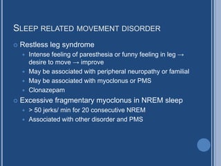


This document provides information about myoclonus, which are sudden, shock-like contractions of muscles. It describes different types of myoclonus including focal, cortical, brainstem, spinal, peripheral, multifocal, generalized, essential, and childhood myoclonic epilepsies. Diagnostic tests like EMG and EEG are discussed. Various causes and treatment options are also mentioned.


