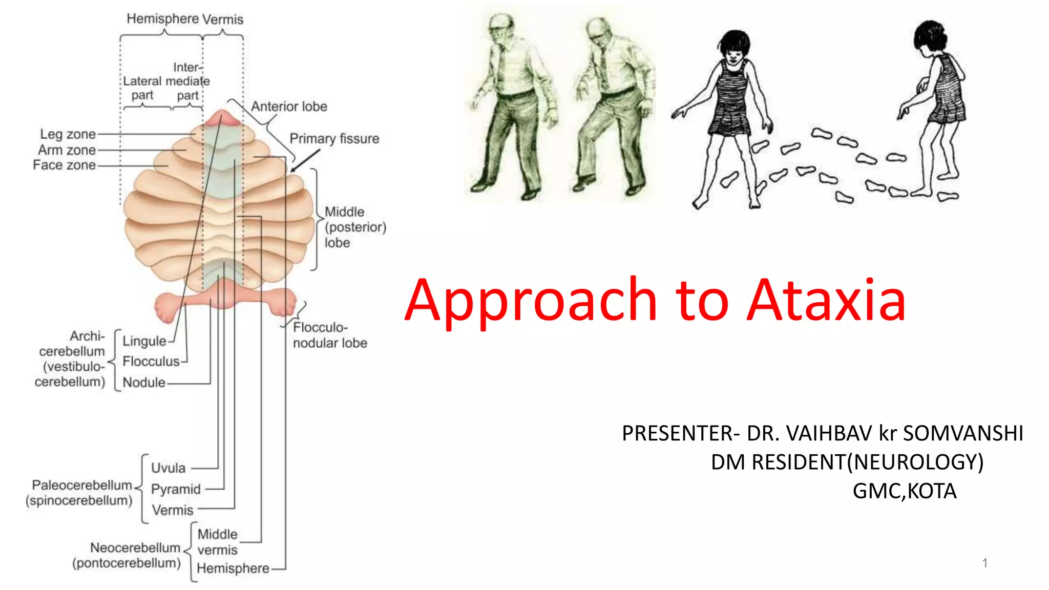
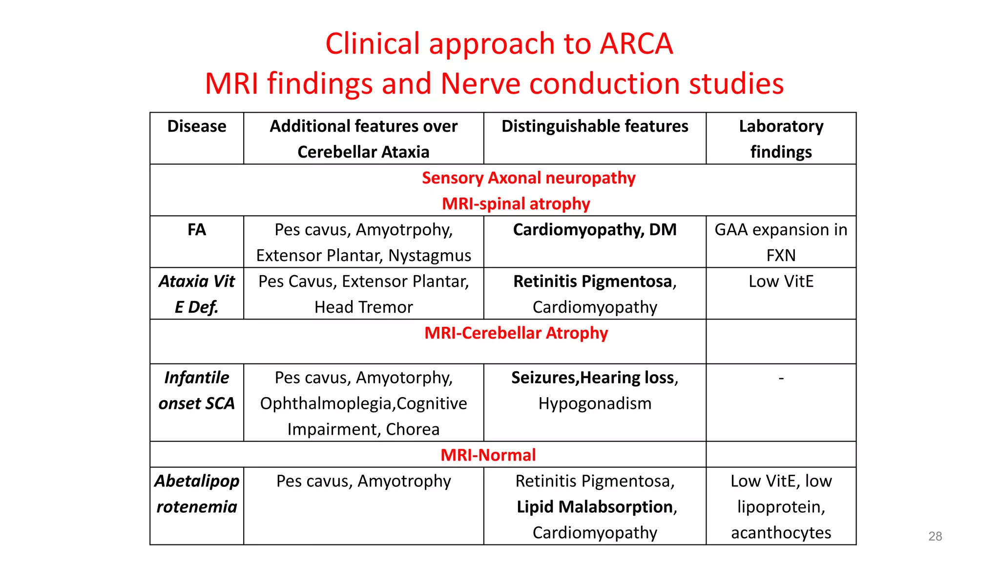
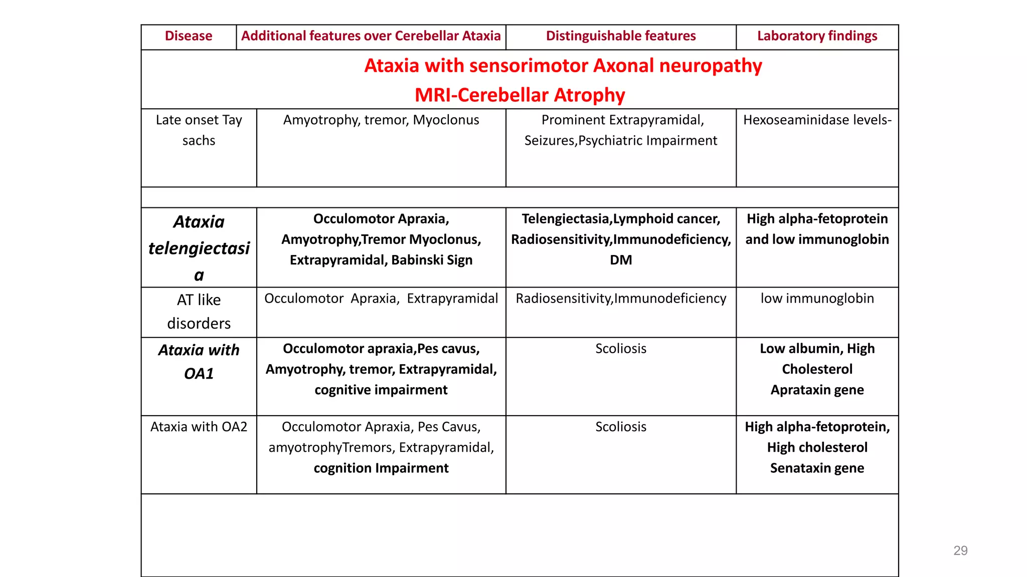
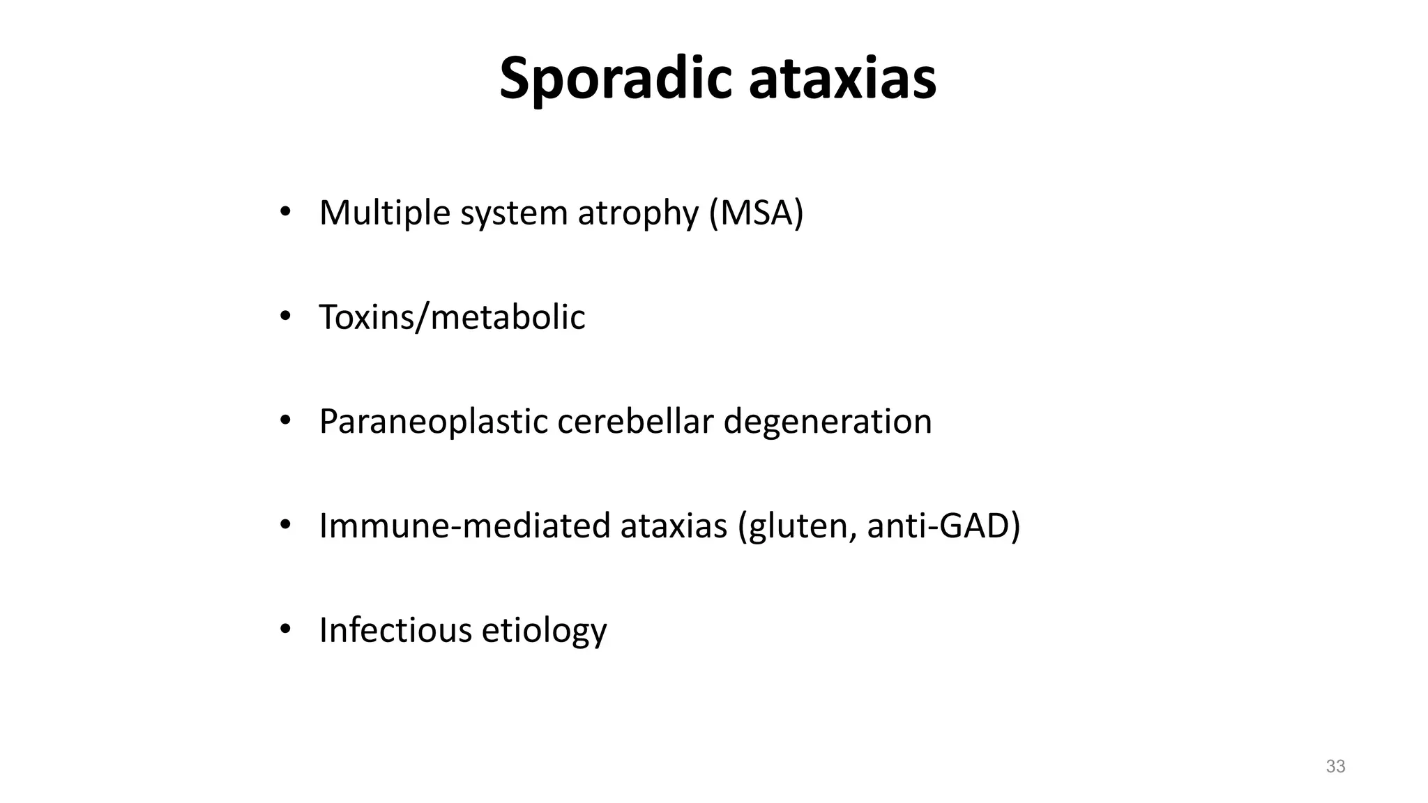
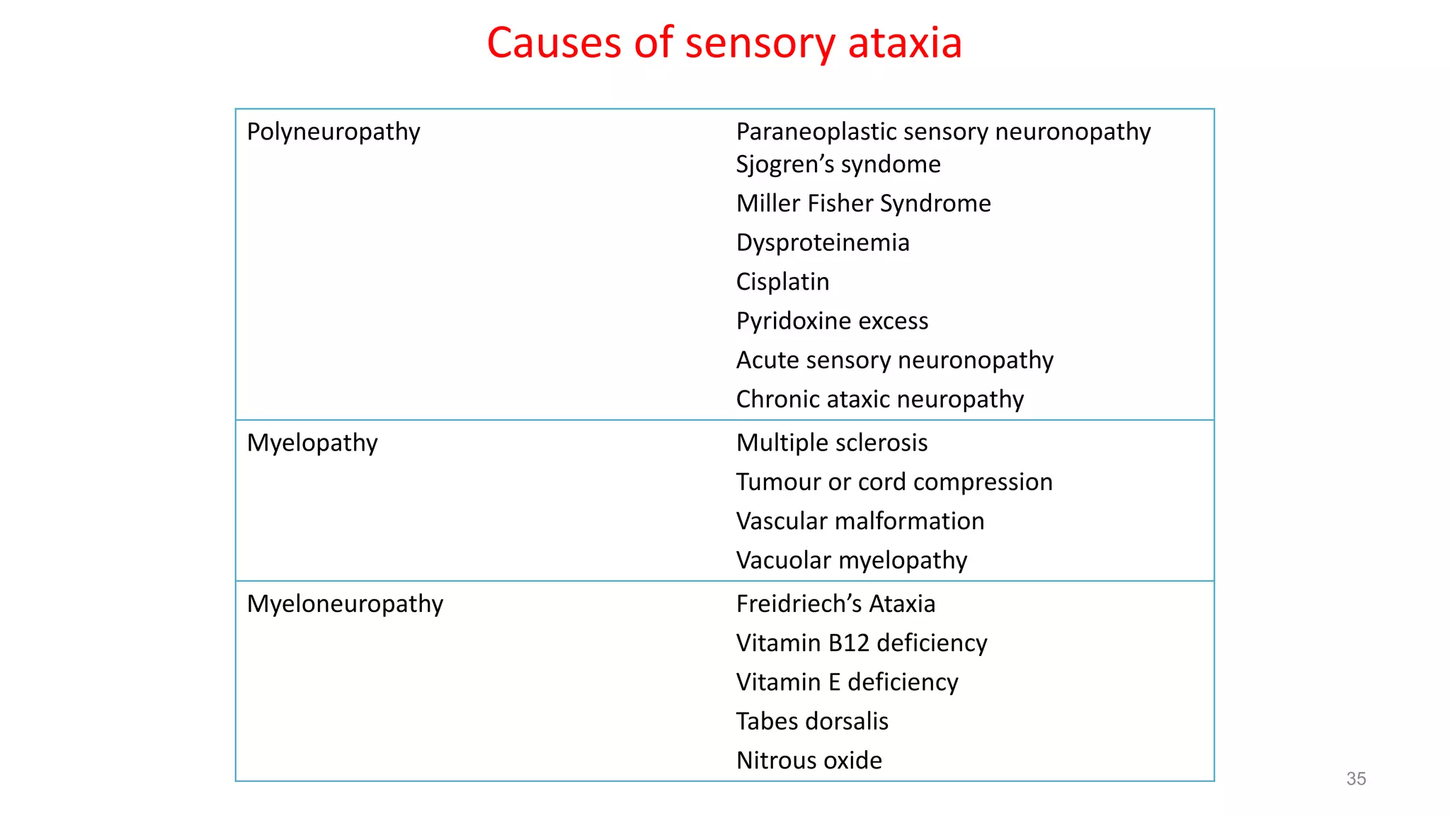
1
PRESENTER- DR. VAIHBAV kr SOMVANSHI
DM RESIDENT(NEUROLOGY)
GMC,KOTA
Approach to Ataxia
Introduction
• Ataxia = from Greek- a- [lack of]+ taxia [order]
• Rate, rhythm and force of contraction of voluntary movements
• Disorganized, poorly coordinated, or clumsy movements
Traditionally used specifically for lesions involving
• Cerebellum or it’s pathways
• Proprioceptive sensory pathways
Localisation
• Cerebellum (most common)
• Sensory pathways (Sensory Ataxia)-posterior columns, dorsal root ganglia,
peripheral nerves
• Vestibular dysfunction
Localization of cerebellar lesions
Gait ataxia
Limb ataxia
Dysarthria
Titubation
Action tremor
4
Anterior vermis
Lateral hemispheres
Posterior left hemisphere & vermis
Ant. Vermis & associated deep nuclei
Dentate nuclei, or cerebellar outflow to
ventral thalamus
Localization of cerebellar lesions
Palatal tremor
Saccadic dysmetria
Square wave jerks
Gaze evoked nystagmus
Higher cognitive changes
5
Dentate nucleus, Guillain Mollaret
triangle
Dorsal vermis
Cerebellar outflow
Flocculus & paraflocculus
Lateral hemispheres
Neuroanatomy
6
Sensory Ataxia
• Loss of distal joint, position sense
• Absence of cerebellar signs such as dysarthria or nystagmus
• Loss of tendon reflexes
• Corrective effects of vision on sensory ataxia
• Romberg sign
Vestibular Dysfunction
• Vertigo is prominent
• Consistent fall to one side
• Nystagmus
• Limb ataxia is absent
• Speech is normal
• Joint position sense is normal
Approach to ataxic patient
Meticulous evaluation of History
 Age at Onset
 Course of disease
 Drug intake
 Family History
 Personal Social & Occupational information
 Distribution of ataxia
 History of other system illness
Neurological evaluation
Ancillary tests
9
History
• Age at onset
 Childhood (congenital, metabolic, infectious, posterior fossa tumors,
hereditary ataxias - more common)
 Adult (sporadic ataxias, hereditary ataxias)
• Course of illness/progression
 Acute (metabolic/toxic, infectious, inflammatory, traumatic)
 Subacute (metabolic/toxic, infectious, inflammatory, paraneoplastic,
tumor)
 Chronic (more likely genetic, degenerative, tumor, paraneoplastic)
10
• Drug intake
– Phenytoin, barbiturates, lithium, immunosuppressants (methotrexate,
cyclosporine), chemotherapy (fluorouracil, cytarabine)
• Family history
– Study at least 3 generations
– Consanguinity
– Ethnicity
• Social/Occupational History
– Alcohol and drug use, toxins (heavy metals, solvents, thallium), smoking
(Vascular)
History
11
Distribution of ataxia
• Symmetric - Acquired, Hereditary, degenerative ataxias
• Asymmetric- Vascular, Tumors, demyelinating, Infection, congenital causes
Other system illness
• Gastrointestinal symptoms- gluten ataxia
• Mass lesion- paraneoplastic ataxias
History
12
Children
• Refusal to walk or with a wide-based, "drunken" gait.
• Vertigo, dizziness and vomiting
• Personality and behavioral changes.
• Abnormal mental status
• A history of head trauma ,neck trauma
• Patients with a recent infection or vaccination
• Previous similar episodes of acute ataxia.
• Children with family members with ataxia
13
Examination
Neurological examination
• Ataxia (appendicular or axial)
• Dysmetria
• Dysdiadochokinesia
• Rebound Phenomenon
• Dysarthria
• Tremor
• Titubation and increased postural sway
• Hypotonia
• Nystagmus
• Other system evaluation
 Breast Lump, mass per-abdomen etc.
14
Neuro-ophthalmologic evaluation in ataxia
Retinal pigmentosa Refsum disease, mitochondrial disorders
Retinal/Macular degeneration SCA 7, aceruloplasminemia
Optic atrophy/visual loss MS,FA
Square wave jerks FA
Occulomotor apraxia AT, AOA1, AOA2
Slow saccades
Downbeat Nystagmus
SCA2,SCA 7
SCA 6,EA2,anti GAD ataxia
15
Other Specific signs
Focal and lateralized brainstem
deficits(hemiparesis,facial palsy)
Posterior circulation stroke, tumour, MS
Paplidema, headache Posterior fossa tumours
INO Posterior circulation stroke,MS
Spasticity, UMN signs SCA 1,3,7,8 ,Strokes,tumour compressing
brainstem
Basal ganglia deficits SCA 1,2,12,17, MJD,MSA,Wilson
Tremor SCA 12, 15, FAXTS
16
17
Deafness Mitochondrial, superficial himosiderosis
Myoclonus Mitochondrial ,ceroid lipofuschinosis,SCA
7(early onset),SCA 14
Palatal myoclonus Alexander disease,SCA20
Cognitive decline Alcohol,MS,CJD,HIV,DRPLA,SCA12,13,
superficial siderosis
Psychiatric features SCA 12,17,27
Autonomic failure MSA,FXTAS
Other Specific signs
Ataxia with Neuropathy
• Friedreich ataxia
• AOA2
• Fragile X syndrome
• Vit E deficiency ataxia
• Anti gliadin ataxia
• SCA 2,3,4,12, 18,25,27
• Refsum disease
18
Ataxia with Dementia
• Anti gliadin ataxia
• FXTAS syndrme
• SREAT
• SCA 17, 19, 21, 2, 1, 6
• HIV/AIDS
• Mitochondrial disease
• Amylodosis
19
Ataxia with seizures
• Anti GAD
• Anti gliadin
• Mitochondrial ataxia
• Episodic ataxia 2, 4
• DRPLA
• SCA 10, SCA 17
• CJD
• SREAT
20
Investigations
• Neuro imaging
• Electro diagnostic tests
• Ophthalmologic examination- Pigmentary retinopathy, macular degeneration,
cataracts, Kayser-Fleischer rings
• Genetic tests
• Metabolic – Thyroid function, vitamins B12, E, and B1, serum cholesterol &
plasma lipoprotein profile, phytanic acid, toxicology screen(mercury, bismuth,
lead, ethanol)
• Immune function - Immunoglobulin levels, Antigliadin antibodies, GAD
antibodies, paraneoplastic antibodies, Anti TPO.
Laboratory studies
• Mitochondrial( Serum lactate and pyruvate)
• Heavy metals,PBF for acanthocytes, VLCF, hexosaminidase A/B,
alpha fetoprotein & immunoglobulins, serum ceruloplasmin & 24 hour
urinary copper
• CSF studies - Cell count, glucose and protein, oligoclonal bands, 14-
3-3 protein, GAD antibodies, paraneoplastic antibodies,
lactate/pyruvate
• Diagnostic
workflow
Continuum movement disorder 2019 pgno.161
prabhakar_sudesh_singh_gagandeep_eds_differential_diagnosis chapter 22 pg.no.251
Genetic Testing Protocol of ataxias
Spinocerebellar Ataxia
Aut.Dominant Aut.Recessive
Sporadic
LOCA (>25) EOCA (<25)
SCA 1
SCA 2
SAC 3
SCA 7
SCA 12
FRDA
NO YES
YES NO
SCA 6
SCA 8
SCA 17
DRPLA
YES NO
Rare types of SCAs
(ADCA) screening
Investigation for
other ARCA genes
Level 20
Level 10
Level 30
features suggestive of SCA
LOCA-Late onset cerebellar ataxia
EOCA-Early onset cerebellar ataxia
SCA27
SCA28
Age at
Onset (Yrs)
10-30 >30 Variable
SCA11
SCA14
SCA23
SCA5
SCA13
SCA14
SCA15
SCA28 26
Signs that Distinguishes SCA subtypes
Benign course SCA 6
UMN signs SCA 1,7,8 and 3
Akinetic rigid syndrome SCA 3,2,17 & 12,21
Chorea SCA 2,1,3
Action tremor SCA 12,16
Slow saccades SCA 2 & 7 may be in 1,3
Downbeat nystagmus SCA 6
Hyporeflexia/Areflexia SCA 2,4,3 & 19,21
Vision loss SCA 7
Seizure SCA 10
Myoclonus SCA14 or SCA19
Cognitive impairment SCA2,14,19,21,23
27
Disease Additional features over
Cerebellar Ataxia
Distinguishable features Laboratory
findings
Sensory Axonal neuropathy
MRI-spinal atrophy
FA Pes cavus, Amyotrpohy,
Extensor Plantar, Nystagmus
Cardiomyopathy, DM GAA expansion in
FXN
Ataxia Vit
E Def.
Pes Cavus, Extensor Plantar,
Head Tremor
Retinitis Pigmentosa,
Cardiomyopathy
Low VitE
MRI-Cerebellar Atrophy
Infantile
onset SCA
Pes cavus, Amyotorphy,
Ophthalmoplegia,Cognitive
Impairment, Chorea
Seizures,Hearing loss,
Hypogonadism
-
MRI-Normal
Abetalipop
rotenemia
Pes cavus, Amyotrophy Retinitis Pigmentosa,
Lipid Malabsorption,
Cardiomyopathy
Low VitE, low
lipoprotein,
acanthocytes
Clinical approach to ARCA
MRI findings and Nerve conduction studies
28
Disease Additional features over Cerebellar Ataxia Distinguishable features Laboratory findings
Ataxia with sensorimotor Axonal neuropathy
MRI-Cerebellar Atrophy
Late onset Tay
sachs
Amyotrophy, tremor, Myoclonus Prominent Extrapyramidal,
Seizures,Psychiatric Impairment
Hexoseaminidase levels-
Ataxia
telengiectasi
a
Occulomotor Apraxia,
Amyotrophy,Tremor Myoclonus,
Extrapyramidal, Babinski Sign
Telengiectasia,Lymphoid cancer,
Radiosensitivity,Immunodeficiency,
DM
High alpha-fetoprotein
and low immunoglobin
AT like
disorders
Occulomotor Apraxia, Extrapyramidal Radiosensitivity,Immunodeficiency low immunoglobin
Ataxia with
OA1
Occulomotor apraxia,Pes cavus,
Amyotrophy, tremor, Extrapyramidal,
cognitive impairment
Scoliosis Low albumin, High
Cholesterol
Aprataxin gene
Ataxia with OA2 Occulomotor Apraxia, Pes Cavus,
amyotrophyTremors, Extrapyramidal,
cognition Impairment
Scoliosis High alpha-fetoprotein,
High cholesterol
Senataxin gene
29
Disease Additional features over Cerebellar
Ataxia
Distinguishable features Laboratory findings
MRI-Spinal +Cerebellar Atrophy
AR ataxia
Charlevoix-
Saguenay
Pes Cavus, Amyotrophy, Spasticity,
extensor Plantar, cogitive Impairment
Chromosome 13
MRI-Cerebellar Atrophy + WMH
Cerebrotendinou
s xanthomatosis
Pes Caus Amyotrophy, Spasticity,
myoclonus, Parkinsonism
Psychiatric Impairment,Tendon
Xanthomas,Seizures,Cataract,Liver
failure
CYP27A1
30
31
32
Sporadic ataxias
• Multiple system atrophy (MSA)
• Toxins/metabolic
• Paraneoplastic cerebellar degeneration
• Immune-mediated ataxias (gluten, anti-GAD)
• Infectious etiology
33
Antibody Condition
Anti-Yo (Purkinje cell antobody type1) Breast and ovarian Ca
Anti-Hu (Anti neuronal nuclear antibody
type1) Small cell lung Ca (SCLC)
Anti-Tr Hodgkin Lymphoma
Anti-mGluR1 (metabotrpin glutamate
receptor) Hodgkin Lymphoma
Anti-CRMP5 (Collapsin receptor mediated
protein)/Anti-CV2 SCLC
Anti-ZIC4 (zinc finger protein) SCLC
Paraneoplastic ataxia associated antibodies
Causes of sensory ataxia
Polyneuropathy Paraneoplastic sensory neuronopathy
Sjogren’s syndome
Miller Fisher Syndrome
Dysproteinemia
Cisplatin
Pyridoxine excess
Acute sensory neuronopathy
Chronic ataxic neuropathy
Myelopathy Multiple sclerosis
Tumour or cord compression
Vascular malformation
Vacuolar myelopathy
Myeloneuropathy Freidriech’s Ataxia
Vitamin B12 deficiency
Vitamin E deficiency
Tabes dorsalis
Nitrous oxide
35
36
Cerebral Sensory frontal
Base of support Wide base Looks down Wide base
Velocity Variable Slow Very slow
Initiation Normal Normal Hesitant
Turns Unsteady Unstaedy Hesitant,multiple steps
Postural instability + + +++++
Falls Late event More in night Frequent
Heel shin Abnormal Abnormal,difficulty in
point of initiation
Normal
Types of Ataxia
Management
• Corrective measures for deficiencies – Vitamin E, Thiamine
• Specific diet – Gluten free diet, ketogenic diet
• Immunologic disorder – IVIG, Plasmapharesis
• Miglustat for niemann pick disease
• Riluzole – hereditary and degenerative ataxia and dysarthria
• Varenicline – SCA3
• 4- aminopyridine, Acetazolamide – Episodic ataxia type 2
• Nicotinamide, deferiprone, idebenon – friedreich ataxia, mitochondrial ataxia
• Rehabilitation, exercise help in SCA type 1
• Zinc supplementation and DBS also help in SCA 2
• Actimmune – IFN-γ analogue morbidity benefit for friedreich ataxia
• Antisense oigonuceotide shown to knock down toxic protein level in SCA 2 and 3 mouse model
38
Case Scenario
55-year-old man presented with progressive gait difficulty. He had initially noted difficulty
walking downstairs, upstairs and running at the age of 47. His imbalance problems became
progressively worse over the years, and he developed slurred speech, transient double vision
while turning his head quickly, and loss of hand dexterity. He had frequent falls and needed to
use a walker. He had an extensive family history of cerebellar ataxia, affecting his mother and
brother. On examination, he had slurred speech and slow saccadic eye movements without
nystagmus or hypermetric or hypometric saccades. He had dysmetria on finger-nose-finger
tests and overshoot in finger chase tests. He also had impaired rapid alternating movements
with absent reflexes in upper limb and hyporelexia in lower limb. He had a hypomimic facial
expression and bradykinesia left side with retropusion test positive. His gait showed variable
stride length and was wide based.
39
Patient Video
40
Genetic tests - 38 repeats CAG expansions of the ATXN2 gene (normal <32),
Diagnosed as SCA 2
The patient treated with riluzole 50 mg 2 times a day which
provided modest benefits for his speech. Physical therapy helped with
his balance. Carbidopa/levodopa 25 mg/100 mg, 3 times a day, improved
his parkinsonism by increasing the speed of her movements.
41
Conclusion
An approach to ataxia is based on knowledge of its symptoms and
causes
 Knowledge of differentiating clinical features takes clinicians closer to
the etiological diagnosis which help in investigational decisions.
Treatable causes must be identified and ruled out
 Genetic testing is prudent for providing better insight into the
management.
• On going clinical trials for management of hereditary and degenerative
ataxias will help in better morbidity outcome of these diseases.
42
• Bradley’s Neurology in clinical Practice,eighth edition
• Handbook of Clinical Neurology, Vol. 103 (3rd series), Ataxic Disorders
• http://www.ataxia.org -National Ataxia Foundation web site
• http://www.ncbi.nlm.nih.gov/books/NBK1138/ Detailed information about ataxias
• http://www.clinicaltrials.gov –clinical trials information
• Pubmed-with the searchterms “spinocerebellar ataxia”,“Friedreich’s ataxia”, “sporadic ataxia”,“sensory
ataxia”, “approach to ataxia”, “ataxiadiagnosis”
• The Cochrane Library
• Continuum movement disorder 2016
• Continuum movement disorder 2019
• prabhakar_sudesh_singh_gagandeep_eds_differential_diagnosis chapter 22 pg.no.251
References
44
Genotype-Phenotype correlations in SCA2
Higher repeats are associated with earlier onset
Homozygous expansion- no increase in severity
Allelic variations of RAI 1 and CACNA1A influences age at onset
Disease duration X CAG length affects occurrence of slow saccades,
Fasciculation, Amyotrophy, Areflexia and Vibration senses
Small disease alleles (32-37): Postural Tremors and Parkinsonism, late onset disease
Medium Size alleles (38-44) : Ataxia, areflexia and slowing of saccades
Large Size Alleles (>45) : Onset <20 years, Chorea and dementia
Higher Size Alleles (>91) : Ataxia, Dystonia, Myoclonus, Cardiac failure, optic atrophy
Genotype-Phenotype correlations in
SCA1
Higher repeats are associated with earlier onset and severe disease
Homozygous expansion- no increase in severity
Small disease alleles (39-44) interrupted: Mild Phenotype, Ataxic/non ataxic
features
Medium Size alleles (39-50) Pure CAG: Ataxia and Pyramidal
syndrome
Large Size Alleles (>50) Pure
CAG:
Ataxia and Pyramidal
syndrome &
Amytrophic Lateral sclerosis
Higher Size Alleles
(>91):
Juvenile
disease
Genotype-Phenotype correlations in
SCA3
Earlier onset with Higher repeats and inverse
correlation
Homozygous expansion- confers increasing
severity
Small disease alleles (52-73): Axonal Neuropathy and Parkinsonism (Type-III MJD)
Medium Size alleles (73-80) : Ataxia and Diplopia (Type-II MJD)
Large Size Alleles (80-86) : Ataxia, Dystonia and spasticity (Type-I MJD)
Higher Size Alleles (>86) : Rare cases predominant Dystonia (Type-IV)
Genotype-Phenotype correlations in
SCA7
Earlier onset with higher repeats and anticipation
Greater expandability during transmission of alleles
Recurrent denovo expansions
Small disease alleles (36-41): Cerebellar ataxia without Retinal involvement
Medium Size alleles (42-49) : Ataxia preceedes Vision diminution Large Size
Alleles (49-60) : Vision loss preceedes Ataxia Higher Size Alleles (>80)
: Juvenile Onset
Extreme High Length Alleles : Infantile Onset, Developmental failure, Multisystem
involvement (>200)

Approach to Ataxia

  • 1.
    1 PRESENTER- DR. VAIHBAVkr SOMVANSHI DM RESIDENT(NEUROLOGY) GMC,KOTA Approach to Ataxia
  • 2.
    Introduction • Ataxia =from Greek- a- [lack of]+ taxia [order] • Rate, rhythm and force of contraction of voluntary movements • Disorganized, poorly coordinated, or clumsy movements Traditionally used specifically for lesions involving • Cerebellum or it’s pathways • Proprioceptive sensory pathways
  • 3.
    Localisation • Cerebellum (mostcommon) • Sensory pathways (Sensory Ataxia)-posterior columns, dorsal root ganglia, peripheral nerves • Vestibular dysfunction
  • 4.
    Localization of cerebellarlesions Gait ataxia Limb ataxia Dysarthria Titubation Action tremor 4 Anterior vermis Lateral hemispheres Posterior left hemisphere & vermis Ant. Vermis & associated deep nuclei Dentate nuclei, or cerebellar outflow to ventral thalamus
  • 5.
    Localization of cerebellarlesions Palatal tremor Saccadic dysmetria Square wave jerks Gaze evoked nystagmus Higher cognitive changes 5 Dentate nucleus, Guillain Mollaret triangle Dorsal vermis Cerebellar outflow Flocculus & paraflocculus Lateral hemispheres
  • 6.
  • 7.
    Sensory Ataxia • Lossof distal joint, position sense • Absence of cerebellar signs such as dysarthria or nystagmus • Loss of tendon reflexes • Corrective effects of vision on sensory ataxia • Romberg sign
  • 8.
    Vestibular Dysfunction • Vertigois prominent • Consistent fall to one side • Nystagmus • Limb ataxia is absent • Speech is normal • Joint position sense is normal
  • 9.
    Approach to ataxicpatient Meticulous evaluation of History  Age at Onset  Course of disease  Drug intake  Family History  Personal Social & Occupational information  Distribution of ataxia  History of other system illness Neurological evaluation Ancillary tests 9
  • 10.
    History • Age atonset  Childhood (congenital, metabolic, infectious, posterior fossa tumors, hereditary ataxias - more common)  Adult (sporadic ataxias, hereditary ataxias) • Course of illness/progression  Acute (metabolic/toxic, infectious, inflammatory, traumatic)  Subacute (metabolic/toxic, infectious, inflammatory, paraneoplastic, tumor)  Chronic (more likely genetic, degenerative, tumor, paraneoplastic) 10
  • 11.
    • Drug intake –Phenytoin, barbiturates, lithium, immunosuppressants (methotrexate, cyclosporine), chemotherapy (fluorouracil, cytarabine) • Family history – Study at least 3 generations – Consanguinity – Ethnicity • Social/Occupational History – Alcohol and drug use, toxins (heavy metals, solvents, thallium), smoking (Vascular) History 11
  • 12.
    Distribution of ataxia •Symmetric - Acquired, Hereditary, degenerative ataxias • Asymmetric- Vascular, Tumors, demyelinating, Infection, congenital causes Other system illness • Gastrointestinal symptoms- gluten ataxia • Mass lesion- paraneoplastic ataxias History 12
  • 13.
    Children • Refusal towalk or with a wide-based, "drunken" gait. • Vertigo, dizziness and vomiting • Personality and behavioral changes. • Abnormal mental status • A history of head trauma ,neck trauma • Patients with a recent infection or vaccination • Previous similar episodes of acute ataxia. • Children with family members with ataxia 13
  • 14.
    Examination Neurological examination • Ataxia(appendicular or axial) • Dysmetria • Dysdiadochokinesia • Rebound Phenomenon • Dysarthria • Tremor • Titubation and increased postural sway • Hypotonia • Nystagmus • Other system evaluation  Breast Lump, mass per-abdomen etc. 14
  • 15.
    Neuro-ophthalmologic evaluation inataxia Retinal pigmentosa Refsum disease, mitochondrial disorders Retinal/Macular degeneration SCA 7, aceruloplasminemia Optic atrophy/visual loss MS,FA Square wave jerks FA Occulomotor apraxia AT, AOA1, AOA2 Slow saccades Downbeat Nystagmus SCA2,SCA 7 SCA 6,EA2,anti GAD ataxia 15
  • 16.
    Other Specific signs Focaland lateralized brainstem deficits(hemiparesis,facial palsy) Posterior circulation stroke, tumour, MS Paplidema, headache Posterior fossa tumours INO Posterior circulation stroke,MS Spasticity, UMN signs SCA 1,3,7,8 ,Strokes,tumour compressing brainstem Basal ganglia deficits SCA 1,2,12,17, MJD,MSA,Wilson Tremor SCA 12, 15, FAXTS 16
  • 17.
    17 Deafness Mitochondrial, superficialhimosiderosis Myoclonus Mitochondrial ,ceroid lipofuschinosis,SCA 7(early onset),SCA 14 Palatal myoclonus Alexander disease,SCA20 Cognitive decline Alcohol,MS,CJD,HIV,DRPLA,SCA12,13, superficial siderosis Psychiatric features SCA 12,17,27 Autonomic failure MSA,FXTAS Other Specific signs
  • 18.
    Ataxia with Neuropathy •Friedreich ataxia • AOA2 • Fragile X syndrome • Vit E deficiency ataxia • Anti gliadin ataxia • SCA 2,3,4,12, 18,25,27 • Refsum disease 18
  • 19.
    Ataxia with Dementia •Anti gliadin ataxia • FXTAS syndrme • SREAT • SCA 17, 19, 21, 2, 1, 6 • HIV/AIDS • Mitochondrial disease • Amylodosis 19
  • 20.
    Ataxia with seizures •Anti GAD • Anti gliadin • Mitochondrial ataxia • Episodic ataxia 2, 4 • DRPLA • SCA 10, SCA 17 • CJD • SREAT 20
  • 21.
    Investigations • Neuro imaging •Electro diagnostic tests • Ophthalmologic examination- Pigmentary retinopathy, macular degeneration, cataracts, Kayser-Fleischer rings
  • 22.
    • Genetic tests •Metabolic – Thyroid function, vitamins B12, E, and B1, serum cholesterol & plasma lipoprotein profile, phytanic acid, toxicology screen(mercury, bismuth, lead, ethanol) • Immune function - Immunoglobulin levels, Antigliadin antibodies, GAD antibodies, paraneoplastic antibodies, Anti TPO.
  • 23.
    Laboratory studies • Mitochondrial(Serum lactate and pyruvate) • Heavy metals,PBF for acanthocytes, VLCF, hexosaminidase A/B, alpha fetoprotein & immunoglobulins, serum ceruloplasmin & 24 hour urinary copper • CSF studies - Cell count, glucose and protein, oligoclonal bands, 14- 3-3 protein, GAD antibodies, paraneoplastic antibodies, lactate/pyruvate
  • 24.
  • 25.
  • 26.
    Genetic Testing Protocolof ataxias Spinocerebellar Ataxia Aut.Dominant Aut.Recessive Sporadic LOCA (>25) EOCA (<25) SCA 1 SCA 2 SAC 3 SCA 7 SCA 12 FRDA NO YES YES NO SCA 6 SCA 8 SCA 17 DRPLA YES NO Rare types of SCAs (ADCA) screening Investigation for other ARCA genes Level 20 Level 10 Level 30 features suggestive of SCA LOCA-Late onset cerebellar ataxia EOCA-Early onset cerebellar ataxia SCA27 SCA28 Age at Onset (Yrs) 10-30 >30 Variable SCA11 SCA14 SCA23 SCA5 SCA13 SCA14 SCA15 SCA28 26
  • 27.
    Signs that DistinguishesSCA subtypes Benign course SCA 6 UMN signs SCA 1,7,8 and 3 Akinetic rigid syndrome SCA 3,2,17 & 12,21 Chorea SCA 2,1,3 Action tremor SCA 12,16 Slow saccades SCA 2 & 7 may be in 1,3 Downbeat nystagmus SCA 6 Hyporeflexia/Areflexia SCA 2,4,3 & 19,21 Vision loss SCA 7 Seizure SCA 10 Myoclonus SCA14 or SCA19 Cognitive impairment SCA2,14,19,21,23 27
  • 28.
    Disease Additional featuresover Cerebellar Ataxia Distinguishable features Laboratory findings Sensory Axonal neuropathy MRI-spinal atrophy FA Pes cavus, Amyotrpohy, Extensor Plantar, Nystagmus Cardiomyopathy, DM GAA expansion in FXN Ataxia Vit E Def. Pes Cavus, Extensor Plantar, Head Tremor Retinitis Pigmentosa, Cardiomyopathy Low VitE MRI-Cerebellar Atrophy Infantile onset SCA Pes cavus, Amyotorphy, Ophthalmoplegia,Cognitive Impairment, Chorea Seizures,Hearing loss, Hypogonadism - MRI-Normal Abetalipop rotenemia Pes cavus, Amyotrophy Retinitis Pigmentosa, Lipid Malabsorption, Cardiomyopathy Low VitE, low lipoprotein, acanthocytes Clinical approach to ARCA MRI findings and Nerve conduction studies 28
  • 29.
    Disease Additional featuresover Cerebellar Ataxia Distinguishable features Laboratory findings Ataxia with sensorimotor Axonal neuropathy MRI-Cerebellar Atrophy Late onset Tay sachs Amyotrophy, tremor, Myoclonus Prominent Extrapyramidal, Seizures,Psychiatric Impairment Hexoseaminidase levels- Ataxia telengiectasi a Occulomotor Apraxia, Amyotrophy,Tremor Myoclonus, Extrapyramidal, Babinski Sign Telengiectasia,Lymphoid cancer, Radiosensitivity,Immunodeficiency, DM High alpha-fetoprotein and low immunoglobin AT like disorders Occulomotor Apraxia, Extrapyramidal Radiosensitivity,Immunodeficiency low immunoglobin Ataxia with OA1 Occulomotor apraxia,Pes cavus, Amyotrophy, tremor, Extrapyramidal, cognitive impairment Scoliosis Low albumin, High Cholesterol Aprataxin gene Ataxia with OA2 Occulomotor Apraxia, Pes Cavus, amyotrophyTremors, Extrapyramidal, cognition Impairment Scoliosis High alpha-fetoprotein, High cholesterol Senataxin gene 29
  • 30.
    Disease Additional featuresover Cerebellar Ataxia Distinguishable features Laboratory findings MRI-Spinal +Cerebellar Atrophy AR ataxia Charlevoix- Saguenay Pes Cavus, Amyotrophy, Spasticity, extensor Plantar, cogitive Impairment Chromosome 13 MRI-Cerebellar Atrophy + WMH Cerebrotendinou s xanthomatosis Pes Caus Amyotrophy, Spasticity, myoclonus, Parkinsonism Psychiatric Impairment,Tendon Xanthomas,Seizures,Cataract,Liver failure CYP27A1 30
  • 31.
  • 32.
  • 33.
    Sporadic ataxias • Multiplesystem atrophy (MSA) • Toxins/metabolic • Paraneoplastic cerebellar degeneration • Immune-mediated ataxias (gluten, anti-GAD) • Infectious etiology 33
  • 34.
    Antibody Condition Anti-Yo (Purkinjecell antobody type1) Breast and ovarian Ca Anti-Hu (Anti neuronal nuclear antibody type1) Small cell lung Ca (SCLC) Anti-Tr Hodgkin Lymphoma Anti-mGluR1 (metabotrpin glutamate receptor) Hodgkin Lymphoma Anti-CRMP5 (Collapsin receptor mediated protein)/Anti-CV2 SCLC Anti-ZIC4 (zinc finger protein) SCLC Paraneoplastic ataxia associated antibodies
  • 35.
    Causes of sensoryataxia Polyneuropathy Paraneoplastic sensory neuronopathy Sjogren’s syndome Miller Fisher Syndrome Dysproteinemia Cisplatin Pyridoxine excess Acute sensory neuronopathy Chronic ataxic neuropathy Myelopathy Multiple sclerosis Tumour or cord compression Vascular malformation Vacuolar myelopathy Myeloneuropathy Freidriech’s Ataxia Vitamin B12 deficiency Vitamin E deficiency Tabes dorsalis Nitrous oxide 35
  • 36.
    36 Cerebral Sensory frontal Baseof support Wide base Looks down Wide base Velocity Variable Slow Very slow Initiation Normal Normal Hesitant Turns Unsteady Unstaedy Hesitant,multiple steps Postural instability + + +++++ Falls Late event More in night Frequent Heel shin Abnormal Abnormal,difficulty in point of initiation Normal Types of Ataxia
  • 38.
    Management • Corrective measuresfor deficiencies – Vitamin E, Thiamine • Specific diet – Gluten free diet, ketogenic diet • Immunologic disorder – IVIG, Plasmapharesis • Miglustat for niemann pick disease • Riluzole – hereditary and degenerative ataxia and dysarthria • Varenicline – SCA3 • 4- aminopyridine, Acetazolamide – Episodic ataxia type 2 • Nicotinamide, deferiprone, idebenon – friedreich ataxia, mitochondrial ataxia • Rehabilitation, exercise help in SCA type 1 • Zinc supplementation and DBS also help in SCA 2 • Actimmune – IFN-γ analogue morbidity benefit for friedreich ataxia • Antisense oigonuceotide shown to knock down toxic protein level in SCA 2 and 3 mouse model 38
  • 39.
    Case Scenario 55-year-old manpresented with progressive gait difficulty. He had initially noted difficulty walking downstairs, upstairs and running at the age of 47. His imbalance problems became progressively worse over the years, and he developed slurred speech, transient double vision while turning his head quickly, and loss of hand dexterity. He had frequent falls and needed to use a walker. He had an extensive family history of cerebellar ataxia, affecting his mother and brother. On examination, he had slurred speech and slow saccadic eye movements without nystagmus or hypermetric or hypometric saccades. He had dysmetria on finger-nose-finger tests and overshoot in finger chase tests. He also had impaired rapid alternating movements with absent reflexes in upper limb and hyporelexia in lower limb. He had a hypomimic facial expression and bradykinesia left side with retropusion test positive. His gait showed variable stride length and was wide based. 39
  • 40.
  • 41.
    Genetic tests -38 repeats CAG expansions of the ATXN2 gene (normal <32), Diagnosed as SCA 2 The patient treated with riluzole 50 mg 2 times a day which provided modest benefits for his speech. Physical therapy helped with his balance. Carbidopa/levodopa 25 mg/100 mg, 3 times a day, improved his parkinsonism by increasing the speed of her movements. 41
  • 42.
    Conclusion An approach toataxia is based on knowledge of its symptoms and causes  Knowledge of differentiating clinical features takes clinicians closer to the etiological diagnosis which help in investigational decisions. Treatable causes must be identified and ruled out  Genetic testing is prudent for providing better insight into the management. • On going clinical trials for management of hereditary and degenerative ataxias will help in better morbidity outcome of these diseases. 42
  • 44.
    • Bradley’s Neurologyin clinical Practice,eighth edition • Handbook of Clinical Neurology, Vol. 103 (3rd series), Ataxic Disorders • http://www.ataxia.org -National Ataxia Foundation web site • http://www.ncbi.nlm.nih.gov/books/NBK1138/ Detailed information about ataxias • http://www.clinicaltrials.gov –clinical trials information • Pubmed-with the searchterms “spinocerebellar ataxia”,“Friedreich’s ataxia”, “sporadic ataxia”,“sensory ataxia”, “approach to ataxia”, “ataxiadiagnosis” • The Cochrane Library • Continuum movement disorder 2016 • Continuum movement disorder 2019 • prabhakar_sudesh_singh_gagandeep_eds_differential_diagnosis chapter 22 pg.no.251 References 44
  • 45.
    Genotype-Phenotype correlations inSCA2 Higher repeats are associated with earlier onset Homozygous expansion- no increase in severity Allelic variations of RAI 1 and CACNA1A influences age at onset Disease duration X CAG length affects occurrence of slow saccades, Fasciculation, Amyotrophy, Areflexia and Vibration senses Small disease alleles (32-37): Postural Tremors and Parkinsonism, late onset disease Medium Size alleles (38-44) : Ataxia, areflexia and slowing of saccades Large Size Alleles (>45) : Onset <20 years, Chorea and dementia Higher Size Alleles (>91) : Ataxia, Dystonia, Myoclonus, Cardiac failure, optic atrophy
  • 46.
    Genotype-Phenotype correlations in SCA1 Higherrepeats are associated with earlier onset and severe disease Homozygous expansion- no increase in severity Small disease alleles (39-44) interrupted: Mild Phenotype, Ataxic/non ataxic features Medium Size alleles (39-50) Pure CAG: Ataxia and Pyramidal syndrome Large Size Alleles (>50) Pure CAG: Ataxia and Pyramidal syndrome & Amytrophic Lateral sclerosis Higher Size Alleles (>91): Juvenile disease
  • 47.
    Genotype-Phenotype correlations in SCA3 Earlieronset with Higher repeats and inverse correlation Homozygous expansion- confers increasing severity Small disease alleles (52-73): Axonal Neuropathy and Parkinsonism (Type-III MJD) Medium Size alleles (73-80) : Ataxia and Diplopia (Type-II MJD) Large Size Alleles (80-86) : Ataxia, Dystonia and spasticity (Type-I MJD) Higher Size Alleles (>86) : Rare cases predominant Dystonia (Type-IV)
  • 48.
    Genotype-Phenotype correlations in SCA7 Earlieronset with higher repeats and anticipation Greater expandability during transmission of alleles Recurrent denovo expansions Small disease alleles (36-41): Cerebellar ataxia without Retinal involvement Medium Size alleles (42-49) : Ataxia preceedes Vision diminution Large Size Alleles (49-60) : Vision loss preceedes Ataxia Higher Size Alleles (>80) : Juvenile Onset Extreme High Length Alleles : Infantile Onset, Developmental failure, Multisystem involvement (>200)

Editor's Notes

  • #6 Red nucleus Infeior olivary nucleus Contralateral detate nuceus
  • #7 Organ of 3
  • #8 30 seconds
  • #9 Peripheral vestibular – fatiguable nystagmus- vertigo better with affected ear up while sleeping Central vestibular – fast component away from the eccentric side
  • #10 Symmetrical/ unilateral
  • #12 SCA 12 north india 1,2,3,7 SCA 6 japanes
  • #13 Congenital – Arnold chiari malformation
  • #15 Intention, action Cerebellar Hypotonia aka gamma spasticity (preserved reflexes) Gamma alpha coordination failure Occular flutter – to and froth saccading movement like square wave jerk without pause Opsoclonus – chaotic continuous multidirectional rapid eye movement Suare wave jerk- opposite direction saccadic eye movement more than 0.3degree with no movement in between ( >10 movement – abnormal)
  • #16 Ataxia with oculomotor apraxia
  • #23 Repeat sequencing WGS Anti GQ1 ab - MFS
  • #24 Murcury, bismuth, lead Low CSF glucose – glucose transporter type 1 deficiency AFP – AOA 1, 2
  • #25 AD = autosomal dominant; AR = autosomal recessive; FXTAS = fragile X tremor-ataxia syndrome; GAD = glutamic acid decarboxylase; ILOCA = idiopathic late-onset cerebellar ataxia; MRI = magnetic resonance imaging; MSA = multiple system atrophy; REM = rapid eye movement; SCA = spinocerebellar ataxia.
  • #26 HD, Hodgkin's disease; OPCA, olivopontocerebellar atrophy; RD, registered dietitions; DRPLA, dentatorubral-pallidoluysian atrophy; ARSAS, autosomal recessive spastic ataxia of Charlevoix Saguenay; CTX, cerebrotendinous xanthomatosis; MIRAS, mitochondrial recessive ataxia syndrome. MSS – marinesco sjogren syndrome
  • #27 Genetic analyses should be directed according to the frequency of genetic subtypes in the relevant ethnic background (figure 1) and with regard to clinical features.
  • #32 Axial T2-weighted MRI sequence shows the hot cross bun sign - multiple system atrophy selective loss of myelinated transverse pontocerebellar fibers and neurons in the pontine raphe with preservation of the pontine tegmentum and corticospinal tracts. May be seen in SCA 2 SCA 3 Vcjd HIV related PML Axial T2-weighted brain MRI demonstrates hyperintensities in the bilateral inferior olivary nuclei - POLG ataxia. SANDO syndrome – POLG mutation - sensory ataxia neuropathy dysarthria ophthalamoplegia Sagittal T1-weighted brain MRI spinal cord atrophy but no cerebellar atrophy - Friedreich ataxia (GRE) axial brain MRI show hypointensity surrounding the brainstem, cerebellum, and spinal cord - superficial siderosis. Fragile X tremor-ataxia syndrome. Axial T2- weighted MRI symmetric increased signal within the MCP.
  • #33 axial T2-weighted MRI slices demonstrate linear hypointensity in the pons. autosomal recessive spastic ataxia of Charlevoix-Saguenay showing degeneration of the corticospinal tract Joubert syndrome. delayed milestones, a prominent forehead and low-set ears, and cerebellar ataxia, hypotonia, and hyperreflexia, showing dysgenesis of the isthmus (the part of the brainstem between the pons and inferior colliculus), thick superior cerebellar peduncles, and hypoplasia of the vermis- molar tooth sign. Sign also seen – hepatic fibrosis, nephronopthisis, cogan’s syndrome, pontine tegmental cap dysplasia
  • #35 Anti-Ri
  • #39 Loss of frataxin function in mitochondria leads to iron-sulfur cluster deficits, impaired oxidation, and iron accumulation. Nicotinamide has been shown to increase frataxin but no clinical benefit iron chelator deferiprone and the antioxidant idebenone are unclear on long-term benefit with side effect of heart muscle injury. Idebanon – Co-enzyme Q analogue
  • #40 Brain MRI showed pontocerebellar atrophy Axial(SPECT) - SCA 2 shows a marked symmetrical reduction of dopamine transporter binding in the caudate nucleus and putamen. 18-FDG PET - SCA2. Representative sagittal (top), coronal (mid) and axial (bottom) images of 18F-fluorodeoxyglucose uptake show diffusely decreased hypometabolism in the cerebellar cortex and dentate nuclei (mid and bottom images). Note in the SCA2 patients, the decreased tracer uptake in the basis pontis and medulla oblongata as well
  • #41 Dorsal spooning Pseudoathetosis