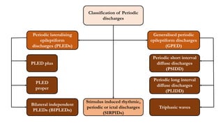
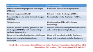
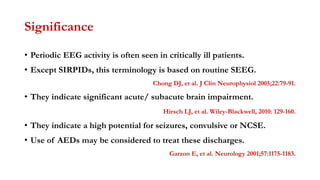
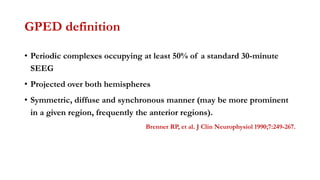
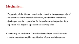
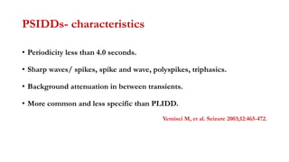
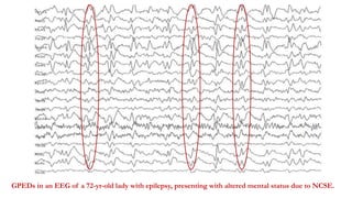
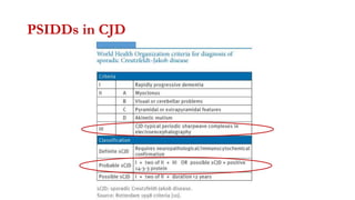
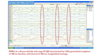
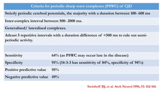
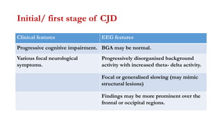
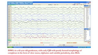
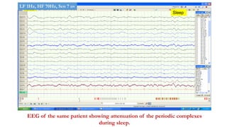
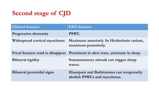
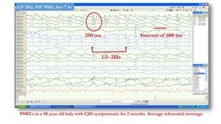
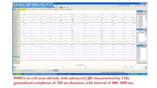
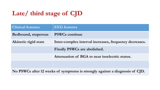
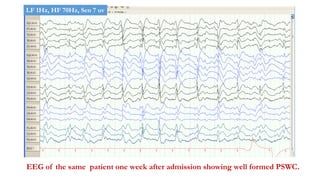
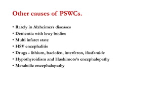
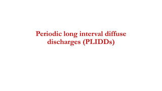
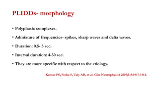
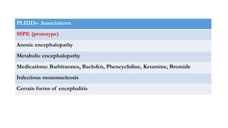
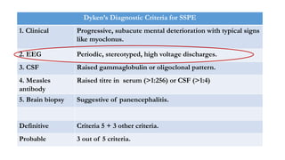
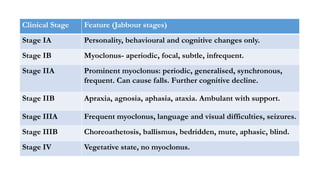
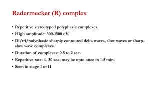
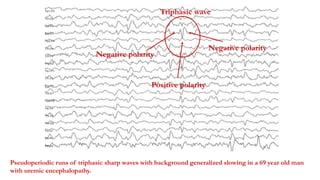
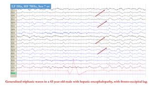
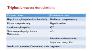
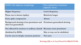
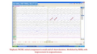
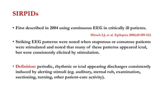
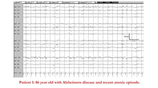
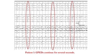
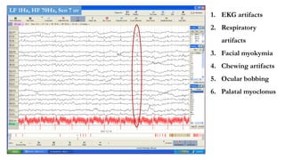
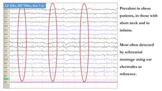
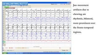
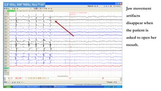
Generalised Periodic
Discharges
Dr. Pramod Krishnan
Consultant Neurologist and Epileptologist
Manipal Hospital, Bengaluru
Definition of ‘periodicity’
• The term “periodic” was first used by Cobb in 1950, in pts with SSPE.
Cobb W, Hill D. Brain 1950;73:392-404.
• Stereotyped EEG pattern consisting of waves or complexes.
• Larger than the background activity.
• Definable duration and separated by predictable ‘fixed’ intervals.
• Approximately ‘regular’ rate.
• Constant repetition rate in a patient during a particular time period.
Chong DJ, Hirsch LJ. J Clin Neurophysiol 2005;22:79-91. 3.
Hirsch LJ, Brenner RP. Atlas of EEG in critical care. 2010: 129-160.
Periodic short interval
diffuse discharges
(PSIDD)
Periodic long interval
diffuse discharges
(PLIDD)
Triphasic waves
Generalised periodic
epileptiform discharges
(GPED)
Periodic lateralising
epileptiform
discharges (PLEDs)
PLED plus
PLED
proper
Bilateral independent
PLEDs (BIPLEDs)
Classification of Periodic
discharges
Stimulus induced rhythmic,
periodic or ictal discharges
(SIRPIDs)
Old terminology New terminology
Periodic lateralised epileptiform discharges
(PLEDs)
Lateralised periodic discharges (LPDs).
Bilateral independent PLEDs Bilateral periodic discharges (BPDs).
Generalised periodic epileptiform discharges
(GPEDs)
Generalised periodic discharges (GPDs).
Triphasic waves Continuous 2/s GPDs with triphasic
morphology.
Stimulus induced rhythmic, periodic or ictal
discharges (SIRPIDs) with focal evolving
rhythmic delta activity.
Stimulus induced evolving lateralised rhythmic
delta activity (SI evolving LRDA).
Coma with lateralised epileptiform discharges Coma with lateralised periodic discharges.
Coma with generalised epileptiform
discharges.
Coma with generalised periodic discharges.
Hirsch LJ, et al. American Clinical Neurophysiology Society's Standardized Critical Care EEG
Terminology: 2012 version. J Clin Neurophysiol 2013;30(1):1–27.
Significance
• Periodic EEG activity is often seen in critically ill patients.
• Except SIRPIDs, this terminology is based on routine SEEG.
Chong DJ, et al. J Clin Neurophysiol 2005;22:79-91.
• They indicate significant acute/ subacute brain impairment.
Hirsch LJ, et al. Wiley-Blackwell, 2010: 129-160.
• They indicate a high potential for seizures, convulsive or NCSE.
• Use of AEDs may be considered to treat these discharges.
Garzon E, et al. Neurology 2001;57:1175-1183.
GPED definition
• Periodic complexes occupying at least 50% of a standard 30-minute
SEEG
• Projected over both hemispheres
• Symmetric, diffuse and synchronous manner (may be more prominent
in a given region, frequently the anterior regions).
Brenner RP, et al. J Clin Neurophysiol 1990;7:249-267.
Mechanism
• Periodicity of the discharges might be related to the recovery cycle of
both cortical and subcortical structures, and that the subcortical
discharges may be responsible for the surface discharges, but their
repetition rate depends upon cortical recovery time.
• There may be an abnormal functional state in the central nervous
system, permitting rapid generalization of neuronal discharges.
Periodic short interval diffuse
discharges (PSIDDs)
PSIDDs- characteristics
• Periodicity less than 4.0 seconds.
• Sharp waves/ spikes, spike and wave, polyspikes, triphasics.
• Background attenuation in between transients.
• More common and less specific than PLIDD.
Yemisci M, et al. Seizure 2003;12:465-472.
PSIDDs- Associations
Common Uncommon
Metabolic encephalopathy Hashimotos encephalopathy
Anoxic-hypoxemic encephalopathy Ceroid lipidosis
Toxic- CNS depressants, Baclofen,
Lithium
Lennox Gastaut syndrome
NCSE Continuous spike and wave in sleep (CSWS)
CJD- prototype Meningeal Carcinomatosis.
GPEDs in an EEG of a 72-yr-old lady with epilepsy, presenting with altered mental status due to NCSE.
PSIDDs in CJD
PSWCs in a 58 year old lady with stage II CJD characterised by 1.5Hz generalised complexes
of 200 ms duration, with interval of 600 ms. Longitudinal montage.
LF 1Hz, HF 70Hz, Sen 7 uv
Criteria for periodic sharp wave complexes (PSWC) of CJD
Strictly periodic cerebral potentials, the majority with a duration between 100- 600 ms
Inter-complex interval between 500- 2000 ms.
Generalised/ lateralised complexes.
Atleast 5 repetitive intervals with a duration difference of <500 ms to rule out semi-
periodic activity.
Sensitivity 64% (as PSWC may occur late in the disease)
Specificity 91% (14-3-3 has sensitivity of 84%, specificity of 94%)
Positive predictive value 95%
Negative predictive value 49%
Steinhoff BJ, et al. Arch Neurol 1996; 53: 162-166
Initial/ first stage of CJD
Clinical features EEG features
Progressive cognitive impairment. BGA may be normal.
Various focal neurological
symptoms.
Progressively disorganised background
activity with increased theta- delta activity.
Focal or generalised slowing (may mimic
structural lesions)
Findings may be more prominent over the
frontal or occipital regions.
PSWCs in a 65 year old gentleman, with early CJD with poorly formed morphology of
complexes in the form of slow waves, triphasics and variable periodicity, slow BGA.
LF 1Hz, HF 70Hz, Sen 7 uv
Sleep
EEG of the same patient showing attenuation of the periodic complexes
during sleep.
LF 1Hz, HF 70Hz, Sen 7 uv
Second stage of CJD
Clinical features EEG features
Progressive dementia PSWC.
Widespread cortical myoclonus Maximum anteriorly. In Heidenhain variant,
maximum posteriorly.
Focal features tend to disappear Prominent in alert state, attenuate in sleep.
Bilateral rigidity Somatosensory stimuli can trigger sharp
waves.
Bilateral pyramidal signs Diazepam and Barbiturates can temporarily
abolish PSWCs and myoclonus.
250 ms
1.5- 2Hz
Interval of 600 ms
PSWCs in a 58 year old lady with CJD symptomatic for 2 months. Average referential montage.
LF 1Hz, HF 70Hz, Sen 7 uv
PSWCs in a 62 year old lady with advanced CJD characterised by 1 Hz
generalised complexes of 250 ms duration, with interval of 800- 1000 ms.
LF 1Hz, HF 70Hz, Sen 7 uv
PSWCs in the same patient. Average referential montage.
LF 1Hz, HF 70Hz, Sen 7 uv
Late/ third stage of CJD
Clinical features EEG features
Bedbound, stuperous PSWCs continue
Akinetic rigid state Inter-complex interval increases, frequency decreases.
Finally PSWCs are abolished.
Attenuation of BGA to near isoelectric status.
No PSWCs after 12 weeks of symptoms is strongly against a diagnosis of CJD.
PSWCs in a 65 year old man with advanced CJD, more in the form of large amplitude slow
waves, suggesting more advanced disease. Average referential montage.
LF 1Hz, HF 70Hz, Sen 7 uv
55 year old lady with CJD, symptomatic since 1 month, MRI showing diffusion
restriction in cortical regions and caudate nucleus.
EEG of the same patient at admission showed predominantly left sided periodic
complexes resembling PLEDs
LF 1Hz, HF 70Hz, Sen 7 uv
EEG of the same patient, average referential montage, resembling left
sided PLEDs.
LF 1Hz, HF 70Hz, Sen 7 uv
EEG of the same patient one week after admission showing well formed PSWC.
LF 1Hz, HF 70Hz, Sen 7 uv
Other causes of PSWCs.
• Rarely in Alzheimers diseases
• Dementia with lewy bodies
• Multi infarct state
• HSV encephalitis
• Drugs - lithium, baclofen, interferon, ifosfamide
• Hypothyroidism and Hashimoto’s encephalopathy
• Metabolic encephalopathy
Periodic long interval diffuse
discharges (PLIDDs)
PLIDDs- morphology
• Polyphasic complexes.
• Admixture of frequencies- spikes, sharp waves and delta waves.
• Duration: 0.5- 3 sec.
• Interval duration: 4-30 sec.
• They are more specific with respect to the etiology.
Kumar PS, Sinha S, Taly AB, et al. Clin Neurophysiol 2007;118:1947-1954.
PLIDDs- Associations
SSPE (prototype)
Anoxic encephalopathy
Metabolic encephalopathy
Medications: Barbiturates, Baclofen, Phencyclidine, Ketamine, Bromide
Infectious mononucleosis
Certain forms of encephalitis
Dyken’s Diagnostic Criteria for SSPE
1. Clinical Progressive, subacute mental deterioration with typical signs
like myoclonus.
2. EEG Periodic, stereotyped, high voltage discharges.
3. CSF Raised gammaglobulin or oligoclonal pattern.
4. Measles
antibody
Raised titre in serum (>1:256) or CSF (>1:4)
5. Brain biopsy Suggestive of panencephalitis.
Definitive Criteria 5 + 3 other criteria.
Probable 3 out of 5 criteria.
Clinical Stage Feature (Jabbour stages)
Stage IA Personality, behavioural and cognitive changes only.
Stage IB Myoclonus- aperiodic, focal, subtle, infrequent.
Stage IIA Prominent myoclonus: periodic, generalised, synchronous,
frequent. Can cause falls. Further cognitive decline.
Stage IIB Apraxia, agnosia, aphasia, ataxia. Ambulant with support.
Stage IIIA Frequent myoclonus, language and visual difficulties, seizures.
Stage IIIB Choreoathetosis, ballismus, bedridden, mute, aphasic, blind.
Stage IV Vegetative state, no myoclonus.
Radermecker (R) complex
• Repetitive stereotyped polyphasic complexes.
• High amplitude: 300-1500 uV.
• Di/tri/polyphasic sharply contoured delta waves, slow waves or sharp-
slow wave complexes.
• Duration of complexes: 0.5 to 2 sec.
• Repetitive rate: 4- 30 sec, may be upto once in 1-5 min.
• Seen in stage I or II
R complexes in a 10 year old boy with SSPE. Duration of 1 sec. Interval of 6 sec.
High amplitude polyphasic delta waves.
Interval of 6 sec
LF 1Hz, HF 70Hz, Sen 7 uv
Radermecker’s complex
• 1:1 relation with slow myoclonus or dystonic myoclonus. May precede or
follow.
• Periodic discharges better seen if paper speed in increased
• During sleep:
• Complexes may be present only during sleep
• Pattern persists
• Clinical myoclonus disappears
• Normal sleep rhythms are suppressed
EEG of the same patient at 20 sec/ page. Periodicity is noted to vary slightly.
LF 1Hz, HF 70Hz, Sen 7 uv
Myoclonus on EMG
R complex
EEG of a 11 year old boy with SSPE showing myoclonus recorded on
EMG, immediately after the R complex.
LF 1Hz, HF 70Hz, Sen 7 uv
During sleep
During sleep, R complexes continue, myoclonus subsides, normal sleep
rhythms are suppressed.
LF 1Hz, HF 70Hz, Sen 7 uv
Initial Stage
• Normal BGA.
• Complexes irregular or sometimes lateralized.
• Diazepam abolishes myoclonus but periodic complexes persists.
• Complexes can be evoked by external stimuli.
• Once established, periodic complexes are no longer influenced by sleep,
drugs or external stimuli.
10 year old boy with SSPE showing R complexes and relatively preserved BGA.
LF 1Hz, HF 70Hz, Sen 7 uv
Stage II
• BGA - slows, disorganization or asymmetry of BGA.
• Polymorphic delta may be seen.
• Complexes are not evoked by external stimuli.
• Shortening of interval between complexes.
• Focal or generalized spikes, sharp waves or spike wave complexes may
be seen.
15 year old boy with advanced SSPE with broader R complexes with shorter
interval, poorly formed BGA.
LF 1Hz, HF 70Hz, Sen 7 uv
EEG of the same patient- average referential montage. Duration of R complex is 2 sec,
and interval is 2 sec.
LF 1Hz, HF 70Hz, Sen 7 uv
EEG of the same patient- at 20sec/page. Average referential montage.
LF 1Hz, HF 70Hz, Sen 7 uv
Late stage (III, IV)
• BGA attenuation
• Burst suppression pattern
• Periodic patterns disappear.
• Finally EEG becomes flat.
Atypical EEG patterns in SSPE
• Prolonged and variable interval between complexes.
• FIRDA
• Focal spike wave discharges.
• PLEDS
• Asymmetric periodic complexes.
11 year old boy with SSPE showing asymmetric (Rt> Lt) delta range
slowing and asymmetric complexes.
LF 1Hz, HF 70Hz, Sen 7 uv
Triphasic waves
Triphasic wave morphology
Generalised sharp waves or sharply contoured delta waves
Triphasic (negative/ positive/
negative)
Each phase longer than the prior
Moderate to high amplitude 100- 300 mv
Frequency 1-3 Hz
Anterior dominant 60 %
Diffuse or posterior dominant 40 %
Antero-posterior lag 20- 120 ms (Average of 60 ms)
Slow background activity
Pseudoperiodic runs of triphasic sharp waves with background generalized slowing in a 69 year old man
with uremic encephalopathy.
Triphasic wave
Negative polarity
Positive polarity
Negative polarity
Generalised triphasic waves in a 43 year old male with hepatic encephalopathy, with fronto-occipital lag.
LF 1Hz, HF 70Hz, Sen 7 uv
Triphasic waves: Associations
Common causes Uncommon causes
Hepatic encephalopathy (first described) Hashimoto encephalopathy
Uremic encephalopathy Hypothyroidism
Anoxic encephalopathy CJD
Toxic encephalopathy- lithium,
Metrizamide
AD
Posterior circulation stroke
Major head injury, SDH
Seen in mild alteration of sensorium, not in deep coma.
NCSE with triphasic morphology Non-epileptiform rhythmic
triphasic waves
Higher frequency Lower frequency
Phase one is shorter (spiky) Relatively longer
Extra spike components Absent
Background slowing is less prominent and
often not generalised.
Prominent generalised slowing
Not affected by noxious or auditory stimuli Becomes more prominent.
Abolished by BZDs May or may not be abolished.
Can be seen in deeply comatose patients. Not seen.
Boulanger JM, et al. Can J Neurol Sci 2006;33:175-180.
Triphasic NCSE in a 40 year old male, rhythmic, 2Hz, maximum frontal, with subtle
eye movements.
LF 1Hz, HF 70Hz, Sen 7 uv
Triphasic NCSE: initial component is small and of short duration. Abolished by BZD, with
improvement in responsiveness.
LF 1Hz, HF 70Hz, Sen 7 uv
SIRPIDs
• First described in 2004 using continuous EEG in critically ill patients.
Hirsch LJ, et al. Epilepsia 2004;45:109-123.
• Striking EEG patterns were noted when stuporous or comatose patients
were stimulated and noted that many of these patterns appeared ictal,
but were consistently elicited by stimulation.
• Definition: periodic, rhythmic or ictal appearing discharges consistently
induced by alerting stimuli (eg. auditory, sternal rub, examination,
suctioning, turning, other patient-care activity).
Patient 1: 86 year old with Alzheimers disease and recent anoxic episode.
Patient 1: Sternal rub results in generalised periodic epileptiform discharges.
Patient 1: GPEDs continue for several seconds.
Patient 2: 76 year old with Hunt and Hess Grade III SAH and b/l medial frontal
lobe injury.
Stimulation- someone at bedside
Patient 2: Stimulation consistently generated left hemispheric ical appearing discharges.
Patient 2: Discharges continue for several seconds.
Discharges subside
Patient 3: A 61-year-old with recurrent right frontal GBM. Stimulation consistently elicited
PLEDs, maximal at C4, with some underlying rhythmic delta and bilateral spread.
PLEDs
Patient 3: After vigorous or repetitive stimulation, this pattern often became a focal ictal-
appearing pattern.
SIRPIDs
• Some patients have clinical seizures with SIRPIDs, especially focal
motor seizures, but this pattern is usually a purely electrographic
change, with no obvious clinical manifestations.
• Pathophysiology, clinical, therapeutic and prognostic significance of
SIRPIDs is still undefined.
Hirsch LJ, et al. Epilepsia 2004;45:109-123.
Chong DJ, et al. J Clin Neurophysiol 2005;22:79-91.
Burst suppression pattern
Definition
• Electrographic bursts containing theta and/or delta waves, spikes,
sharp waves or any other morphology.
• Intervening periods of low amplitude (below 10 µV).
• Isoelectric or low-amplitude inter-burst interval of at least 1 second.
• Bilateral synchronous.
• Inter-ictal phenomenon.
• No stimulus sensitivity.
• May be associated with myoclonus, subtle eye movements.
Burst suppression pattern in a 28 year old lady with status epilepticus on IV anesthetic agents.
Burst Suppression
LF 1Hz, HF 70Hz, Sen 7 uv
Causes of burst suppression pattern
Hypoxic encephalopathy
Toxic encephalopathy- CNS depressants like barbiturates.
Iatrogenic- management of status epilepticus, use of anesthetic agents.
Hypothermia
Hypsarrhythmia variant
Terminal stage of SSPE
With clinical deterioration, bursts become shorter, simpler in morphology and
low amplitude
LF 1Hz, HF 70Hz, Sen 7 uv
With clinical improvement, bursts become longer with shorter inter-burst interval,
and physiological rhythms return.
LF 1Hz, HF 70Hz, Sen 7 uv
Prognosis
• Precedes electrocerebral silence
• It indicates severe insult to brain.
• When this pattern occurs, prognosis is grave with mortality rate of
>90%.
• The grave prognosis of suppression bursts does not apply to infants
and premature babies.
Non cerebral causes of GPD
1. EKG artifacts
2. Respiratory
artifacts
3. Facial myokymia
4. Chewing artifacts
5. Ocular bobbing
6. Palatal myoclonus
LF 1Hz, HF 70Hz, Sen 7 uv
Prevalent in obese
patients, in those with
short neck and in
infants.
Most often detected
by referential
montage using ear
electrodes as
reference.
LF 1Hz, HF 70Hz, Sen 7 uv
Jaw movement
artifacts due to
chewing are
rhythmic, bilateral,
more prominent over
the fronto temporal
regions.
LF 1Hz, HF 70Hz, Sen 7 uv
Jaw movement
artifacts
disappear when
the patient is
asked to open her
mouth.
LF 1Hz, HF 70Hz, Sen 7 uv
Conclusion
• GPEDs have varied etiology and are electrographically heterogenous.
• Some have diagnostic and prognostic importance.
• Some types are unequivocally epileptogenic, but approach to treatment
with AEDs is still unclear in other cases.
• A clinical trial with AEDs to treat a possible NCSE is indicated in a
majority of cases although most patients will not respond clinically or
electroencephalographically.
THANK YOU

Generalised periodic epileptiform discharges

  • 1.
    Generalised Periodic Discharges Dr. PramodKrishnan Consultant Neurologist and Epileptologist Manipal Hospital, Bengaluru
  • 2.
    Definition of ‘periodicity’ •The term “periodic” was first used by Cobb in 1950, in pts with SSPE. Cobb W, Hill D. Brain 1950;73:392-404. • Stereotyped EEG pattern consisting of waves or complexes. • Larger than the background activity. • Definable duration and separated by predictable ‘fixed’ intervals. • Approximately ‘regular’ rate. • Constant repetition rate in a patient during a particular time period. Chong DJ, Hirsch LJ. J Clin Neurophysiol 2005;22:79-91. 3. Hirsch LJ, Brenner RP. Atlas of EEG in critical care. 2010: 129-160.
  • 3.
    Periodic short interval diffusedischarges (PSIDD) Periodic long interval diffuse discharges (PLIDD) Triphasic waves Generalised periodic epileptiform discharges (GPED) Periodic lateralising epileptiform discharges (PLEDs) PLED plus PLED proper Bilateral independent PLEDs (BIPLEDs) Classification of Periodic discharges Stimulus induced rhythmic, periodic or ictal discharges (SIRPIDs)
  • 4.
    Old terminology Newterminology Periodic lateralised epileptiform discharges (PLEDs) Lateralised periodic discharges (LPDs). Bilateral independent PLEDs Bilateral periodic discharges (BPDs). Generalised periodic epileptiform discharges (GPEDs) Generalised periodic discharges (GPDs). Triphasic waves Continuous 2/s GPDs with triphasic morphology. Stimulus induced rhythmic, periodic or ictal discharges (SIRPIDs) with focal evolving rhythmic delta activity. Stimulus induced evolving lateralised rhythmic delta activity (SI evolving LRDA). Coma with lateralised epileptiform discharges Coma with lateralised periodic discharges. Coma with generalised epileptiform discharges. Coma with generalised periodic discharges. Hirsch LJ, et al. American Clinical Neurophysiology Society's Standardized Critical Care EEG Terminology: 2012 version. J Clin Neurophysiol 2013;30(1):1–27.
  • 5.
    Significance • Periodic EEGactivity is often seen in critically ill patients. • Except SIRPIDs, this terminology is based on routine SEEG. Chong DJ, et al. J Clin Neurophysiol 2005;22:79-91. • They indicate significant acute/ subacute brain impairment. Hirsch LJ, et al. Wiley-Blackwell, 2010: 129-160. • They indicate a high potential for seizures, convulsive or NCSE. • Use of AEDs may be considered to treat these discharges. Garzon E, et al. Neurology 2001;57:1175-1183.
  • 6.
    GPED definition • Periodiccomplexes occupying at least 50% of a standard 30-minute SEEG • Projected over both hemispheres • Symmetric, diffuse and synchronous manner (may be more prominent in a given region, frequently the anterior regions). Brenner RP, et al. J Clin Neurophysiol 1990;7:249-267.
  • 7.
    Mechanism • Periodicity ofthe discharges might be related to the recovery cycle of both cortical and subcortical structures, and that the subcortical discharges may be responsible for the surface discharges, but their repetition rate depends upon cortical recovery time. • There may be an abnormal functional state in the central nervous system, permitting rapid generalization of neuronal discharges.
  • 8.
    Periodic short intervaldiffuse discharges (PSIDDs)
  • 9.
    PSIDDs- characteristics • Periodicityless than 4.0 seconds. • Sharp waves/ spikes, spike and wave, polyspikes, triphasics. • Background attenuation in between transients. • More common and less specific than PLIDD. Yemisci M, et al. Seizure 2003;12:465-472.
  • 10.
    PSIDDs- Associations Common Uncommon Metabolicencephalopathy Hashimotos encephalopathy Anoxic-hypoxemic encephalopathy Ceroid lipidosis Toxic- CNS depressants, Baclofen, Lithium Lennox Gastaut syndrome NCSE Continuous spike and wave in sleep (CSWS) CJD- prototype Meningeal Carcinomatosis.
  • 11.
    GPEDs in anEEG of a 72-yr-old lady with epilepsy, presenting with altered mental status due to NCSE.
  • 12.
  • 13.
    PSWCs in a58 year old lady with stage II CJD characterised by 1.5Hz generalised complexes of 200 ms duration, with interval of 600 ms. Longitudinal montage. LF 1Hz, HF 70Hz, Sen 7 uv
  • 14.
    Criteria for periodicsharp wave complexes (PSWC) of CJD Strictly periodic cerebral potentials, the majority with a duration between 100- 600 ms Inter-complex interval between 500- 2000 ms. Generalised/ lateralised complexes. Atleast 5 repetitive intervals with a duration difference of <500 ms to rule out semi- periodic activity. Sensitivity 64% (as PSWC may occur late in the disease) Specificity 91% (14-3-3 has sensitivity of 84%, specificity of 94%) Positive predictive value 95% Negative predictive value 49% Steinhoff BJ, et al. Arch Neurol 1996; 53: 162-166
  • 15.
    Initial/ first stageof CJD Clinical features EEG features Progressive cognitive impairment. BGA may be normal. Various focal neurological symptoms. Progressively disorganised background activity with increased theta- delta activity. Focal or generalised slowing (may mimic structural lesions) Findings may be more prominent over the frontal or occipital regions.
  • 16.
    PSWCs in a65 year old gentleman, with early CJD with poorly formed morphology of complexes in the form of slow waves, triphasics and variable periodicity, slow BGA. LF 1Hz, HF 70Hz, Sen 7 uv
  • 17.
    Sleep EEG of thesame patient showing attenuation of the periodic complexes during sleep. LF 1Hz, HF 70Hz, Sen 7 uv
  • 18.
    Second stage ofCJD Clinical features EEG features Progressive dementia PSWC. Widespread cortical myoclonus Maximum anteriorly. In Heidenhain variant, maximum posteriorly. Focal features tend to disappear Prominent in alert state, attenuate in sleep. Bilateral rigidity Somatosensory stimuli can trigger sharp waves. Bilateral pyramidal signs Diazepam and Barbiturates can temporarily abolish PSWCs and myoclonus.
  • 19.
    250 ms 1.5- 2Hz Intervalof 600 ms PSWCs in a 58 year old lady with CJD symptomatic for 2 months. Average referential montage. LF 1Hz, HF 70Hz, Sen 7 uv
  • 20.
    PSWCs in a62 year old lady with advanced CJD characterised by 1 Hz generalised complexes of 250 ms duration, with interval of 800- 1000 ms. LF 1Hz, HF 70Hz, Sen 7 uv
  • 21.
    PSWCs in thesame patient. Average referential montage. LF 1Hz, HF 70Hz, Sen 7 uv
  • 22.
    Late/ third stageof CJD Clinical features EEG features Bedbound, stuperous PSWCs continue Akinetic rigid state Inter-complex interval increases, frequency decreases. Finally PSWCs are abolished. Attenuation of BGA to near isoelectric status. No PSWCs after 12 weeks of symptoms is strongly against a diagnosis of CJD.
  • 23.
    PSWCs in a65 year old man with advanced CJD, more in the form of large amplitude slow waves, suggesting more advanced disease. Average referential montage. LF 1Hz, HF 70Hz, Sen 7 uv
  • 24.
    55 year oldlady with CJD, symptomatic since 1 month, MRI showing diffusion restriction in cortical regions and caudate nucleus.
  • 25.
    EEG of thesame patient at admission showed predominantly left sided periodic complexes resembling PLEDs LF 1Hz, HF 70Hz, Sen 7 uv
  • 26.
    EEG of thesame patient, average referential montage, resembling left sided PLEDs. LF 1Hz, HF 70Hz, Sen 7 uv
  • 27.
    EEG of thesame patient one week after admission showing well formed PSWC. LF 1Hz, HF 70Hz, Sen 7 uv
  • 28.
    Other causes ofPSWCs. • Rarely in Alzheimers diseases • Dementia with lewy bodies • Multi infarct state • HSV encephalitis • Drugs - lithium, baclofen, interferon, ifosfamide • Hypothyroidism and Hashimoto’s encephalopathy • Metabolic encephalopathy
  • 29.
    Periodic long intervaldiffuse discharges (PLIDDs)
  • 30.
    PLIDDs- morphology • Polyphasiccomplexes. • Admixture of frequencies- spikes, sharp waves and delta waves. • Duration: 0.5- 3 sec. • Interval duration: 4-30 sec. • They are more specific with respect to the etiology. Kumar PS, Sinha S, Taly AB, et al. Clin Neurophysiol 2007;118:1947-1954.
  • 31.
    PLIDDs- Associations SSPE (prototype) Anoxicencephalopathy Metabolic encephalopathy Medications: Barbiturates, Baclofen, Phencyclidine, Ketamine, Bromide Infectious mononucleosis Certain forms of encephalitis
  • 32.
    Dyken’s Diagnostic Criteriafor SSPE 1. Clinical Progressive, subacute mental deterioration with typical signs like myoclonus. 2. EEG Periodic, stereotyped, high voltage discharges. 3. CSF Raised gammaglobulin or oligoclonal pattern. 4. Measles antibody Raised titre in serum (>1:256) or CSF (>1:4) 5. Brain biopsy Suggestive of panencephalitis. Definitive Criteria 5 + 3 other criteria. Probable 3 out of 5 criteria.
  • 33.
    Clinical Stage Feature(Jabbour stages) Stage IA Personality, behavioural and cognitive changes only. Stage IB Myoclonus- aperiodic, focal, subtle, infrequent. Stage IIA Prominent myoclonus: periodic, generalised, synchronous, frequent. Can cause falls. Further cognitive decline. Stage IIB Apraxia, agnosia, aphasia, ataxia. Ambulant with support. Stage IIIA Frequent myoclonus, language and visual difficulties, seizures. Stage IIIB Choreoathetosis, ballismus, bedridden, mute, aphasic, blind. Stage IV Vegetative state, no myoclonus.
  • 34.
    Radermecker (R) complex •Repetitive stereotyped polyphasic complexes. • High amplitude: 300-1500 uV. • Di/tri/polyphasic sharply contoured delta waves, slow waves or sharp- slow wave complexes. • Duration of complexes: 0.5 to 2 sec. • Repetitive rate: 4- 30 sec, may be upto once in 1-5 min. • Seen in stage I or II
  • 35.
    R complexes ina 10 year old boy with SSPE. Duration of 1 sec. Interval of 6 sec. High amplitude polyphasic delta waves. Interval of 6 sec LF 1Hz, HF 70Hz, Sen 7 uv
  • 36.
    Radermecker’s complex • 1:1relation with slow myoclonus or dystonic myoclonus. May precede or follow. • Periodic discharges better seen if paper speed in increased • During sleep: • Complexes may be present only during sleep • Pattern persists • Clinical myoclonus disappears • Normal sleep rhythms are suppressed
  • 37.
    EEG of thesame patient at 20 sec/ page. Periodicity is noted to vary slightly. LF 1Hz, HF 70Hz, Sen 7 uv
  • 38.
    Myoclonus on EMG Rcomplex EEG of a 11 year old boy with SSPE showing myoclonus recorded on EMG, immediately after the R complex. LF 1Hz, HF 70Hz, Sen 7 uv
  • 39.
    During sleep During sleep,R complexes continue, myoclonus subsides, normal sleep rhythms are suppressed. LF 1Hz, HF 70Hz, Sen 7 uv
  • 40.
    Initial Stage • NormalBGA. • Complexes irregular or sometimes lateralized. • Diazepam abolishes myoclonus but periodic complexes persists. • Complexes can be evoked by external stimuli. • Once established, periodic complexes are no longer influenced by sleep, drugs or external stimuli.
  • 41.
    10 year oldboy with SSPE showing R complexes and relatively preserved BGA. LF 1Hz, HF 70Hz, Sen 7 uv
  • 42.
    Stage II • BGA- slows, disorganization or asymmetry of BGA. • Polymorphic delta may be seen. • Complexes are not evoked by external stimuli. • Shortening of interval between complexes. • Focal or generalized spikes, sharp waves or spike wave complexes may be seen.
  • 43.
    15 year oldboy with advanced SSPE with broader R complexes with shorter interval, poorly formed BGA. LF 1Hz, HF 70Hz, Sen 7 uv
  • 44.
    EEG of thesame patient- average referential montage. Duration of R complex is 2 sec, and interval is 2 sec. LF 1Hz, HF 70Hz, Sen 7 uv
  • 45.
    EEG of thesame patient- at 20sec/page. Average referential montage. LF 1Hz, HF 70Hz, Sen 7 uv
  • 46.
    Late stage (III,IV) • BGA attenuation • Burst suppression pattern • Periodic patterns disappear. • Finally EEG becomes flat.
  • 47.
    Atypical EEG patternsin SSPE • Prolonged and variable interval between complexes. • FIRDA • Focal spike wave discharges. • PLEDS • Asymmetric periodic complexes.
  • 48.
    11 year oldboy with SSPE showing asymmetric (Rt> Lt) delta range slowing and asymmetric complexes. LF 1Hz, HF 70Hz, Sen 7 uv
  • 49.
  • 50.
    Triphasic wave morphology Generalisedsharp waves or sharply contoured delta waves Triphasic (negative/ positive/ negative) Each phase longer than the prior Moderate to high amplitude 100- 300 mv Frequency 1-3 Hz Anterior dominant 60 % Diffuse or posterior dominant 40 % Antero-posterior lag 20- 120 ms (Average of 60 ms) Slow background activity
  • 51.
    Pseudoperiodic runs oftriphasic sharp waves with background generalized slowing in a 69 year old man with uremic encephalopathy. Triphasic wave Negative polarity Positive polarity Negative polarity
  • 52.
    Generalised triphasic wavesin a 43 year old male with hepatic encephalopathy, with fronto-occipital lag. LF 1Hz, HF 70Hz, Sen 7 uv
  • 53.
    Triphasic waves: Associations Commoncauses Uncommon causes Hepatic encephalopathy (first described) Hashimoto encephalopathy Uremic encephalopathy Hypothyroidism Anoxic encephalopathy CJD Toxic encephalopathy- lithium, Metrizamide AD Posterior circulation stroke Major head injury, SDH Seen in mild alteration of sensorium, not in deep coma.
  • 54.
    NCSE with triphasicmorphology Non-epileptiform rhythmic triphasic waves Higher frequency Lower frequency Phase one is shorter (spiky) Relatively longer Extra spike components Absent Background slowing is less prominent and often not generalised. Prominent generalised slowing Not affected by noxious or auditory stimuli Becomes more prominent. Abolished by BZDs May or may not be abolished. Can be seen in deeply comatose patients. Not seen. Boulanger JM, et al. Can J Neurol Sci 2006;33:175-180.
  • 55.
    Triphasic NCSE ina 40 year old male, rhythmic, 2Hz, maximum frontal, with subtle eye movements. LF 1Hz, HF 70Hz, Sen 7 uv
  • 56.
    Triphasic NCSE: initialcomponent is small and of short duration. Abolished by BZD, with improvement in responsiveness. LF 1Hz, HF 70Hz, Sen 7 uv
  • 57.
    SIRPIDs • First describedin 2004 using continuous EEG in critically ill patients. Hirsch LJ, et al. Epilepsia 2004;45:109-123. • Striking EEG patterns were noted when stuporous or comatose patients were stimulated and noted that many of these patterns appeared ictal, but were consistently elicited by stimulation. • Definition: periodic, rhythmic or ictal appearing discharges consistently induced by alerting stimuli (eg. auditory, sternal rub, examination, suctioning, turning, other patient-care activity).
  • 58.
    Patient 1: 86year old with Alzheimers disease and recent anoxic episode.
  • 59.
    Patient 1: Sternalrub results in generalised periodic epileptiform discharges.
  • 60.
    Patient 1: GPEDscontinue for several seconds.
  • 61.
    Patient 2: 76year old with Hunt and Hess Grade III SAH and b/l medial frontal lobe injury. Stimulation- someone at bedside
  • 62.
    Patient 2: Stimulationconsistently generated left hemispheric ical appearing discharges.
  • 63.
    Patient 2: Dischargescontinue for several seconds. Discharges subside
  • 64.
    Patient 3: A61-year-old with recurrent right frontal GBM. Stimulation consistently elicited PLEDs, maximal at C4, with some underlying rhythmic delta and bilateral spread. PLEDs
  • 65.
    Patient 3: Aftervigorous or repetitive stimulation, this pattern often became a focal ictal- appearing pattern.
  • 66.
    SIRPIDs • Some patientshave clinical seizures with SIRPIDs, especially focal motor seizures, but this pattern is usually a purely electrographic change, with no obvious clinical manifestations. • Pathophysiology, clinical, therapeutic and prognostic significance of SIRPIDs is still undefined. Hirsch LJ, et al. Epilepsia 2004;45:109-123. Chong DJ, et al. J Clin Neurophysiol 2005;22:79-91.
  • 67.
  • 68.
    Definition • Electrographic burstscontaining theta and/or delta waves, spikes, sharp waves or any other morphology. • Intervening periods of low amplitude (below 10 µV). • Isoelectric or low-amplitude inter-burst interval of at least 1 second. • Bilateral synchronous. • Inter-ictal phenomenon. • No stimulus sensitivity. • May be associated with myoclonus, subtle eye movements.
  • 69.
    Burst suppression patternin a 28 year old lady with status epilepticus on IV anesthetic agents. Burst Suppression LF 1Hz, HF 70Hz, Sen 7 uv
  • 70.
    Causes of burstsuppression pattern Hypoxic encephalopathy Toxic encephalopathy- CNS depressants like barbiturates. Iatrogenic- management of status epilepticus, use of anesthetic agents. Hypothermia Hypsarrhythmia variant Terminal stage of SSPE
  • 71.
    With clinical deterioration,bursts become shorter, simpler in morphology and low amplitude LF 1Hz, HF 70Hz, Sen 7 uv
  • 72.
    With clinical improvement,bursts become longer with shorter inter-burst interval, and physiological rhythms return. LF 1Hz, HF 70Hz, Sen 7 uv
  • 73.
    Prognosis • Precedes electrocerebralsilence • It indicates severe insult to brain. • When this pattern occurs, prognosis is grave with mortality rate of >90%. • The grave prognosis of suppression bursts does not apply to infants and premature babies.
  • 74.
  • 75.
    1. EKG artifacts 2.Respiratory artifacts 3. Facial myokymia 4. Chewing artifacts 5. Ocular bobbing 6. Palatal myoclonus LF 1Hz, HF 70Hz, Sen 7 uv
  • 76.
    Prevalent in obese patients,in those with short neck and in infants. Most often detected by referential montage using ear electrodes as reference. LF 1Hz, HF 70Hz, Sen 7 uv
  • 77.
    Jaw movement artifacts dueto chewing are rhythmic, bilateral, more prominent over the fronto temporal regions. LF 1Hz, HF 70Hz, Sen 7 uv
  • 78.
    Jaw movement artifacts disappear when thepatient is asked to open her mouth. LF 1Hz, HF 70Hz, Sen 7 uv
  • 79.
    Conclusion • GPEDs havevaried etiology and are electrographically heterogenous. • Some have diagnostic and prognostic importance. • Some types are unequivocally epileptogenic, but approach to treatment with AEDs is still unclear in other cases. • A clinical trial with AEDs to treat a possible NCSE is indicated in a majority of cases although most patients will not respond clinically or electroencephalographically.
  • 80.