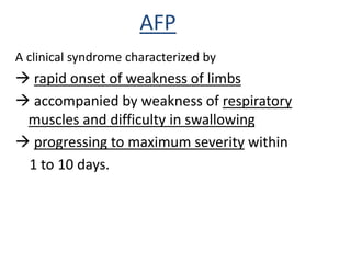
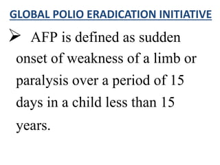
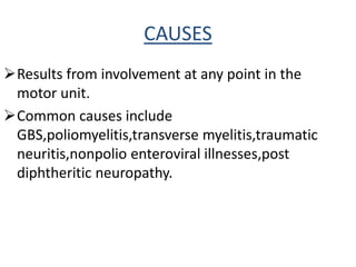
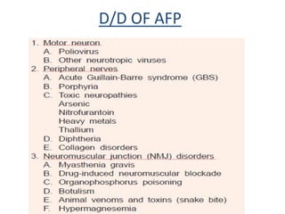
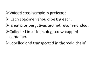
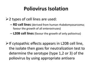
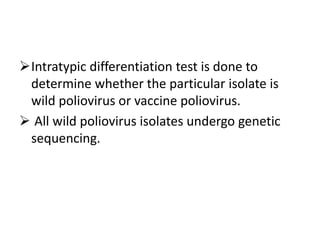
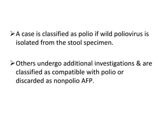
AFP AND SURVEILLANCE
AFP
A clinical syndrome characterized by
 rapid onset of weakness of limbs
 accompanied by weakness of respiratory
muscles and difficulty in swallowing
 progressing to maximum severity within
1 to 10 days.
 AFP is defined as sudden
onset of weakness of a limb or
paralysis over a period of 15
days in a child less than 15
years.
GLOBAL POLIO ERADICATION INITIATIVE
CAUSES
Results from involvement at any point in the
motor unit.
Common causes include
GBS,poliomyelitis,transverse myelitis,traumatic
neuritis,nonpolio enteroviral illnesses,post
diphtheritic neuropathy.
D/D OF AFP
POLIOMYELITIS
 Highly infectious enteroviral disease.
 Virus transmitted through faeco-oral route 
multiplies in the intestine  invades the nervous
system  causes paralysis.
 Age at onset <5 years.
 Presents as acute febrile viral meningitis syndrome
with meningeal signs,headache,malaise,GI symptoms.
 Motor signs develop within 1 to 4 days
-start with severe myalgias.
-asymmetric.
-lumbar involvement > cervical.
-spinal cord involvement > brainstem.
-no sensory deficit.
-paralysis within 1 – 2 weeks.
-atrophy appears rapidly,
progresses over several weeks.
Diagnosis by isolation of pathogen from
stools.
CSF – neutrophilic pleocytosis
lymphocytes and monocytes
Protein content raises over time,becomes
normal by 6 weeks.
Electrophysiology studies to differentiate polio
from GBS (acute denervation,reduced compound
action potentials.)
MRI shows increased T2 weighted signal in
anterior horns and cord swelling.
Polio like illness due to non polio
viruses
Non-polio entero viruses like coxsackie
A7,enterovirus 71,japanese B virus,westnile
virus,tick borne encephalitis cause damage to
anterior horn cells resulting in AFP.
GUILLAIN-BARRE SYNDROME
 Immune attack (IgM,IgG,macrophages,complement
activation) directed at schwann cells,myelin
demyelination.
 Axonal injury is secondary to pathological events of
demyelination ( bystander injury )
 Synonymous with acute inflammatory
demyelinating polyneuropathy ( AIDP )
Two patterns of axonal involvement
- Involving both sensory and motor neurons
(acute motor-sensory axonal neuropathy)
- Involving only motor neurons
(acute motor axonal neuropathy)
Age at presentation - >3years .
M>F .
An antecedent infectious disease with
CMV,EBV,HIV,vaccinia virus,
campylobacter jejuni diarrhoea,
vaccinations precede AIDP.
(3days – 6 weeks before the onset of symptoms)
Motor + sensory symptoms
(hypo/hyperesthesia)+ ataxia.
Autonomic symptoms in 30%.
Present with pain in the back,thighs and legs
along with motor weakness starting in legs and
ascending upwards.
Maximal weakness within 2 weeks.
Areflexia/hyporeflexia.
Atonia/hypotonia.
CSF shows albumino-cytological dissociation.
Electrophysiology shows demyelination of
peripheral nerves.
Slow or block of Nerve conduction velocity.
MRI is normal.
Clinical variants
1–Polyneuritis cranialis
Cranial nerve involvement
2–Miller fisher syndrome
Ophthalmoplegia, ataxia, areflexia
3–Chronic progressive GBS
Symptoms persisting more than 6 weeks
4- Chronic relapsing GB
Differentiation from spinal cord
syndrome
Absence of sensory level
Lack of spinal tenderness
Normal bowel and bladder function
Treatment
 IVIG given (2g/kg/day over 2 days).
-As early as the diagnosis is made.
-A/E : mild flu like symptoms,nausea,headache,malaise.
 Plasmapheresis is equally effective.
- 5 exchanges of 50 ml plasma/ kg on alternate days (10
days course).
-Cannot be done in patients with cardiovascular
compromise.
 Trial of immunosuppressives,corticosteroids.
 Supportive treatment and treatment of complications.
Outcome
• Regressive : 90 % of patients make a good
recovery
Recovery begins 2 to 4 weeks after
progression stops starting from the last
muscles affected till lower limb ( descending
pattern .)
• Chronic Relapsing: Less than 5% of patients.
• Mortality: 3% die from complications.
TRANSVERSE MYELITIS
Caused by inflammation of the spinal cord.
In 60% unknown
In 40% , associated with autoimmune
disorders such as:
– multiple sclerosis
– neuromyelitis optica
– systemic lupus erythematous
– Sjogren’s syndrome
– sarcoidosis
In children <3 years and between 5-14 years.
H/O vaccination with OPV,MMR,Hep
A,DPT,influenza,varicella,Jap B,HiB in the
preceding month.
Earliest symptom is sensory loss or pain in the
back,thighs,legs.
Sudden onset of progressive weakness of legs.
Ascends leading to flaccid quadriplegia.
Bowel bladder involvement in 70% .
 Maximal weakness by 48hrs.
85% showed a sensory level.
Cranial nerves not involved.
Normal EMG,NCV.
MRI shows focal areas of >> T2 signals in the
cord.
Elevated wbc’s , high IgG index in CSF.
Diagnostic criteria
Treatment
Methyl prednisolone (1g/1.73 sq m) iv daily
for a period of 3-5days
Oral prednisolone tapered over 2-3 weeks.
IVIG,immunosuppressants in non responders.
Outcome
 Residual disabilities of
gait,numbness,bladder dysfunction persist in
40-75% of the cases even after many years.
 Better outcome if early time of diagnosis,
lower anatomic level,
involvement of only few spinal segments,
older age at onset,
lack of leucocytes in CSF.
TRAUMATIC NEURITIS
• Inflammation of a nerve following an injury.
• Sciatic nerve injury leading to paralysis of the
foot and permanent sequelae following
intramuscular injection in the gluteal region is
common.
POST DIPHTHERITIC POLYNEUROPATHY
 Vaccine preventable illness caused by
exotoxin producing strains of
Corynebacterium diphtheriae.
Initial symptoms of low grade fever,sore
throat,neck swelling,nasal twang,ipsilateral
palatal paralysis.
Polyneuropathy occurs as a complication in
20% of patients.
Due to higher release of exotoxin.
The time between initial symptoms and onset
of polyneuropathy is termed as latency
period.
Classic features include acute flaccid
paralysis,reduced/absent DTR,sensory
symptoms.
Onset of paralysis is 4 – 6 weeks after the
onset of bulbar signs and symptoms.
Low fatality rate.
Paralysis resolves with time.
Early administration of antitoxin(im/iv)
neutralizes the exotoxin.
AFP SURVEILLANCE
Poliomyelitis is targeted for eradication.
Highly sensitive surveillance including
immediate case investigation and specimen
collection are critical for the detection of wild
poliovirus circulation and for documenting the
absence of poliovirus circulation for polio-free
certification.
 All patients with acute flaccid paralysis should be reported to
Surveillance Medical Officer of World Health Organization.
 Every case of AFP within the last 6 months has to be reported.
 Additionally, other conditions which need notification are:
• Isolated facial palsy
• Isolated bulbar palsy
• Unproved hypokalemia
• Neck flop
• Floppy baby
• Flaccid hemiplegia
• Encephalitis
• Postictal weakness (Todd’s paralysis)
• Postdiphretic polyneuritis.
Cases are investigated
immediately, usually
within 48 hrs of
notification, by a trained
medical officer
-Detailed medical history,
examination & investigations are
done
- Collection & transportation of
stool specimens
- Search for additional cases &
outbreak response in affected
community
- 60 days follow up examination
- Analysis of laboratory results
- Case classification
AfterconfirmingthecasesasAFP
Stool Specimen Collection
For every cases of AFP,
2 stool specimens are collected (at least 24
hours apart)
Ideally within 14 days of onset of paralysis
(optimal time period for detection of polio virus)
Should also be collected in any late-reported AFP
case upto 60 days from the day of onset of
paralysis.
Voided stool sample is preferred.
 Each specimen should be 8 g each.
 Enema or purgatives are not recommended.
Collected in a clean, dry, screw-capped
container.
Labelled and transported in the ‘cold chain’
Poliovirus Isolation
2 types of cell lines are used:
– RD cell lines (derived from human rhabdomyosarcoma;
favour the growth of all enteroviruses)
– L20B cell lines (favour the growth of only poliovirus)
If cytopathic effects appears in L20B cell line,
the isolate then goes for neutralization test to
determine the serotype (type 1,2 or 3) of the
poliovirus by using appropriate antisera
Intratypic differentiation test is done to
determine whether the particular isolate is
wild poliovirus or vaccine poliovirus.
 All wild poliovirus isolates undergo genetic
sequencing.
A case is classified as polio if wild poliovirus is
isolated from the stool specimen.
Others undergo additional investigations & are
classified as compatible with polio or
discarded as nonpolio AFP.
THANK YOU !

acute flaccid paralysis and surveillance

  • 1.
  • 2.
    AFP A clinical syndromecharacterized by  rapid onset of weakness of limbs  accompanied by weakness of respiratory muscles and difficulty in swallowing  progressing to maximum severity within 1 to 10 days.
  • 3.
     AFP isdefined as sudden onset of weakness of a limb or paralysis over a period of 15 days in a child less than 15 years. GLOBAL POLIO ERADICATION INITIATIVE
  • 4.
    CAUSES Results from involvementat any point in the motor unit. Common causes include GBS,poliomyelitis,transverse myelitis,traumatic neuritis,nonpolio enteroviral illnesses,post diphtheritic neuropathy.
  • 5.
  • 7.
    POLIOMYELITIS  Highly infectiousenteroviral disease.  Virus transmitted through faeco-oral route  multiplies in the intestine  invades the nervous system  causes paralysis.  Age at onset <5 years.  Presents as acute febrile viral meningitis syndrome with meningeal signs,headache,malaise,GI symptoms.
  • 8.
     Motor signsdevelop within 1 to 4 days -start with severe myalgias. -asymmetric. -lumbar involvement > cervical. -spinal cord involvement > brainstem. -no sensory deficit. -paralysis within 1 – 2 weeks. -atrophy appears rapidly, progresses over several weeks.
  • 9.
    Diagnosis by isolationof pathogen from stools. CSF – neutrophilic pleocytosis lymphocytes and monocytes Protein content raises over time,becomes normal by 6 weeks.
  • 10.
    Electrophysiology studies todifferentiate polio from GBS (acute denervation,reduced compound action potentials.) MRI shows increased T2 weighted signal in anterior horns and cord swelling.
  • 11.
    Polio like illnessdue to non polio viruses Non-polio entero viruses like coxsackie A7,enterovirus 71,japanese B virus,westnile virus,tick borne encephalitis cause damage to anterior horn cells resulting in AFP.
  • 12.
    GUILLAIN-BARRE SYNDROME  Immuneattack (IgM,IgG,macrophages,complement activation) directed at schwann cells,myelin demyelination.  Axonal injury is secondary to pathological events of demyelination ( bystander injury )  Synonymous with acute inflammatory demyelinating polyneuropathy ( AIDP )
  • 13.
    Two patterns ofaxonal involvement - Involving both sensory and motor neurons (acute motor-sensory axonal neuropathy) - Involving only motor neurons (acute motor axonal neuropathy)
  • 14.
    Age at presentation- >3years . M>F . An antecedent infectious disease with CMV,EBV,HIV,vaccinia virus, campylobacter jejuni diarrhoea, vaccinations precede AIDP. (3days – 6 weeks before the onset of symptoms)
  • 15.
    Motor + sensorysymptoms (hypo/hyperesthesia)+ ataxia. Autonomic symptoms in 30%. Present with pain in the back,thighs and legs along with motor weakness starting in legs and ascending upwards. Maximal weakness within 2 weeks. Areflexia/hyporeflexia. Atonia/hypotonia.
  • 17.
    CSF shows albumino-cytologicaldissociation. Electrophysiology shows demyelination of peripheral nerves. Slow or block of Nerve conduction velocity. MRI is normal.
  • 18.
    Clinical variants 1–Polyneuritis cranialis Cranialnerve involvement 2–Miller fisher syndrome Ophthalmoplegia, ataxia, areflexia 3–Chronic progressive GBS Symptoms persisting more than 6 weeks 4- Chronic relapsing GB
  • 19.
    Differentiation from spinalcord syndrome Absence of sensory level Lack of spinal tenderness Normal bowel and bladder function
  • 20.
    Treatment  IVIG given(2g/kg/day over 2 days). -As early as the diagnosis is made. -A/E : mild flu like symptoms,nausea,headache,malaise.  Plasmapheresis is equally effective. - 5 exchanges of 50 ml plasma/ kg on alternate days (10 days course). -Cannot be done in patients with cardiovascular compromise.  Trial of immunosuppressives,corticosteroids.  Supportive treatment and treatment of complications.
  • 22.
    Outcome • Regressive :90 % of patients make a good recovery Recovery begins 2 to 4 weeks after progression stops starting from the last muscles affected till lower limb ( descending pattern .) • Chronic Relapsing: Less than 5% of patients. • Mortality: 3% die from complications.
  • 23.
    TRANSVERSE MYELITIS Caused byinflammation of the spinal cord. In 60% unknown In 40% , associated with autoimmune disorders such as: – multiple sclerosis – neuromyelitis optica – systemic lupus erythematous – Sjogren’s syndrome – sarcoidosis
  • 25.
    In children <3years and between 5-14 years. H/O vaccination with OPV,MMR,Hep A,DPT,influenza,varicella,Jap B,HiB in the preceding month. Earliest symptom is sensory loss or pain in the back,thighs,legs. Sudden onset of progressive weakness of legs. Ascends leading to flaccid quadriplegia. Bowel bladder involvement in 70% .
  • 26.
     Maximal weaknessby 48hrs. 85% showed a sensory level. Cranial nerves not involved. Normal EMG,NCV. MRI shows focal areas of >> T2 signals in the cord. Elevated wbc’s , high IgG index in CSF.
  • 27.
  • 28.
    Treatment Methyl prednisolone (1g/1.73sq m) iv daily for a period of 3-5days Oral prednisolone tapered over 2-3 weeks. IVIG,immunosuppressants in non responders.
  • 29.
    Outcome  Residual disabilitiesof gait,numbness,bladder dysfunction persist in 40-75% of the cases even after many years.  Better outcome if early time of diagnosis, lower anatomic level, involvement of only few spinal segments, older age at onset, lack of leucocytes in CSF.
  • 30.
    TRAUMATIC NEURITIS • Inflammationof a nerve following an injury. • Sciatic nerve injury leading to paralysis of the foot and permanent sequelae following intramuscular injection in the gluteal region is common.
  • 31.
    POST DIPHTHERITIC POLYNEUROPATHY Vaccine preventable illness caused by exotoxin producing strains of Corynebacterium diphtheriae. Initial symptoms of low grade fever,sore throat,neck swelling,nasal twang,ipsilateral palatal paralysis. Polyneuropathy occurs as a complication in 20% of patients. Due to higher release of exotoxin.
  • 32.
    The time betweeninitial symptoms and onset of polyneuropathy is termed as latency period. Classic features include acute flaccid paralysis,reduced/absent DTR,sensory symptoms. Onset of paralysis is 4 – 6 weeks after the onset of bulbar signs and symptoms.
  • 33.
    Low fatality rate. Paralysisresolves with time. Early administration of antitoxin(im/iv) neutralizes the exotoxin.
  • 34.
    AFP SURVEILLANCE Poliomyelitis istargeted for eradication. Highly sensitive surveillance including immediate case investigation and specimen collection are critical for the detection of wild poliovirus circulation and for documenting the absence of poliovirus circulation for polio-free certification.
  • 35.
     All patientswith acute flaccid paralysis should be reported to Surveillance Medical Officer of World Health Organization.  Every case of AFP within the last 6 months has to be reported.  Additionally, other conditions which need notification are: • Isolated facial palsy • Isolated bulbar palsy • Unproved hypokalemia • Neck flop • Floppy baby • Flaccid hemiplegia • Encephalitis • Postictal weakness (Todd’s paralysis) • Postdiphretic polyneuritis.
  • 36.
    Cases are investigated immediately,usually within 48 hrs of notification, by a trained medical officer -Detailed medical history, examination & investigations are done - Collection & transportation of stool specimens - Search for additional cases & outbreak response in affected community - 60 days follow up examination - Analysis of laboratory results - Case classification AfterconfirmingthecasesasAFP
  • 37.
    Stool Specimen Collection Forevery cases of AFP, 2 stool specimens are collected (at least 24 hours apart) Ideally within 14 days of onset of paralysis (optimal time period for detection of polio virus) Should also be collected in any late-reported AFP case upto 60 days from the day of onset of paralysis.
  • 38.
    Voided stool sampleis preferred.  Each specimen should be 8 g each.  Enema or purgatives are not recommended. Collected in a clean, dry, screw-capped container. Labelled and transported in the ‘cold chain’
  • 39.
    Poliovirus Isolation 2 typesof cell lines are used: – RD cell lines (derived from human rhabdomyosarcoma; favour the growth of all enteroviruses) – L20B cell lines (favour the growth of only poliovirus) If cytopathic effects appears in L20B cell line, the isolate then goes for neutralization test to determine the serotype (type 1,2 or 3) of the poliovirus by using appropriate antisera
  • 40.
    Intratypic differentiation testis done to determine whether the particular isolate is wild poliovirus or vaccine poliovirus.  All wild poliovirus isolates undergo genetic sequencing.
  • 41.
    A case isclassified as polio if wild poliovirus is isolated from the stool specimen. Others undergo additional investigations & are classified as compatible with polio or discarded as nonpolio AFP.
  • 42.

Editor's Notes

  • #13 aidp
  • #16 Labile htn arrythmias bowelbladder gi motility disorders
  • #21 Parenteral nutrition,bed sores,physiotherapy,nsaids for pain,prevent dvt
  • #22 Vital capacity