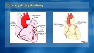
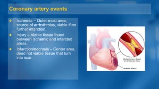
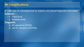
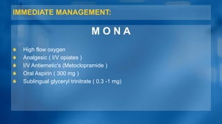
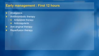
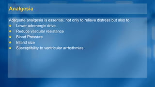
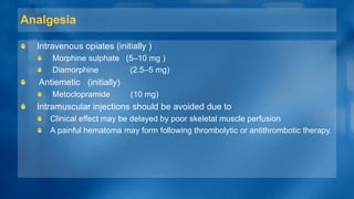
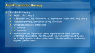
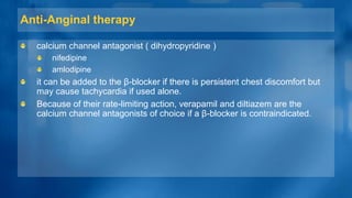
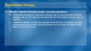
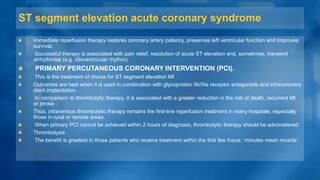
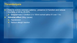
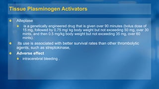
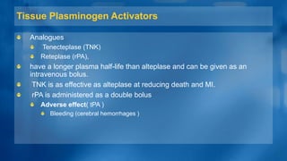
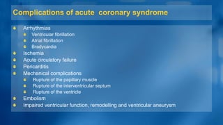
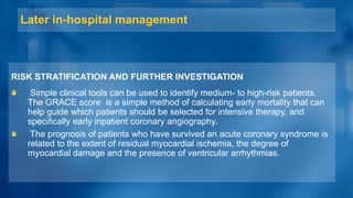
DEPARTMENT OF MEDICINE
MUHAMMAD MEDICAL COLLEGE
MYOCARDIAL INFARCTION
PRESENTED TO:
Prof. Dr. Muhammad Ali
(Dept. of Medicine Muhammad
Medical college)
PRESENTED BY:
o Khalid Jamal
o Muhammad Nouman
o Amir Ali
o Muhammad Ihsan
o Muhammad Idrees
o Anza Tariq
o Natasha Maryam
o Namra Jabeen
ACUTE CORONARY SYNDROME
Acute coronary syndrome includes
 ST-elevation myocardial infarction (STEMI)
 Non- ST-elevation myocardial infarction (NSTEMI)
 Unstable Angina (UA)
The difference UA and STEMI is that in the latter there is occluding
thrombus which leads to myocardial necrosis and rise in serum troponin or
CK-MB.
Myocardial infarction when cardiac myocytes die due to myocardial
ischemia and can be diagnose on the basis of appropriate clinical history,
12 leads ECG and elevated biochemical marker-troponin I and T, CK-MB
MYOCARDIAL INFARCTION
MI is almost due to formation of occlusive thrombus at the site of rupture or
erosion of a atheromatous plaque in a coronary artery most patient present
when it is still possible to salvage myocardium and improve outcome.
Types
 Types 1- spontaneous MI with ischemia due to a primary coronary event, e.g.
plaque, erosion/rupture, fissuring or dissection
 Type 2 - MI secondary to ischemia due to increased oxygen demand or
decreased supply.
 Type 3,4,5 - Diagnosis of MI in sudden cardiac death, after percutaneous
coronary intervention(PCI) and after coronary artery bypass graft (CABG)
Respectively
Coronary Artery Anatomy
Coronary artery events
Ischemia – Outer most area,
source of arrhythmias, viable if no
further infarction.
Injury – Viable tissue found
between ischemic and infarcted
areas.
Infarction/necrosis – Center area,
dead not viable tissue that turn
into scar.
MI Classifications
MI’s can be subcategorized by anatomy and clinical diagnostic information.
Anatomic
Trans mural
Subendocardial
Diagnostic
ST- elevations (STEMI)
non ST- elevations (NSTEMI).
INTRODUCTION (CONTINUE)
Criteria for acute myocardial infarction
The term acute myocardial infarction (MI) should be used when there is
evidence of myocardial necrosis in the clinical setting consistent with acute
myocardial ischemia. Under these conditions, any one of the following criteria
meets for the diagnosis of MI:
Detection of a rise and/or fall of cardiac biomarker values
(preferably cardiac troponin (cTn)), with at least one value above the 99th
centile upper reference limit (URL) and with at least one of the following:
1. Symptoms of ischemia
2. New or presumed new significant ST segment-T wave (ST-T) changes or
new left bundle branch block (LBBB)
3. Development of pathological Q waves in the ECG
4. Imaging evidence of new loss of viable myocardium or new regional wall
motion abnormality
5. Identification of an intracoronary thrombus by angiography or post-mortem
CRITERIA FOR PRIOR MYOCARDIAL INFARCTION
Any of the following criteria meets the diagnosis for prior MI:
Pathological Q waves with or without symptoms in the absence of non-
ischemic causes
Imaging evidence of a region of loss of viable myocardium that is thinned
and fails to contract, in the absence of a non-ischemic cause
Risk Factors
The presence of any risk factor is
associated with doubling the risk
of an MI.
Non Modifiable
Age
Gender
Family history
Risk Factors (cont.…)
Modifiable
Smoking
Diabetes Control
Hypertension
Hyperlipidemia
Obesity
Physical Inactivity
Smoking
Tobacco use increases the risk of
coronary artery disease two to six
times more than non smokers.
Nicotine increases platelet
thrombus adhesion and vessel
inflammation.
Diabetes & Hypertension
Diabetes not only increases the
rate of atherosclerotic formation in
vascular vessels but also at an
earlier age.
The constant stress of high blood
pressure has been associated
with the increased rate of plaque
formation.
Shearing Stress and inflammation
of endothelial lining begins the
process.
Hyperlipidemia
Elevated levels of cholesterol,
LDL’s or triglycerides are
associated with the increased risk
of coronary plaque formation and
MI.
Almost 50% of the U.S.
population has some
form of dyslipidemia.
Obesity and Physical Inactivity
Mortality rate from CAD is higher
in those who are obese.
Some evidence shows that those
who carry their weight in their
abdomen have a higher incidence
of CAD
Physically inactive people have
lower HDL levels with higher LDL
levels and an increase in clot
formation.
Pathophysiology
Ischemia develops when there is an increased demand for oxygen or a
decreased supply of oxygen.
Ischemia can develop within 10 seconds and if it lasts longer than 20
minutes, irreversible cell and tissue death occurs.
Myocardial cell death begins at the endocardium.
The area most distal to the arterial blood supply.
Pathophysiology (cont.…)
As vessel occlusion continues cell death spreads to the myocardium and
eventually to the epicardium.
Severity of the MI depends on three factors.
Level of occlusion
Length of time of occlusion
Presence or absence of collateral circulation
Pathophysiology (cont.…)
CLINICAL FEATURES
SYMPTOMS
Prolonged cardiac pain:
chest, throat. arm, epigastric
or back
Anxiety and fear of
impending death
Nausea and vomiting
Breathlessness
Collapse/syncope
CLINICAL FEATURES (CONTINUE…)
PHYSICAL SIGNS
Sign of sympathetic
activation:
• Pallor,
• Sweating,
• Tachycardia
Sign of vagal activation:
• Vomiting,
• Bradycardia
CLINICAL FEATURES (CONTINUE…)
PHYSICAL SIGNS
Sign of impaired myocardial
function
Hypotension, oliguria, cold
peripheries
Narrow pulse pressure
Raised JVP
Third heart sound
Quiet first heart sound
Diffuse apical impulse
Lung crepitation
CLINICAL FEATURES (CONTINUE…)
PHYSICAL SIGNS
Sign of tissue damage:
• Fever
Sign of complication: e.g.
• Mitral regurgitation,
• Pericarditis
INVESTIGATIONS
Electrocardiography
Show a characteristic series of changes
The earliest change is usually ST elevation followed by
diminution in the Size of the R wave, and development of a Q
wave (indicating full thickness Infarction).
Subsequently, the T wave becomes inverted and this change
persists after the ST segment has returned to normal.
ECG changes are best seen in the lead that ‘face’ the infarcted
area.
INVESTIGATIONS
Electrocardiography
With anteroseptal Infarction, abnormalities are found in
one or more leads from V1 to V4.
Anterolateral infarction produces changes from V4 to
V6, in aVL and lead 1.
Inferior infarction is best shown in leads II, III and aVF.
Anterolateral infarction produces changes from V4 to V6,
in aVL and lead 1.
INVESTIGATIONS (cont.…)
Electrocardiography
Inferior infarction is best shown in leads II, III and aVF.
Infarction of the posterior wall of the left ventricle does
not cause ST elevation or Q waves in the standard
leads, but can be diagnosed by the presence of
reciprocal changes (ST depression and a tall R wave in
leads V1-V4).
Occasionally, new onset LBBB is the only CCG change.
INVESTIGATION (Continue)
Plasma cardiac biomarkers
The plasma concentration of enzymes and proteins normally
concentrated within cardiac cells is increased in MI.
Creatine kinase (CK) and CK-MB (a cardiaospecific isoform)
start to rise at 4-6 hrs.
Peak at about 12hrs and fall to normal with in 48-74hrs the most
sensitive marker of myocardial cell damage are the cardiac
troponins T and I.
Which are released within 4-6hrs and remain elevated for up to
2 wks.
Investigation (cont.…)
Other blood tests
A leukocytosis is usual, reaching a peak on the first day.
The erythrocyte sedimentation rate (ESR) and C-reactive protein (CRP) are
also elevated.
Chest X-ray
This may demonstrate pulmonary edema that is not evident on clinical
examination.
The heart size is often normal but there may be cardiomegaly due to pre-
existing myocardial damage.
Investigation (cont.…)
Echocardiography
This is useful for assessing ventricular function and for detecting important
complications, such as
 Mural thrombus,
 Cardiac rupture,
 Ventricular septal defect,
 Mitral regurgitation &
 Pericardial effusion.
MANAGEMENT OF
MYOCARDIAL INFARCTION
PRESENTED BY
o Mirza Sikander Baig
o Simra Arshad
o Samra Jabeen
o Rana farrukh Irshad
o Mamoona
o Erum Sharif
o Mohsin Abbas
o Anum Ashraf
o Sidra
IMMEDIATE MANAGEMENT:
M O N A
High flow oxygen
Analgesic ( I/V opiates )
I/V Antiemetic's (Metoclopramide )
Oral Aspirin ( 300 mg )
Sublingual glyceryl trinitrate ( 0.3 -1 mg)
Early management : First 12 hours
Analgesics
Antithrombotic therapy
Antiplatelet therapy
Anticoagulants
Anti-anginal therapy
Reperfusion therapy
Analgesia
Adequate analgesia is essential, not only to relieve distress but also to
Lower adrenergic drive
Reduce vascular resistance
Blood Pressure
Infarct size
Susceptibility to ventricular arrhythmias.
Analgesia
Intravenous opiates (initially )
Morphine sulphate (5–10 mg )
Diamorphine (2.5–5 mg)
Antiemetic (initially)
Metoclopramide (10 mg)
Intramuscular injections should be avoided due to
Clinical effect may be delayed by poor skeletal muscle perfusion
A painful hematoma may form following thrombolytic or antithrombotic therapy.
Anti-Thrombotic therapy
Antiplatelet therapy
Aspirin (75–325 mg )
Clopidogrel (600 mg, followed by 150 mg daily for 1 week and 75 mg daily )
Ticagrelor (180 mg, followed by 90 mg twice daily)
Glycoprotein receptor antagonists
Tirofiban
Abciximab
(Glycoprotein are of particular benefit in patients with acute coronary
syndromes who undergo PCI , those with recurrent ischemia and those at
particularly high risk, such as patients with diabetes mellitus or an elevated
troponin concentration.)
Anticoagulants
Anticoagulation reduces the risk of thromboembolic complications, and
prevents re-infarction in the absence of reperfusion therapy or after
successful thrombolysis
Unfractionated heparin,
Fractioned (low-molecular weight) heparin
Pent saccharide.
Pent saccharides (s/c fondaparinux 2.5 mg daily) have the best safety and
efficacy profile, with compare to low-molecular-weight heparin (s/c
enoxaparin 1 mg/kg twice daily) being a reasonable alternative
Anticoagulants (cont.…)
A period of treatment with warfarin should be considered if there is
persistent
Atrial fibrillation
Evidence of extensive anterior infarction,
If echocardiography shows mobile mural thrombus,
( because these patients are at increased risk of systemic thromboembolism. )
Anti-Anginal therapy
Sublingual glyceryl trinitrate (300–500 µg)
intravenous nitrates
Glyceryl trinitrate 0.6–1.2 mg/hr
Isosorbide dinitrate 1–2 mg/hr
Sublingual glyceryl trinitrate is a valuable first-aid measure in
Unstable angina
Threatened infarction
Intravenous nitrates are useful for the treatment of
left ventricular failure
The relief of recurrent or persistent ischemic pain.
Anti-Anginal therapy (cont.…..)
Intravenous β-blockers
• Atenolol (5–10 mg)
• Metoprolo ( 5–15 mg given over 5 mints)
o Relieve pain, reduce arrhythmias and improve short-term mortality in
patients who present within 12 hours of the onset of symptoms .
o Contraindicated in heart failure (pulmonary edema), hypotension (systolic
BP < 105 mmHg) or bradycardia (heart rate < 65/min).
Anti-Anginal therapy
calcium channel antagonist ( dihydropyridine )
nifedipine
amlodipine
it can be added to the β-blocker if there is persistent chest discomfort but
may cause tachycardia if used alone.
Because of their rate-limiting action, verapamil and diltiazem are the
calcium channel antagonists of choice if a β-blocker is contraindicated.
Reperfusion therapy
Non-ST segment elevation acute coronary syndrome
Immediate emergency reperfusion therapy has no demonstrable benefit in
patients with non-ST segment elevation MI and thrombolytic therapy may be
harmful.
Selected medium- to high-risk patients do benefit from in-hospital coronary
angiography and coronary revascularization but this does not need to take
place in the first 12 hours.
ST segment elevation acute coronary syndrome
Immediate reperfusion therapy restores coronary artery patency, preserves left ventricular function and improves
survival.
Successful therapy is associated with pain relief, resolution of acute ST elevation and, sometimes, transient
arrhythmias (e.g. idioventricular rhythm).
PRIMARY PERCUTANEOUS CORONARY INTERVENTION (PCI).
This is the treatment of choice for ST segment elevation MI
Outcomes are best when it is used in combination with glycoprotein IIb/IIIa receptor antagonists and intracoronary
stent implantation.
In comparison to thrombolytic therapy, it is associated with a greater reduction in the risk of death, recurrent MI
or stroke .
Thus, intravenous thrombolytic therapy remains the first-line reperfusion treatment in many hospitals, especially
those in rural or remote areas.
When primary PCI cannot be achieved within 2 hours of diagnosis, thrombolytic therapy should be administered.
Thrombolysis .
The benefit is greatest in those patients who receive treatment within the first few hours: ‘minutes mean muscle’.
Thrombolysis
Help to restore coronary patency , preserve LV function and reduce
mortality of MI by 25-50% .
Streptokinase (1.5million U in 100ml normal saline I/V over 1 hr)
Adverse effect (May cause)
Hypotension
Serious allergic reaction
Tissue Plasminogen Activators
Alteplase
is a genetically engineered drug that is given over 90 minutes (bolus dose of
15 mg, followed by 0.75 mg/ kg body weight but not exceeding 50 mg, over 30
mints, and then 0.5 mg/kg body weight but not exceeding 35 mg, over 60
mints).
Its use is associated with better survival rates than other thrombolytic
agents, such as streptokinase,
Adverse effect
intracerebral bleeding .
Tissue Plasminogen Activators
Analogues
Tenecteplase (TNK)
Reteplase (rPA),
have a longer plasma half-life than alteplase and can be given as an
intravenous bolus.
TNK is as effective as alteplase at reducing death and MI.
rPA is administered as a double bolus
Adverse effect( tPA )
Bleeding (cerebral hemorrhages )
Complications of acute coronary syndrome
Arrhythmias
Ventricular fibrillation
Atrial fibrillation
Bradycardia
Ischemia
Acute circulatory failure
Pericarditis
Mechanical complications
Rupture of the papillary muscle
Rupture of the interventricular septum
Rupture of the ventricle
Embolism
Impaired ventricular function, remodelling and ventricular aneurysm
Later in-hospital management
RISK STRATIFICATION AND FURTHER INVESTIGATION
Simple clinical tools can be used to identify medium- to high-risk patients.
The GRACE score is a simple method of calculating early mortality that can
help guide which patients should be selected for intensive therapy, and
specifically early inpatient coronary angiography.
The prognosis of patients who have survived an acute coronary syndrome is
related to the extent of residual myocardial ischemia, the degree of
myocardial damage and the presence of ventricular arrhythmias.
LEFT VENTRICULAR FUNCTION
The degree of left ventricular dysfunction can be crudely assessed from
physical findings
Tachycardia,
Third heart sound,
Crackles at the lung bases,
Elevated venous pressure a,
ECG changes
chest X-ray (size of the heart and presence of pulmonary edema).
Echocardiography
Early ischemia
Acute coronary
syndrome
Angiography
Low risk patients
ETT
ETT Positive ETT Negative
15-25% risk
(12 months)
1-4% risk
(12
months)
ischaemia
Arrhythmias
The presence of ventricular arrhythmias during the convalescent phase of
acute coronary syndrome may be a marker of poor ventricular function and
may herald sudden death.
Although empirical anti-arrhythmic treatment is of no value and is even
hazardous, selected patients may benefit from electrophysiological testing and
specific anti-arrhythmic therapy (including implantable cardiac defibrillators.
Lifestyle and risk factor modification
Smoking
The 5-year mortality of patients who continue to smoke cigarettes is double
that of those who quit smoking at the time of their acute coronary
syndrome.
Giving up smoking is the single most effective contribution a patient can
make to his or her future.
The success of smoking cessation can be increased by supportive advice
and pharmacological therapy
Hyperlipidemia
The importance of lowering serum cholesterol following acute coronary
syndrome has been demonstrated in large-scale randomized trials.
Irrespective of serum cholesterol concentrations, all patients should receive
statin therapy after acute coronary syndrome, but those with serum LDL
cholesterol concentrations above 3.2 mmol/L (~120 mg/dL) benefit from
more intensive therapy, such as atorvastatin 80 mg daily.
Other risk factors
Maintaining an ideal
Body weight,
Eating a Mediterranean-style diet,
Taking regular exercise,
Achieving good control of hypertension
Diabetes mellitus
Secondary prevention drug therapy
Aspirin and clopidogrel
Low-dose aspirin therapy (approximately 25%)
Clopidogrel should be given in combination with aspirin for at least 3
months.
If patients are intolerant of long-term aspirin, clopidogrel is a suitable
alternative.
Secondary prevention drug therapy (continue)
Beta-blockers
Continuous treatment with an oral β-blocker reduces long-term mortality by
approximately 25% among the survivors of acute MI
Unfortunately, a minority of patients do not tolerate β-blockers because of
bradycardia, AV block, hypotension or asthma.
The secondary preventative role of β-blockers in patients with unstable
angina is unknown.
Secondary prevention drug therapy (continue)
ACE inhibitors
Several clinical trials have shown that long-term treatment with an ACE inhibitor
(e.g. enalapril 10 mg twice daily or ramipril 2.5–5 mg twice daily) can counteract
ventricular remodelling, prevent the onset of heart failure, improve survival, reduce
recurrent MI and avoid rehospitalisation.
introduction of an ACE inhibitor may exacerbate
Hypotension
Impair coronary perfusion.
In patients intolerant of ACE inhibitors, angiotensin receptor blockers (e.g.
valsartan 40–160 mg twice daily or candesartan 4–16 mg daily) are alternatives
and are better tolerated.
Patients with acute MI and left ventricular dysfunction (ejection fraction < 35%)
and either pulmonary edema or diabetes mellitus further benefit from additional
mineralocorticoid receptor antagonism (e.g. eplerenone 25–50 mg daily).
Secondary prevention drug therapy (continue)
Coronary Revascularization
Most low-risk patients stabilize with aspirin, clopidogrel, anticoagulation and
anti-anginal therapy, and can be rapidly mobilized.
In the absence of recurrent symptoms, low-risk patients do not benefit from
routine coronary angiography.
Coronary angiography should be considered with a view to
revascularization in all patients at moderate or high risk, including those
who fail to settle on medical therapy, those with extensive ECG changes,
those with an elevated plasma troponin and those with severe pre-existing
stable angina.
This often reveals disease that is amenable to PCI or urgent CABG.
In these cases, coronary revascularization is associated with short- and
long-term benefits, including reductions in MI and death.
Secondary prevention drug therapy (continue)
Device therapy
Implantable cardiac defibrillators are of benefit in preventing sudden
cardiac death in patients who have severe left ventricular impairment
(ejection fraction ≤ 30%) after MI .
Later in-hospital management
Prognosis
In almost one-quarter of all cases of MI, death occurs within a few minutes without
medical care.
Half the deaths occur within 24 hours of the onset of symptoms and about 40% of
all affected patients die within the first month.
The prognosis of those who survive to reach hospital is much better, with a 28-
day survival of more than 85%.
Patients with unstable angina have a mortality of approximately half that of those
patients with MI.
Early death is usually due to an arrhythmia and is independent of the extent of MI.
However, late outcomes are determined by the extent of myocardial damage, and
unfavorable features include poor left ventricular function, AV block and persistent
ventricular arrhythmias.
Prognosis (continue)
The prognosis is worse for anterior than for inferior infarcts.
Bundle branch block and high cardiac marker levels both indicate extensive
myocardial damage.
Old age, depression and social isolation are also associated with a higher
mortality.
Of those who survive an acute attack, more than 80% live for a further year,
about 75% for 5 years, 50% for 10 years and 25% for 20 years
Thank You…

myocardial infarction

  • 1.
    DEPARTMENT OF MEDICINE MUHAMMADMEDICAL COLLEGE MYOCARDIAL INFARCTION
  • 2.
    PRESENTED TO: Prof. Dr.Muhammad Ali (Dept. of Medicine Muhammad Medical college) PRESENTED BY: o Khalid Jamal o Muhammad Nouman o Amir Ali o Muhammad Ihsan o Muhammad Idrees o Anza Tariq o Natasha Maryam o Namra Jabeen
  • 3.
    ACUTE CORONARY SYNDROME Acutecoronary syndrome includes  ST-elevation myocardial infarction (STEMI)  Non- ST-elevation myocardial infarction (NSTEMI)  Unstable Angina (UA) The difference UA and STEMI is that in the latter there is occluding thrombus which leads to myocardial necrosis and rise in serum troponin or CK-MB. Myocardial infarction when cardiac myocytes die due to myocardial ischemia and can be diagnose on the basis of appropriate clinical history, 12 leads ECG and elevated biochemical marker-troponin I and T, CK-MB
  • 4.
    MYOCARDIAL INFARCTION MI isalmost due to formation of occlusive thrombus at the site of rupture or erosion of a atheromatous plaque in a coronary artery most patient present when it is still possible to salvage myocardium and improve outcome. Types  Types 1- spontaneous MI with ischemia due to a primary coronary event, e.g. plaque, erosion/rupture, fissuring or dissection  Type 2 - MI secondary to ischemia due to increased oxygen demand or decreased supply.  Type 3,4,5 - Diagnosis of MI in sudden cardiac death, after percutaneous coronary intervention(PCI) and after coronary artery bypass graft (CABG) Respectively
  • 5.
  • 6.
    Coronary artery events Ischemia– Outer most area, source of arrhythmias, viable if no further infarction. Injury – Viable tissue found between ischemic and infarcted areas. Infarction/necrosis – Center area, dead not viable tissue that turn into scar.
  • 7.
    MI Classifications MI’s canbe subcategorized by anatomy and clinical diagnostic information. Anatomic Trans mural Subendocardial Diagnostic ST- elevations (STEMI) non ST- elevations (NSTEMI).
  • 8.
  • 9.
    Criteria for acutemyocardial infarction The term acute myocardial infarction (MI) should be used when there is evidence of myocardial necrosis in the clinical setting consistent with acute myocardial ischemia. Under these conditions, any one of the following criteria meets for the diagnosis of MI: Detection of a rise and/or fall of cardiac biomarker values (preferably cardiac troponin (cTn)), with at least one value above the 99th centile upper reference limit (URL) and with at least one of the following: 1. Symptoms of ischemia 2. New or presumed new significant ST segment-T wave (ST-T) changes or new left bundle branch block (LBBB) 3. Development of pathological Q waves in the ECG 4. Imaging evidence of new loss of viable myocardium or new regional wall motion abnormality 5. Identification of an intracoronary thrombus by angiography or post-mortem
  • 10.
    CRITERIA FOR PRIORMYOCARDIAL INFARCTION Any of the following criteria meets the diagnosis for prior MI: Pathological Q waves with or without symptoms in the absence of non- ischemic causes Imaging evidence of a region of loss of viable myocardium that is thinned and fails to contract, in the absence of a non-ischemic cause
  • 11.
    Risk Factors The presenceof any risk factor is associated with doubling the risk of an MI. Non Modifiable Age Gender Family history
  • 12.
    Risk Factors (cont.…) Modifiable Smoking DiabetesControl Hypertension Hyperlipidemia Obesity Physical Inactivity
  • 13.
    Smoking Tobacco use increasesthe risk of coronary artery disease two to six times more than non smokers. Nicotine increases platelet thrombus adhesion and vessel inflammation.
  • 14.
    Diabetes & Hypertension Diabetesnot only increases the rate of atherosclerotic formation in vascular vessels but also at an earlier age. The constant stress of high blood pressure has been associated with the increased rate of plaque formation. Shearing Stress and inflammation of endothelial lining begins the process.
  • 15.
    Hyperlipidemia Elevated levels ofcholesterol, LDL’s or triglycerides are associated with the increased risk of coronary plaque formation and MI. Almost 50% of the U.S. population has some form of dyslipidemia.
  • 16.
    Obesity and PhysicalInactivity Mortality rate from CAD is higher in those who are obese. Some evidence shows that those who carry their weight in their abdomen have a higher incidence of CAD Physically inactive people have lower HDL levels with higher LDL levels and an increase in clot formation.
  • 17.
    Pathophysiology Ischemia develops whenthere is an increased demand for oxygen or a decreased supply of oxygen. Ischemia can develop within 10 seconds and if it lasts longer than 20 minutes, irreversible cell and tissue death occurs. Myocardial cell death begins at the endocardium. The area most distal to the arterial blood supply.
  • 18.
    Pathophysiology (cont.…) As vesselocclusion continues cell death spreads to the myocardium and eventually to the epicardium. Severity of the MI depends on three factors. Level of occlusion Length of time of occlusion Presence or absence of collateral circulation
  • 19.
  • 20.
    CLINICAL FEATURES SYMPTOMS Prolonged cardiacpain: chest, throat. arm, epigastric or back Anxiety and fear of impending death Nausea and vomiting Breathlessness Collapse/syncope
  • 21.
    CLINICAL FEATURES (CONTINUE…) PHYSICALSIGNS Sign of sympathetic activation: • Pallor, • Sweating, • Tachycardia Sign of vagal activation: • Vomiting, • Bradycardia
  • 22.
    CLINICAL FEATURES (CONTINUE…) PHYSICALSIGNS Sign of impaired myocardial function Hypotension, oliguria, cold peripheries Narrow pulse pressure Raised JVP Third heart sound Quiet first heart sound Diffuse apical impulse Lung crepitation
  • 23.
    CLINICAL FEATURES (CONTINUE…) PHYSICALSIGNS Sign of tissue damage: • Fever Sign of complication: e.g. • Mitral regurgitation, • Pericarditis
  • 24.
    INVESTIGATIONS Electrocardiography Show a characteristicseries of changes The earliest change is usually ST elevation followed by diminution in the Size of the R wave, and development of a Q wave (indicating full thickness Infarction). Subsequently, the T wave becomes inverted and this change persists after the ST segment has returned to normal. ECG changes are best seen in the lead that ‘face’ the infarcted area.
  • 25.
    INVESTIGATIONS Electrocardiography With anteroseptal Infarction,abnormalities are found in one or more leads from V1 to V4. Anterolateral infarction produces changes from V4 to V6, in aVL and lead 1. Inferior infarction is best shown in leads II, III and aVF. Anterolateral infarction produces changes from V4 to V6, in aVL and lead 1.
  • 26.
    INVESTIGATIONS (cont.…) Electrocardiography Inferior infarctionis best shown in leads II, III and aVF. Infarction of the posterior wall of the left ventricle does not cause ST elevation or Q waves in the standard leads, but can be diagnosed by the presence of reciprocal changes (ST depression and a tall R wave in leads V1-V4). Occasionally, new onset LBBB is the only CCG change.
  • 31.
    INVESTIGATION (Continue) Plasma cardiacbiomarkers The plasma concentration of enzymes and proteins normally concentrated within cardiac cells is increased in MI. Creatine kinase (CK) and CK-MB (a cardiaospecific isoform) start to rise at 4-6 hrs. Peak at about 12hrs and fall to normal with in 48-74hrs the most sensitive marker of myocardial cell damage are the cardiac troponins T and I. Which are released within 4-6hrs and remain elevated for up to 2 wks.
  • 33.
    Investigation (cont.…) Other bloodtests A leukocytosis is usual, reaching a peak on the first day. The erythrocyte sedimentation rate (ESR) and C-reactive protein (CRP) are also elevated. Chest X-ray This may demonstrate pulmonary edema that is not evident on clinical examination. The heart size is often normal but there may be cardiomegaly due to pre- existing myocardial damage.
  • 34.
    Investigation (cont.…) Echocardiography This isuseful for assessing ventricular function and for detecting important complications, such as  Mural thrombus,  Cardiac rupture,  Ventricular septal defect,  Mitral regurgitation &  Pericardial effusion.
  • 35.
  • 36.
    PRESENTED BY o MirzaSikander Baig o Simra Arshad o Samra Jabeen o Rana farrukh Irshad o Mamoona o Erum Sharif o Mohsin Abbas o Anum Ashraf o Sidra
  • 37.
    IMMEDIATE MANAGEMENT: M ON A High flow oxygen Analgesic ( I/V opiates ) I/V Antiemetic's (Metoclopramide ) Oral Aspirin ( 300 mg ) Sublingual glyceryl trinitrate ( 0.3 -1 mg)
  • 38.
    Early management :First 12 hours Analgesics Antithrombotic therapy Antiplatelet therapy Anticoagulants Anti-anginal therapy Reperfusion therapy
  • 39.
    Analgesia Adequate analgesia isessential, not only to relieve distress but also to Lower adrenergic drive Reduce vascular resistance Blood Pressure Infarct size Susceptibility to ventricular arrhythmias.
  • 40.
    Analgesia Intravenous opiates (initially) Morphine sulphate (5–10 mg ) Diamorphine (2.5–5 mg) Antiemetic (initially) Metoclopramide (10 mg) Intramuscular injections should be avoided due to Clinical effect may be delayed by poor skeletal muscle perfusion A painful hematoma may form following thrombolytic or antithrombotic therapy.
  • 41.
    Anti-Thrombotic therapy Antiplatelet therapy Aspirin(75–325 mg ) Clopidogrel (600 mg, followed by 150 mg daily for 1 week and 75 mg daily ) Ticagrelor (180 mg, followed by 90 mg twice daily) Glycoprotein receptor antagonists Tirofiban Abciximab (Glycoprotein are of particular benefit in patients with acute coronary syndromes who undergo PCI , those with recurrent ischemia and those at particularly high risk, such as patients with diabetes mellitus or an elevated troponin concentration.)
  • 42.
    Anticoagulants Anticoagulation reduces therisk of thromboembolic complications, and prevents re-infarction in the absence of reperfusion therapy or after successful thrombolysis Unfractionated heparin, Fractioned (low-molecular weight) heparin Pent saccharide. Pent saccharides (s/c fondaparinux 2.5 mg daily) have the best safety and efficacy profile, with compare to low-molecular-weight heparin (s/c enoxaparin 1 mg/kg twice daily) being a reasonable alternative
  • 43.
    Anticoagulants (cont.…) A periodof treatment with warfarin should be considered if there is persistent Atrial fibrillation Evidence of extensive anterior infarction, If echocardiography shows mobile mural thrombus, ( because these patients are at increased risk of systemic thromboembolism. )
  • 44.
    Anti-Anginal therapy Sublingual glyceryltrinitrate (300–500 µg) intravenous nitrates Glyceryl trinitrate 0.6–1.2 mg/hr Isosorbide dinitrate 1–2 mg/hr Sublingual glyceryl trinitrate is a valuable first-aid measure in Unstable angina Threatened infarction Intravenous nitrates are useful for the treatment of left ventricular failure The relief of recurrent or persistent ischemic pain.
  • 45.
    Anti-Anginal therapy (cont.…..) Intravenousβ-blockers • Atenolol (5–10 mg) • Metoprolo ( 5–15 mg given over 5 mints) o Relieve pain, reduce arrhythmias and improve short-term mortality in patients who present within 12 hours of the onset of symptoms . o Contraindicated in heart failure (pulmonary edema), hypotension (systolic BP < 105 mmHg) or bradycardia (heart rate < 65/min).
  • 46.
    Anti-Anginal therapy calcium channelantagonist ( dihydropyridine ) nifedipine amlodipine it can be added to the β-blocker if there is persistent chest discomfort but may cause tachycardia if used alone. Because of their rate-limiting action, verapamil and diltiazem are the calcium channel antagonists of choice if a β-blocker is contraindicated.
  • 47.
    Reperfusion therapy Non-ST segmentelevation acute coronary syndrome Immediate emergency reperfusion therapy has no demonstrable benefit in patients with non-ST segment elevation MI and thrombolytic therapy may be harmful. Selected medium- to high-risk patients do benefit from in-hospital coronary angiography and coronary revascularization but this does not need to take place in the first 12 hours.
  • 48.
    ST segment elevationacute coronary syndrome Immediate reperfusion therapy restores coronary artery patency, preserves left ventricular function and improves survival. Successful therapy is associated with pain relief, resolution of acute ST elevation and, sometimes, transient arrhythmias (e.g. idioventricular rhythm). PRIMARY PERCUTANEOUS CORONARY INTERVENTION (PCI). This is the treatment of choice for ST segment elevation MI Outcomes are best when it is used in combination with glycoprotein IIb/IIIa receptor antagonists and intracoronary stent implantation. In comparison to thrombolytic therapy, it is associated with a greater reduction in the risk of death, recurrent MI or stroke . Thus, intravenous thrombolytic therapy remains the first-line reperfusion treatment in many hospitals, especially those in rural or remote areas. When primary PCI cannot be achieved within 2 hours of diagnosis, thrombolytic therapy should be administered. Thrombolysis . The benefit is greatest in those patients who receive treatment within the first few hours: ‘minutes mean muscle’.
  • 49.
    Thrombolysis Help to restorecoronary patency , preserve LV function and reduce mortality of MI by 25-50% . Streptokinase (1.5million U in 100ml normal saline I/V over 1 hr) Adverse effect (May cause) Hypotension Serious allergic reaction
  • 50.
    Tissue Plasminogen Activators Alteplase isa genetically engineered drug that is given over 90 minutes (bolus dose of 15 mg, followed by 0.75 mg/ kg body weight but not exceeding 50 mg, over 30 mints, and then 0.5 mg/kg body weight but not exceeding 35 mg, over 60 mints). Its use is associated with better survival rates than other thrombolytic agents, such as streptokinase, Adverse effect intracerebral bleeding .
  • 51.
    Tissue Plasminogen Activators Analogues Tenecteplase(TNK) Reteplase (rPA), have a longer plasma half-life than alteplase and can be given as an intravenous bolus. TNK is as effective as alteplase at reducing death and MI. rPA is administered as a double bolus Adverse effect( tPA ) Bleeding (cerebral hemorrhages )
  • 52.
    Complications of acutecoronary syndrome Arrhythmias Ventricular fibrillation Atrial fibrillation Bradycardia Ischemia Acute circulatory failure Pericarditis Mechanical complications Rupture of the papillary muscle Rupture of the interventricular septum Rupture of the ventricle Embolism Impaired ventricular function, remodelling and ventricular aneurysm
  • 53.
    Later in-hospital management RISKSTRATIFICATION AND FURTHER INVESTIGATION Simple clinical tools can be used to identify medium- to high-risk patients. The GRACE score is a simple method of calculating early mortality that can help guide which patients should be selected for intensive therapy, and specifically early inpatient coronary angiography. The prognosis of patients who have survived an acute coronary syndrome is related to the extent of residual myocardial ischemia, the degree of myocardial damage and the presence of ventricular arrhythmias.
  • 54.
    LEFT VENTRICULAR FUNCTION Thedegree of left ventricular dysfunction can be crudely assessed from physical findings Tachycardia, Third heart sound, Crackles at the lung bases, Elevated venous pressure a, ECG changes chest X-ray (size of the heart and presence of pulmonary edema). Echocardiography
  • 55.
    Early ischemia Acute coronary syndrome Angiography Lowrisk patients ETT ETT Positive ETT Negative 15-25% risk (12 months) 1-4% risk (12 months) ischaemia
  • 56.
    Arrhythmias The presence ofventricular arrhythmias during the convalescent phase of acute coronary syndrome may be a marker of poor ventricular function and may herald sudden death. Although empirical anti-arrhythmic treatment is of no value and is even hazardous, selected patients may benefit from electrophysiological testing and specific anti-arrhythmic therapy (including implantable cardiac defibrillators.
  • 57.
    Lifestyle and riskfactor modification Smoking The 5-year mortality of patients who continue to smoke cigarettes is double that of those who quit smoking at the time of their acute coronary syndrome. Giving up smoking is the single most effective contribution a patient can make to his or her future. The success of smoking cessation can be increased by supportive advice and pharmacological therapy
  • 58.
    Hyperlipidemia The importance oflowering serum cholesterol following acute coronary syndrome has been demonstrated in large-scale randomized trials. Irrespective of serum cholesterol concentrations, all patients should receive statin therapy after acute coronary syndrome, but those with serum LDL cholesterol concentrations above 3.2 mmol/L (~120 mg/dL) benefit from more intensive therapy, such as atorvastatin 80 mg daily.
  • 59.
    Other risk factors Maintainingan ideal Body weight, Eating a Mediterranean-style diet, Taking regular exercise, Achieving good control of hypertension Diabetes mellitus
  • 60.
    Secondary prevention drugtherapy Aspirin and clopidogrel Low-dose aspirin therapy (approximately 25%) Clopidogrel should be given in combination with aspirin for at least 3 months. If patients are intolerant of long-term aspirin, clopidogrel is a suitable alternative.
  • 61.
    Secondary prevention drugtherapy (continue) Beta-blockers Continuous treatment with an oral β-blocker reduces long-term mortality by approximately 25% among the survivors of acute MI Unfortunately, a minority of patients do not tolerate β-blockers because of bradycardia, AV block, hypotension or asthma. The secondary preventative role of β-blockers in patients with unstable angina is unknown.
  • 62.
    Secondary prevention drugtherapy (continue) ACE inhibitors Several clinical trials have shown that long-term treatment with an ACE inhibitor (e.g. enalapril 10 mg twice daily or ramipril 2.5–5 mg twice daily) can counteract ventricular remodelling, prevent the onset of heart failure, improve survival, reduce recurrent MI and avoid rehospitalisation. introduction of an ACE inhibitor may exacerbate Hypotension Impair coronary perfusion. In patients intolerant of ACE inhibitors, angiotensin receptor blockers (e.g. valsartan 40–160 mg twice daily or candesartan 4–16 mg daily) are alternatives and are better tolerated. Patients with acute MI and left ventricular dysfunction (ejection fraction < 35%) and either pulmonary edema or diabetes mellitus further benefit from additional mineralocorticoid receptor antagonism (e.g. eplerenone 25–50 mg daily).
  • 63.
    Secondary prevention drugtherapy (continue) Coronary Revascularization Most low-risk patients stabilize with aspirin, clopidogrel, anticoagulation and anti-anginal therapy, and can be rapidly mobilized. In the absence of recurrent symptoms, low-risk patients do not benefit from routine coronary angiography. Coronary angiography should be considered with a view to revascularization in all patients at moderate or high risk, including those who fail to settle on medical therapy, those with extensive ECG changes, those with an elevated plasma troponin and those with severe pre-existing stable angina. This often reveals disease that is amenable to PCI or urgent CABG. In these cases, coronary revascularization is associated with short- and long-term benefits, including reductions in MI and death.
  • 64.
    Secondary prevention drugtherapy (continue) Device therapy Implantable cardiac defibrillators are of benefit in preventing sudden cardiac death in patients who have severe left ventricular impairment (ejection fraction ≤ 30%) after MI .
  • 65.
  • 66.
    Prognosis In almost one-quarterof all cases of MI, death occurs within a few minutes without medical care. Half the deaths occur within 24 hours of the onset of symptoms and about 40% of all affected patients die within the first month. The prognosis of those who survive to reach hospital is much better, with a 28- day survival of more than 85%. Patients with unstable angina have a mortality of approximately half that of those patients with MI. Early death is usually due to an arrhythmia and is independent of the extent of MI. However, late outcomes are determined by the extent of myocardial damage, and unfavorable features include poor left ventricular function, AV block and persistent ventricular arrhythmias.
  • 67.
    Prognosis (continue) The prognosisis worse for anterior than for inferior infarcts. Bundle branch block and high cardiac marker levels both indicate extensive myocardial damage. Old age, depression and social isolation are also associated with a higher mortality. Of those who survive an acute attack, more than 80% live for a further year, about 75% for 5 years, 50% for 10 years and 25% for 20 years
  • 68.