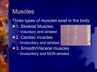
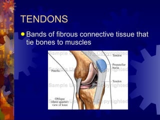
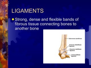
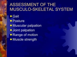
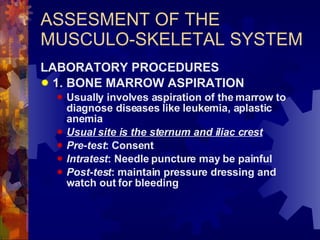
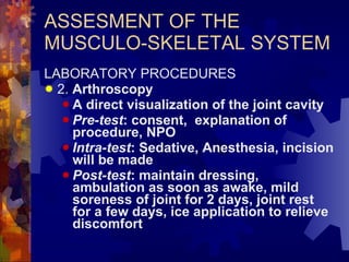
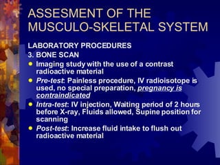
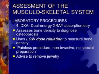
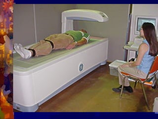
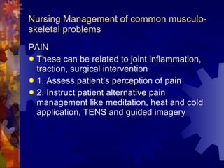
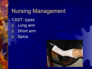
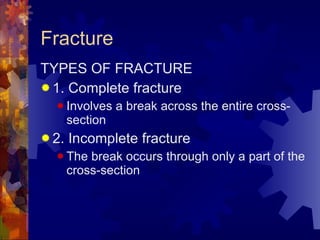
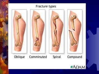
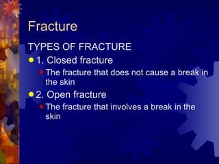
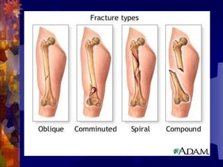
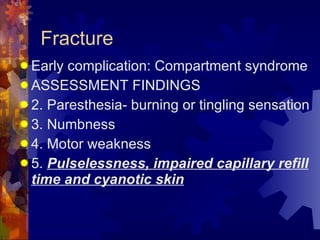
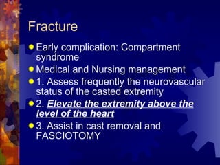
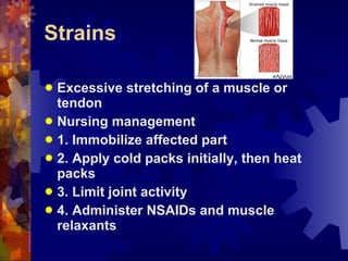
MUSCULO-SKELETAL SYSTEM Nurse Licensure Examination Review pinoynursing.webkotoh.com
Review of Anatomy and Physiology The musculo-skeletal system consists of the muscles, tendons, bones and cartilage together with the joints The primary function of which is to produce skeletal movements
Muscles Three types of muscles exist in the body 1. Skeletal Muscles Voluntary and striated 2. Cardiac muscles Involuntary and striated 3. Smooth/Visceral muscles Involuntary and NON-striated
TENDONS Bands of fibrous connective tissue that tie bones to muscles
LIGAMENTS Strong, dense and flexible bands of fibrous tissue connecting bones to another bone
BONES Variously classified according to shape, location and size Functions 1. Locomotion 2. Protection 3. Support and lever 4. Blood production 5. Mineral deposition
JOINTS The part of the Skeleton where two or more bones are connected
CARTILAGES A dense connective tissue that consists of fibers embedded in a strong gel-like substance
BURSAE Sac containing fluid that are located around the joints to prevent friction
ASSESMENT OF THE MUSCULO-SKELETAL SYSTEM The nurse usually evaluates this small part of the over-all assessment and concentrates on the patient’s posture, body symmetry, gait and muscle and joint function
ASSESMENT OF THE MUSCULO-SKELETAL SYSTEM 1. HISTORY 2. Physical Examination Perform a head to toe assessment Nurses need to inspect and palpate  The special procedure is the assessment of joint and muscle movement Usually, a tape measure and a  protractor are the only instruments
ASSESSMENT OF THE MUSCULO-SKELETAL SYSTEM Gait Posture Muscular palpation Joint palpation Range of motion Muscle strength
ASSESMENT OF THE MUSCULO-SKELETAL SYSTEM LABORATORY PROCEDURES 1. BONE MARROW ASPIRATION Usually involves aspiration of the marrow to diagnose diseases like leukemia, aplastic anemia Usual site is the sternum and iliac crest Pre-test : Consent Intratest : Needle puncture may be painful Post-test : maintain pressure dressing and watch out for bleeding
ASSESMENT OF THE MUSCULO-SKELETAL SYSTEM LABORATORY PROCEDURES 2.  Arthroscopy A direct visualization of the joint cavity Pre-test : consent,  explanation of procedure, NPO Intra-test : Sedative, Anesthesia, incision will be made Post-test : maintain dressing, ambulation as soon as awake, mild soreness of joint for 2 days, joint rest for a few days, ice application to relieve discomfort
ASSESMENT OF THE MUSCULO-SKELETAL SYSTEM LABORATORY PROCEDURES 3. BONE SCAN Imaging study with the use of a contrast radioactive material Pre-test : Painless procedure, IV radioisotope is used, no special preparation,  pregnancy is contraindicated Intra-test : IV injection, Waiting period of 2 hours before X-ray, Fluids allowed, Supine position for scanning Post-test : Increase fluid intake to flush out radioactive material
ASSESMENT OF THE MUSCULO-SKELETAL SYSTEM LABORATORY PROCEDURES 4. DXA- Dual-energy XRAY absorptiometry Assesses bone density to diagnose osteoporosis Uses  LOW dose radiation  to measure bone density Painless procedure, non-invasive, no special preparation Advise to remove jewelry
 
Common musculoskeletal problems The Nursing Management
Nursing Management of common musculo-skeletal problems PAIN These can be related to joint inflammation, traction, surgical intervention 1. Assess patient’s perception of pain 2. Instruct patient alternative pain management like meditation, heat and cold application, TENS and guided imagery
Nursing Management PAIN 3. Administer analgesics as prescribed Usually NSAIDS Meperidine can be given for severe pain 4. Assess the effectiveness of pain measures
Nursing Management IMPAIRED PHYSICAL MOBILITY 1. Instruct patient to perform range of motion exercises, either passive or active 2. Provide support in ambulation with assistive devices 3. Turn and change position every 2 hours 4. Encourage mobility for a short period and provide positive reinforcements for small accomplishments
Nursing Management SELF-CARE DEFICITS 1. Assess functional levels of the patient 2. Provide support for feeding problems  Place patient in Fowler’s position Provide assistive device and supervise mealtime Offer finger foods that can be handled by patient Keep suction equipment ready
Nursing Management SELF-CARE DEFICITS 3. Assist patient with difficulty bathing and hygiene Assist with bath only when patient has difficulty Provide ample time for patient to finish activity
Musculoskeletal Modalities Traction Cast
Nursing Management Traction A method of fracture immobilization by applying equipments to align bone fragments Used for immobilization, bone alignment and relief of muscle spasm
Traction Skin traction Skeletal traction
Traction Pulling force exerted on bones to reduce or immobilize fractures, reduce muscle spasm, correct or prevent deformities
 
 
Nursing Management Traction: General principles 1.  ALWAYS ensure that the weights hang freely and do not touch the floor 2.  NEVER remove the weights 3. Maintain proper body alignment 4. Ensure that the pulleys and ropes are properly functioning and fastened by tying  square knot
Nursing Management Traction: General principles 5. Observe and prevent foot drop Provide foot plate 6. Observe for DVT, skin irritation and breakdown 7. Provide pin care for clients in skeletal traction- use of hydrogen peroxide
Nursing Management CAST Immobilizing tool made of plaster of Paris or fiberglass Provides immobilization of the fracture
Nursing Management CAST: types Long arm Short arm Spica
Casting Materials Plaster of Paris Drying takes 1-3 days If dry, it is SHINY, WHITE, hard and resistant Fiberglass Lightweight and dries in 20-30 minutes Water resistant
Nursing Management CAST: General Nursing Care 1. Allow the cast to dry (usually 24-72 hours) 2. Handle a wet cast  with the PALMS  not the fingertips 3.  Keep the casted extremity ELEVATED using a pillow 4.  Turn the extremity for equal drying.  DO NOT USE DRYER  for plaster cast
Nursing Management CAST: General Nursing Care 5. Petal the edges of the cast to prevent crumbling of the edges 6. Examine the skin for pressure areas and Regularly check the pulses and skin
Nursing Management CAST: General Nursing Care 7. Instruct the patient not to place sticks or small objects inside the cast 8. Monitor for the following:  pain, swelling, discoloration, coolness, tingling or lack of sensation and diminished pulses
Common Musculoskeletal conditions Nursing management
METABOLIC BONE DISORDERS Osteoporosis A disease of the bone characterized by a decrease in the bone mass and density with a change in bone structure
METABOLIC BONE DISORDERS Osteoporosis: Pathophysiology Normal homeostatic bone turnover is altered   rate of bone  RESORPTION is greater than bone FORMATION   reduction in total bone mass   reduction in bone mineral density   prone to  FRACTURE
METABOLIC BONE DISORDERS Osteoporosis: TYPES 1. Primary Osteoporosis- advanced age, post-menopausal 2. Secondary osteoporosis- Steroid overuse, Renal failure
METABOLIC BONE DISORDERS RISK factors for the development of Osteoporosis 1. Sedentary lifestyle 2. Age 3. Diet- caffeine, alcohol, low Ca and Vit D 4. Post-menopausal 5. Genetics-  caucasian and asian 6.  Immobility
METABOLIC DISORDER ASSESSMENT FINDINGS 1. Low stature 2. Fracture Femur 3. Bone pain
METABOLIC DISORDER LABORATORY FINDINGS 1. DEXA-scan Provides information about bone mineral density T-score is at least 2.5 SD below the young adult mean value 2. X-ray studies
METABOLIC DISORDER Medical management of Osteoporosis 1. Diet therapy with calcium and Vitamin D  2. Hormone replacement therapy 3. Biphosphonates- Alendronate, risedronate produce increased bone mass by inhibiting the OSTEOCLAST  4. Moderate weight bearing exercises 5. Management of fractures
METABOLIC DISORDER Osteoporosis Nursing Interventions 1. Promote understanding of osteoporosis and the treatment regimen Provide adequate dietary supplement of calcium and vitamin D Instruct to employ a regular program of moderate exercises and physical activity Manage the constipating side-effect of calcium supplements
METABOLIC DISORDER Osteoporosis Nursing Interventions Take calcium supplements with meals Take alendronate with an EMPTY stomach with water Instruct on intake of Hormonal replacement
METABOLIC DISORDER Osteoporosis Nursing Interventions 2. Relieve the pain Instruct the patient to rest on a firm mattress Suggest that knee flexion will cause relaxation of back muscles Heat application may provide comfort Encourage good posture and body mechanics Instruct to avoid twisting and heavy lifting
METABOLIC DISORDER Osteoporosis Nursing Interventions 3. Improve bowel elimination Constipation is a problem of calcium supplements and immobility Advise intake of HIGH fiber diet and increased fluids
METABOLIC DISORDER Osteoporosis Nursing Interventions 4. Prevent injury Instruct to use isometric exercise to strengthen the trunk muscles AVOID sudden jarring, bending and strenuous lifting Provide a safe environment
Juvenile rheumatoid Arthritis Definition: AUTO-IMMUNE inflammatory joint disorder of UNKNOWN cause SYSTEMIC chronic disorder of connective tissue Diagnosed BEFORE age 16 years old
Juvenile rheumatoid Arthritis PATHOPHYSIOLOGY : unknown Affected by stress, climate and genetics Common in girls 2-5 and 9-12 y.o.
Juvenile rheumatoid Arthritis Poor prognosis Very Good prognosis Anorexia, anemia, fatigue Five or more joints Less than 4 joints Five or more joints Weight Bearing joints IRIDOCYCLITIS Salmon-pink rash Morning joint stiffness and fever MILD joint pain and swelling FEVER Polyarticular Pauci-articular Systemic JRA
JRA Symptoms may decrease as child enters adulthood With periods of remissions and exacerbations
JRA Medical Management ASPIRIN and NSAIDs-  mainstay treatment Slow-acting anti-rheumatic drugs Corticosteroids
JRA Nursing Management Encourage normal performance of daily activities Assist child in ROM exercises Administer medications Encourage social and emotional development
JRA Nursing Management During acute attack: SPLINT the joints NEUTRAL positioning Warm or cold packs
DEGENERATIVE JOINT DISEASE OSTEOARTHRITIS The most common form of degenerative joint disorder
DEGENERATIVE JOINT DISEASE OSTEOARTHRITIS Chronic, NON-systemic disorder of joints
DEGENERATIVE JOINT DISEASE OSTEOARTHRITIS: Pathophysiology Injury, genetic, Previous joint damage,  Obesity ,  Advanced age     Stimulate the chondrocytes to release chemicals    chemicals will cause cartilage degeneration, reactive inflammation of the synovial lining and bone stiffening
DEGENERATIVE JOINT DISEASE OSTEOARTHRITIS: Risk factors 1.  Increased age 2.  Obesity 3. Repetitive use of joints with previous joint damage 4. Anatomical deformity 5. genetic susceptibility
DEGENERATIVE JOINT DISEASE OSTEOARTHRITIS: Assessment findings 1. Joint pain 2. Joint stiffness 3. Functional joint impairment limitation The joint involvement is  ASYMMETRICAL This is not systemic, there is  no FEVER, no severe swelling Atrophy of unused muscles Usual joint are the  WEIGHT bearing joints
DEGENERATIVE JOINT DISEASE OSTEOARTHRITIS: Assessment findings 1. Joint pain Caused by  Inflamed synovium Stretching of the joint capsule Irritation of nerve endings
DEGENERATIVE JOINT DISEASE OSTEOARTHRITIS: Assessment findings 2. Stiffness commonly occurs in the morning after awakening Lasts only for  less than 30 minutes DECREASES with movement Crepitation may be elicited
DEGENERATIVE JOINT DISEASE OSTEOARTHRITIS: Diagnostic findings 1. X-ray Narrowing of joint space Loss of cartilage Osteophytes 2. Blood tests will show  no evidence  of systemic inflammation and are not useful
DEGENERATIVE JOINT DISEASE OSTEOARTHRITIS: Medical management 1. Weight reduction 2. Use of splinting devices to support joints 3. Occupational and physical therapy 4. Pharmacologic management Use of  PARACETAMOL, NSAIDS Use of Glucosamine and chondroitin Topical analgesics Intra-articular steroids to decrease inflam
DEGENERATIVE JOINT DISEASE OSTEOARTHRITIS: Nursing Interventions 1. Provide relief of PAIN Administer prescribed analgesics Application of  heat modalities.  ICE PACKS may be used in the early acute stage!!! Plan daily activities when pain is less severe Pain meds before exercising
DEGENERATIVE JOINT DISEASE OSTEOARTHRITIS: Nursing Interventions 2. Advise patient to reduce weight Aerobic exercise Walking 3. Administer prescribed medications NSAIDS
Rheumatoid arthritis A type of chronic systemic inflammatory arthritis and connective tissue disorder affecting  more women (ages 35-45)  than men
Rheumatoid arthritis FACTORS: Genetic Auto-immune connective tissue disorders Fatigue, emotional stress, cold, infection
Rheumatoid arthritis Pathophysiology Immune reaction  in the synovium    attracts neutrophils     releases enzymes    breakdown of collagen     irritates the synovial lining  causing  synovial inflammation  edema and pannus formation and joint erosions and swelling
Rheumatoid arthritis ASSESSMENT FINDINGS 1.  PAIN 2. Joint swelling and stiffness- SYMMETRICAL, Bilateral 3.  Warmth, erythema and lack of function 4.  Fever ,  weight loss, anemia , fatigue 5. Palpation of join reveals spongy tissue 6. Hesitancy in joint movement
Rheumatoid arthritis ASSESSMENT FINDINGS Joint involvement is  SYMMETRICAL and BILATERAL Characteristically beginning in the hands, wrist and feet Joint  STIFFNESS occurs early morning, lasts MORE than 30 minutes, not relieved by movement, diminishes as the day progresses
Rheumatoid arthritis ASSESSMENT FINDINGS Joints are  swollen and warm Painful when moved Deformities are common in the hands and feet causing misalignment  Rheumatoid nodules may be found in the subcutaneous tissues
Rheumatoid arthritis Diagnostic test 1. X-ray Shows bony erosion 2.  Blood studies reveal (+) rheumatoid factor, elevated ESR and CRP and ANTI-nuclear antibody 3. Arthrocentesis shows synovial fluid that is cloudy, milky or dark yellow containing  numerous WBC  and inflammatory proteins
Rheumatoid arthritis MEDICAL MANAGEMENT 1.  Therapeutic dose of NSAIDS and Aspirin to reduce inflammation 2. Chemotherapy with methotrexate, antimalarials,  gold therapy  and steroid 3. For advanced cases- arthroplasty, synovectomy 4. Nutritional therapy
Rheumatoid arthritis MEDICAL MANAGEMENT GOLD THERAPY: IM or Oral preparation Takes several months (3-6)  before effects can be seen Can damage the kidney and causes bone marrow depression
Rheumatoid arthritis Nursing MANAGEMENT 1. Relieve pain and discomfort USE splints to immobilize the affected extremity during acute stage of the disease and inflammation to REDUCE DEFORMITY Administer prescribed medications Suggest application of  COLD packs during the acute phase of pain, then HEAT application as the inflammation subsides
Rheumatoid arthritis Nursing MANAGEMENT 2. Decrease patient fatigue Schedule activity when pain is less severe Provide adequate periods of rests 3. Promote restorative sleep
Rheumatoid arthritis Nursing Management 4. Increase patient mobility Advise proper posture and body mechanics Support joint in functional position Advise ACTIVE ROME
Rheumatoid arthritis Nursing Management 5. Provide Diet therapy Patients experience anorexia, nausea and weight loss Regular diet with caloric restrictions because steroids may increase appetite Supplements of vitamins, iron and PROTEIN
Rheumatoid arthritis 6. Increase Mobility and prevent deformity: Lie FLAT on a firm mattress Lie PRONE several times to prevent HIP FLEXION contracture Use one pillow under the head because of risk of dorsal kyphosis NO Pillow under the joints because this promotes flexion contractures
Hot versus Cold ACUTE ATTACK After acute attack Use to control inflammation and pain Use to RELIEVE joint stiffness, pain and muscle spasm Cold HOT
Gouty arthritis A systemic disease caused by deposition of uric acid crystals in the joint and body tissues CAUSES: 1. Primary gout-  disorder of Purine metabolism 2. Secondary gout- excessive uric acid in the blood like leukemia
 
 
Gouty arthritis ASSESSMENT FINDINGS 1. Severe pain in the involved joints,  initially the big toe 2. Swelling and inflammation of the joint 3.  TOPHI-  yellowish-whitish, irregular deposits in the skin that break open and reveal a gritty appearance 4. PODAGRA
Gouty arthritis ASSESSMENT FINDINGS 5. Fever, malaise 6. Body weakness and headache 7. Renal stones
Gouty arthritis DIAGNOSTIC TEST Elevated levels of uric acid in the blood Uric acid stones in the kidney
Gouty arthritis Medical management 1. Allupurinol- take it WITH FOOD Rash signifies allergic reaction 2. Colchicine For acute attack
Gouty arthritis Nursing Intervention 1. Provide a diet with  LOW purine Avoid Organ meats, aged and processed foods STRICT dietary restriction is NOT necessary 2. Encourage an  increased fluid intake (2-3L/day) to prevent stone formation 3. Instruct the patient to avoid alcohol 4.  Provide alkaline ash diet to increase urinary pH 5. Provide bed rest during early attack of gout
Gouty arthritis Nursing Intervention 6. Position the affected extremity in mild flexion 7. Administer anti-gout medication and analgesics
Fracture A break in the continuity of the bone and is defined according to its type and extent
Fracture Severe mechanical Stress to bone    bone fracture Direct Blows Crushing forces Sudden twisting motion Extreme muscle contraction
Fracture TYPES OF FRACTURE 1. Complete fracture Involves a break across the entire cross-section 2. Incomplete fracture The break occurs through only a part of the cross-section
 
Fracture TYPES OF FRACTURE 1. Closed fracture The fracture that does not cause a break in the skin 2. Open fracture The fracture that involves a break in the skin
 
Fracture TYPES OF FRACTURE 1. Comminuted fracture A fracture that involves production of several bone fragments 2. Simple fracture A fracture that involves break of bone into two parts or one
Fracture ASSESSMENT FINDINGS 1. Pain or tenderness over the involved area 2. Loss of function 3. Deformity 4. Shortening 5. Crepitus 6. Swelling and discoloration
Fracture ASSESSMENT FINDINGS 1. Pain Continuous and increases in severity  Muscles spasm accompanies the fracture is a reaction of the body to immobilize the fractured bone
Fracture ASSESSMENT FINDINGS 2. Loss of function Abnormal movement and pain can result to this manifestation
Fracture ASSESSMENT FINDINGS 3. Deformity Displacement, angulations or rotation of the fragments Causes deformity
Fracture ASSESSMENT FINDINGS 4. Crepitus A grating sensation produced when the bone fragments rub each other
Fracture DIAGNOSTIC TEST X-ray
Fracture EMERGENCY MANAGEMENT OF FRACTURE 1.  Immobilize any suspected fracture 2. Support the extremity above and below when moving the affected part from a vehicle  3. Suggested temporary splints- hard board, stick, rolled sheets 4. Apply sling if forearm fracture is suspected or the suspected fractured arm maybe bandaged to the chest
Fracture EMERGENCY MANAGEMENT OF FRACTURE 5. Open fracture is managed by covering a clean/sterile gauze to prevent contamination 6. DO NOT attempt to reduce the facture
Fracture MEDICAL MANAGEMENT  1. Reduction of fracture either open or closed, Immobilization and Restoration of function 2. Antibiotics, Muscle relaxants and Pain medications
Fracture General Nursing MANAGEMENT  For CLOSED FRACTURE 1. Assist in reduction and immobilization 2. Administer pain medication and muscle relaxants 3. teach patient to care for the cast 4. Teach patient about potential complication of fracture and to report infection, poor alignment and continuous pain
Fracture General Nursing MANAGEMENT  For OPEN FRACTURE 1. Prevent wound and bone infection Administer prescribed antibiotics Administer tetanus prophylaxis Assist in serial wound debridement 2. Elevate the extremity to prevent edema formation 3. Administer care of traction and cast
Fracture FRACTURE COMPLICATIONS Early 1. Shock 2. Fat embolism 3. Compartment syndrome 4. Infection  5. DVT
Fracture FRACTURE COMPLICATIONS Late 1. Delayed union 2. Avascular necrosis 3. Delayed reaction to fixation devices 4. Complex regional syndrome
Fracture FRACTURE COMPLICATIONS: Fat Embolism Occurs usually in fractures of the long bones Fat globules may move into the blood stream because the marrow pressure is greater than capillary pressure Fat globules occlude the small blood vessels of the lungs, brain kidneys and other organs
Fracture FRACTURE COMPLICATIONS: Fat Embolism Onset is rapid, within 24-72 hours ASSESSMENT FINDINGS 1. Sudden dyspnea and respiratory distress 2. tachycardia 3. Chest pain 4. Crackles, wheezes and cough 5. Petechial rashes over the chest, axilla and hard palate
Fracture FRACTURE COMPLICATIONS: Fat Embolism Nursing Management 1. Support the respiratory function Respiratory failure is the most common cause of death Administer O2 in high concentration Prepare for possible intubation and ventilator support
Fracture FRACTURE COMPLICATIONS: Fat Embolism Nursing Management 2. Administer drugs Corticosteroids Dopamine Morphine
Fracture FRACTURE COMPLICATIONS: Fat Embolism Nursing Management 3. Institute preventive measures Immediate immobilization of fracture Minimal fracture manipulation Adequate support for fractured bone during turning and positioning Maintain adequate hydration and electrolyte balance
Fracture Early complication: Compartment syndrome A complication that develops when tissue perfusion in the muscles is less than required for tissue viability
Fracture Early complication: Compartment syndrome ASSESSMENT FINDINGS 1. Pain- Deep, throbbing and  UNRELIEVED pain by opiods Pain is due to reduction in the size of the muscle compartment by tight cast Pain is due to increased mass in the compartment by edema, swelling or hemorrhage
Fracture Early complication: Compartment syndrome ASSESSMENT FINDINGS 2. Paresthesia- burning or tingling sensation 3. Numbness  4. Motor weakness 5.  Pulselessness, impaired capillary refill time and cyanotic skin
Fracture Early complication: Compartment syndrome Medical and Nursing management 1. Assess frequently the neurovascular status of the casted extremity 2.  Elevate the extremity above the level of the heart 3. Assist in cast removal and FASCIOTOMY
Strains Excessive stretching of a muscle or tendon Nursing management 1. Immobilize affected part 2. Apply cold packs initially, then heat packs 3. Limit joint activity 4. Administer NSAIDs and muscle relaxants
Sprains Excessive stretching of the LIGAMENTS Nursing management 1. Immobilize extremity and advise rest 2. Apply cold packs initially then heat packs 3. Compression bandage may be applied to relieve edema 4. Assist in cast application 5. Administer NSAIDS
End of Musculoskeletal

Musculoskeletal System

  • 1.
    MUSCULO-SKELETAL SYSTEM NurseLicensure Examination Review pinoynursing.webkotoh.com
  • 2.
    Review of Anatomyand Physiology The musculo-skeletal system consists of the muscles, tendons, bones and cartilage together with the joints The primary function of which is to produce skeletal movements
  • 3.
    Muscles Three typesof muscles exist in the body 1. Skeletal Muscles Voluntary and striated 2. Cardiac muscles Involuntary and striated 3. Smooth/Visceral muscles Involuntary and NON-striated
  • 4.
    TENDONS Bands offibrous connective tissue that tie bones to muscles
  • 5.
    LIGAMENTS Strong, denseand flexible bands of fibrous tissue connecting bones to another bone
  • 6.
    BONES Variously classifiedaccording to shape, location and size Functions 1. Locomotion 2. Protection 3. Support and lever 4. Blood production 5. Mineral deposition
  • 7.
    JOINTS The partof the Skeleton where two or more bones are connected
  • 8.
    CARTILAGES A denseconnective tissue that consists of fibers embedded in a strong gel-like substance
  • 9.
    BURSAE Sac containingfluid that are located around the joints to prevent friction
  • 10.
    ASSESMENT OF THEMUSCULO-SKELETAL SYSTEM The nurse usually evaluates this small part of the over-all assessment and concentrates on the patient’s posture, body symmetry, gait and muscle and joint function
  • 11.
    ASSESMENT OF THEMUSCULO-SKELETAL SYSTEM 1. HISTORY 2. Physical Examination Perform a head to toe assessment Nurses need to inspect and palpate The special procedure is the assessment of joint and muscle movement Usually, a tape measure and a protractor are the only instruments
  • 12.
    ASSESSMENT OF THEMUSCULO-SKELETAL SYSTEM Gait Posture Muscular palpation Joint palpation Range of motion Muscle strength
  • 13.
    ASSESMENT OF THEMUSCULO-SKELETAL SYSTEM LABORATORY PROCEDURES 1. BONE MARROW ASPIRATION Usually involves aspiration of the marrow to diagnose diseases like leukemia, aplastic anemia Usual site is the sternum and iliac crest Pre-test : Consent Intratest : Needle puncture may be painful Post-test : maintain pressure dressing and watch out for bleeding
  • 14.
    ASSESMENT OF THEMUSCULO-SKELETAL SYSTEM LABORATORY PROCEDURES 2. Arthroscopy A direct visualization of the joint cavity Pre-test : consent, explanation of procedure, NPO Intra-test : Sedative, Anesthesia, incision will be made Post-test : maintain dressing, ambulation as soon as awake, mild soreness of joint for 2 days, joint rest for a few days, ice application to relieve discomfort
  • 15.
    ASSESMENT OF THEMUSCULO-SKELETAL SYSTEM LABORATORY PROCEDURES 3. BONE SCAN Imaging study with the use of a contrast radioactive material Pre-test : Painless procedure, IV radioisotope is used, no special preparation, pregnancy is contraindicated Intra-test : IV injection, Waiting period of 2 hours before X-ray, Fluids allowed, Supine position for scanning Post-test : Increase fluid intake to flush out radioactive material
  • 16.
    ASSESMENT OF THEMUSCULO-SKELETAL SYSTEM LABORATORY PROCEDURES 4. DXA- Dual-energy XRAY absorptiometry Assesses bone density to diagnose osteoporosis Uses LOW dose radiation to measure bone density Painless procedure, non-invasive, no special preparation Advise to remove jewelry
  • 17.
  • 18.
    Common musculoskeletal problemsThe Nursing Management
  • 19.
    Nursing Management ofcommon musculo-skeletal problems PAIN These can be related to joint inflammation, traction, surgical intervention 1. Assess patient’s perception of pain 2. Instruct patient alternative pain management like meditation, heat and cold application, TENS and guided imagery
  • 20.
    Nursing Management PAIN3. Administer analgesics as prescribed Usually NSAIDS Meperidine can be given for severe pain 4. Assess the effectiveness of pain measures
  • 21.
    Nursing Management IMPAIREDPHYSICAL MOBILITY 1. Instruct patient to perform range of motion exercises, either passive or active 2. Provide support in ambulation with assistive devices 3. Turn and change position every 2 hours 4. Encourage mobility for a short period and provide positive reinforcements for small accomplishments
  • 22.
    Nursing Management SELF-CAREDEFICITS 1. Assess functional levels of the patient 2. Provide support for feeding problems Place patient in Fowler’s position Provide assistive device and supervise mealtime Offer finger foods that can be handled by patient Keep suction equipment ready
  • 23.
    Nursing Management SELF-CAREDEFICITS 3. Assist patient with difficulty bathing and hygiene Assist with bath only when patient has difficulty Provide ample time for patient to finish activity
  • 24.
  • 25.
    Nursing Management TractionA method of fracture immobilization by applying equipments to align bone fragments Used for immobilization, bone alignment and relief of muscle spasm
  • 26.
    Traction Skin tractionSkeletal traction
  • 27.
    Traction Pulling forceexerted on bones to reduce or immobilize fractures, reduce muscle spasm, correct or prevent deformities
  • 28.
  • 29.
  • 30.
    Nursing Management Traction:General principles 1. ALWAYS ensure that the weights hang freely and do not touch the floor 2. NEVER remove the weights 3. Maintain proper body alignment 4. Ensure that the pulleys and ropes are properly functioning and fastened by tying square knot
  • 31.
    Nursing Management Traction:General principles 5. Observe and prevent foot drop Provide foot plate 6. Observe for DVT, skin irritation and breakdown 7. Provide pin care for clients in skeletal traction- use of hydrogen peroxide
  • 32.
    Nursing Management CASTImmobilizing tool made of plaster of Paris or fiberglass Provides immobilization of the fracture
  • 33.
    Nursing Management CAST:types Long arm Short arm Spica
  • 34.
    Casting Materials Plasterof Paris Drying takes 1-3 days If dry, it is SHINY, WHITE, hard and resistant Fiberglass Lightweight and dries in 20-30 minutes Water resistant
  • 35.
    Nursing Management CAST:General Nursing Care 1. Allow the cast to dry (usually 24-72 hours) 2. Handle a wet cast with the PALMS not the fingertips 3. Keep the casted extremity ELEVATED using a pillow 4. Turn the extremity for equal drying. DO NOT USE DRYER for plaster cast
  • 36.
    Nursing Management CAST:General Nursing Care 5. Petal the edges of the cast to prevent crumbling of the edges 6. Examine the skin for pressure areas and Regularly check the pulses and skin
  • 37.
    Nursing Management CAST:General Nursing Care 7. Instruct the patient not to place sticks or small objects inside the cast 8. Monitor for the following: pain, swelling, discoloration, coolness, tingling or lack of sensation and diminished pulses
  • 38.
  • 39.
    METABOLIC BONE DISORDERSOsteoporosis A disease of the bone characterized by a decrease in the bone mass and density with a change in bone structure
  • 40.
    METABOLIC BONE DISORDERSOsteoporosis: Pathophysiology Normal homeostatic bone turnover is altered  rate of bone RESORPTION is greater than bone FORMATION  reduction in total bone mass  reduction in bone mineral density  prone to FRACTURE
  • 41.
    METABOLIC BONE DISORDERSOsteoporosis: TYPES 1. Primary Osteoporosis- advanced age, post-menopausal 2. Secondary osteoporosis- Steroid overuse, Renal failure
  • 42.
    METABOLIC BONE DISORDERSRISK factors for the development of Osteoporosis 1. Sedentary lifestyle 2. Age 3. Diet- caffeine, alcohol, low Ca and Vit D 4. Post-menopausal 5. Genetics- caucasian and asian 6. Immobility
  • 43.
    METABOLIC DISORDER ASSESSMENTFINDINGS 1. Low stature 2. Fracture Femur 3. Bone pain
  • 44.
    METABOLIC DISORDER LABORATORYFINDINGS 1. DEXA-scan Provides information about bone mineral density T-score is at least 2.5 SD below the young adult mean value 2. X-ray studies
  • 45.
    METABOLIC DISORDER Medicalmanagement of Osteoporosis 1. Diet therapy with calcium and Vitamin D 2. Hormone replacement therapy 3. Biphosphonates- Alendronate, risedronate produce increased bone mass by inhibiting the OSTEOCLAST 4. Moderate weight bearing exercises 5. Management of fractures
  • 46.
    METABOLIC DISORDER OsteoporosisNursing Interventions 1. Promote understanding of osteoporosis and the treatment regimen Provide adequate dietary supplement of calcium and vitamin D Instruct to employ a regular program of moderate exercises and physical activity Manage the constipating side-effect of calcium supplements
  • 47.
    METABOLIC DISORDER OsteoporosisNursing Interventions Take calcium supplements with meals Take alendronate with an EMPTY stomach with water Instruct on intake of Hormonal replacement
  • 48.
    METABOLIC DISORDER OsteoporosisNursing Interventions 2. Relieve the pain Instruct the patient to rest on a firm mattress Suggest that knee flexion will cause relaxation of back muscles Heat application may provide comfort Encourage good posture and body mechanics Instruct to avoid twisting and heavy lifting
  • 49.
    METABOLIC DISORDER OsteoporosisNursing Interventions 3. Improve bowel elimination Constipation is a problem of calcium supplements and immobility Advise intake of HIGH fiber diet and increased fluids
  • 50.
    METABOLIC DISORDER OsteoporosisNursing Interventions 4. Prevent injury Instruct to use isometric exercise to strengthen the trunk muscles AVOID sudden jarring, bending and strenuous lifting Provide a safe environment
  • 51.
    Juvenile rheumatoid ArthritisDefinition: AUTO-IMMUNE inflammatory joint disorder of UNKNOWN cause SYSTEMIC chronic disorder of connective tissue Diagnosed BEFORE age 16 years old
  • 52.
    Juvenile rheumatoid ArthritisPATHOPHYSIOLOGY : unknown Affected by stress, climate and genetics Common in girls 2-5 and 9-12 y.o.
  • 53.
    Juvenile rheumatoid ArthritisPoor prognosis Very Good prognosis Anorexia, anemia, fatigue Five or more joints Less than 4 joints Five or more joints Weight Bearing joints IRIDOCYCLITIS Salmon-pink rash Morning joint stiffness and fever MILD joint pain and swelling FEVER Polyarticular Pauci-articular Systemic JRA
  • 54.
    JRA Symptoms maydecrease as child enters adulthood With periods of remissions and exacerbations
  • 55.
    JRA Medical ManagementASPIRIN and NSAIDs- mainstay treatment Slow-acting anti-rheumatic drugs Corticosteroids
  • 56.
    JRA Nursing ManagementEncourage normal performance of daily activities Assist child in ROM exercises Administer medications Encourage social and emotional development
  • 57.
    JRA Nursing ManagementDuring acute attack: SPLINT the joints NEUTRAL positioning Warm or cold packs
  • 58.
    DEGENERATIVE JOINT DISEASEOSTEOARTHRITIS The most common form of degenerative joint disorder
  • 59.
    DEGENERATIVE JOINT DISEASEOSTEOARTHRITIS Chronic, NON-systemic disorder of joints
  • 60.
    DEGENERATIVE JOINT DISEASEOSTEOARTHRITIS: Pathophysiology Injury, genetic, Previous joint damage, Obesity , Advanced age  Stimulate the chondrocytes to release chemicals  chemicals will cause cartilage degeneration, reactive inflammation of the synovial lining and bone stiffening
  • 61.
    DEGENERATIVE JOINT DISEASEOSTEOARTHRITIS: Risk factors 1. Increased age 2. Obesity 3. Repetitive use of joints with previous joint damage 4. Anatomical deformity 5. genetic susceptibility
  • 62.
    DEGENERATIVE JOINT DISEASEOSTEOARTHRITIS: Assessment findings 1. Joint pain 2. Joint stiffness 3. Functional joint impairment limitation The joint involvement is ASYMMETRICAL This is not systemic, there is no FEVER, no severe swelling Atrophy of unused muscles Usual joint are the WEIGHT bearing joints
  • 63.
    DEGENERATIVE JOINT DISEASEOSTEOARTHRITIS: Assessment findings 1. Joint pain Caused by Inflamed synovium Stretching of the joint capsule Irritation of nerve endings
  • 64.
    DEGENERATIVE JOINT DISEASEOSTEOARTHRITIS: Assessment findings 2. Stiffness commonly occurs in the morning after awakening Lasts only for less than 30 minutes DECREASES with movement Crepitation may be elicited
  • 65.
    DEGENERATIVE JOINT DISEASEOSTEOARTHRITIS: Diagnostic findings 1. X-ray Narrowing of joint space Loss of cartilage Osteophytes 2. Blood tests will show no evidence of systemic inflammation and are not useful
  • 66.
    DEGENERATIVE JOINT DISEASEOSTEOARTHRITIS: Medical management 1. Weight reduction 2. Use of splinting devices to support joints 3. Occupational and physical therapy 4. Pharmacologic management Use of PARACETAMOL, NSAIDS Use of Glucosamine and chondroitin Topical analgesics Intra-articular steroids to decrease inflam
  • 67.
    DEGENERATIVE JOINT DISEASEOSTEOARTHRITIS: Nursing Interventions 1. Provide relief of PAIN Administer prescribed analgesics Application of heat modalities. ICE PACKS may be used in the early acute stage!!! Plan daily activities when pain is less severe Pain meds before exercising
  • 68.
    DEGENERATIVE JOINT DISEASEOSTEOARTHRITIS: Nursing Interventions 2. Advise patient to reduce weight Aerobic exercise Walking 3. Administer prescribed medications NSAIDS
  • 69.
    Rheumatoid arthritis Atype of chronic systemic inflammatory arthritis and connective tissue disorder affecting more women (ages 35-45) than men
  • 70.
    Rheumatoid arthritis FACTORS:Genetic Auto-immune connective tissue disorders Fatigue, emotional stress, cold, infection
  • 71.
    Rheumatoid arthritis PathophysiologyImmune reaction in the synovium  attracts neutrophils  releases enzymes  breakdown of collagen  irritates the synovial lining  causing synovial inflammation edema and pannus formation and joint erosions and swelling
  • 72.
    Rheumatoid arthritis ASSESSMENTFINDINGS 1. PAIN 2. Joint swelling and stiffness- SYMMETRICAL, Bilateral 3. Warmth, erythema and lack of function 4. Fever , weight loss, anemia , fatigue 5. Palpation of join reveals spongy tissue 6. Hesitancy in joint movement
  • 73.
    Rheumatoid arthritis ASSESSMENTFINDINGS Joint involvement is SYMMETRICAL and BILATERAL Characteristically beginning in the hands, wrist and feet Joint STIFFNESS occurs early morning, lasts MORE than 30 minutes, not relieved by movement, diminishes as the day progresses
  • 74.
    Rheumatoid arthritis ASSESSMENTFINDINGS Joints are swollen and warm Painful when moved Deformities are common in the hands and feet causing misalignment Rheumatoid nodules may be found in the subcutaneous tissues
  • 75.
    Rheumatoid arthritis Diagnostictest 1. X-ray Shows bony erosion 2. Blood studies reveal (+) rheumatoid factor, elevated ESR and CRP and ANTI-nuclear antibody 3. Arthrocentesis shows synovial fluid that is cloudy, milky or dark yellow containing numerous WBC and inflammatory proteins
  • 76.
    Rheumatoid arthritis MEDICALMANAGEMENT 1. Therapeutic dose of NSAIDS and Aspirin to reduce inflammation 2. Chemotherapy with methotrexate, antimalarials, gold therapy and steroid 3. For advanced cases- arthroplasty, synovectomy 4. Nutritional therapy
  • 77.
    Rheumatoid arthritis MEDICALMANAGEMENT GOLD THERAPY: IM or Oral preparation Takes several months (3-6) before effects can be seen Can damage the kidney and causes bone marrow depression
  • 78.
    Rheumatoid arthritis NursingMANAGEMENT 1. Relieve pain and discomfort USE splints to immobilize the affected extremity during acute stage of the disease and inflammation to REDUCE DEFORMITY Administer prescribed medications Suggest application of COLD packs during the acute phase of pain, then HEAT application as the inflammation subsides
  • 79.
    Rheumatoid arthritis NursingMANAGEMENT 2. Decrease patient fatigue Schedule activity when pain is less severe Provide adequate periods of rests 3. Promote restorative sleep
  • 80.
    Rheumatoid arthritis NursingManagement 4. Increase patient mobility Advise proper posture and body mechanics Support joint in functional position Advise ACTIVE ROME
  • 81.
    Rheumatoid arthritis NursingManagement 5. Provide Diet therapy Patients experience anorexia, nausea and weight loss Regular diet with caloric restrictions because steroids may increase appetite Supplements of vitamins, iron and PROTEIN
  • 82.
    Rheumatoid arthritis 6.Increase Mobility and prevent deformity: Lie FLAT on a firm mattress Lie PRONE several times to prevent HIP FLEXION contracture Use one pillow under the head because of risk of dorsal kyphosis NO Pillow under the joints because this promotes flexion contractures
  • 83.
    Hot versus ColdACUTE ATTACK After acute attack Use to control inflammation and pain Use to RELIEVE joint stiffness, pain and muscle spasm Cold HOT
  • 84.
    Gouty arthritis Asystemic disease caused by deposition of uric acid crystals in the joint and body tissues CAUSES: 1. Primary gout- disorder of Purine metabolism 2. Secondary gout- excessive uric acid in the blood like leukemia
  • 85.
  • 86.
  • 87.
    Gouty arthritis ASSESSMENTFINDINGS 1. Severe pain in the involved joints, initially the big toe 2. Swelling and inflammation of the joint 3. TOPHI- yellowish-whitish, irregular deposits in the skin that break open and reveal a gritty appearance 4. PODAGRA
  • 88.
    Gouty arthritis ASSESSMENTFINDINGS 5. Fever, malaise 6. Body weakness and headache 7. Renal stones
  • 89.
    Gouty arthritis DIAGNOSTICTEST Elevated levels of uric acid in the blood Uric acid stones in the kidney
  • 90.
    Gouty arthritis Medicalmanagement 1. Allupurinol- take it WITH FOOD Rash signifies allergic reaction 2. Colchicine For acute attack
  • 91.
    Gouty arthritis NursingIntervention 1. Provide a diet with LOW purine Avoid Organ meats, aged and processed foods STRICT dietary restriction is NOT necessary 2. Encourage an increased fluid intake (2-3L/day) to prevent stone formation 3. Instruct the patient to avoid alcohol 4. Provide alkaline ash diet to increase urinary pH 5. Provide bed rest during early attack of gout
  • 92.
    Gouty arthritis NursingIntervention 6. Position the affected extremity in mild flexion 7. Administer anti-gout medication and analgesics
  • 93.
    Fracture A breakin the continuity of the bone and is defined according to its type and extent
  • 94.
    Fracture Severe mechanicalStress to bone  bone fracture Direct Blows Crushing forces Sudden twisting motion Extreme muscle contraction
  • 95.
    Fracture TYPES OFFRACTURE 1. Complete fracture Involves a break across the entire cross-section 2. Incomplete fracture The break occurs through only a part of the cross-section
  • 96.
  • 97.
    Fracture TYPES OFFRACTURE 1. Closed fracture The fracture that does not cause a break in the skin 2. Open fracture The fracture that involves a break in the skin
  • 98.
  • 99.
    Fracture TYPES OFFRACTURE 1. Comminuted fracture A fracture that involves production of several bone fragments 2. Simple fracture A fracture that involves break of bone into two parts or one
  • 100.
    Fracture ASSESSMENT FINDINGS1. Pain or tenderness over the involved area 2. Loss of function 3. Deformity 4. Shortening 5. Crepitus 6. Swelling and discoloration
  • 101.
    Fracture ASSESSMENT FINDINGS1. Pain Continuous and increases in severity Muscles spasm accompanies the fracture is a reaction of the body to immobilize the fractured bone
  • 102.
    Fracture ASSESSMENT FINDINGS2. Loss of function Abnormal movement and pain can result to this manifestation
  • 103.
    Fracture ASSESSMENT FINDINGS3. Deformity Displacement, angulations or rotation of the fragments Causes deformity
  • 104.
    Fracture ASSESSMENT FINDINGS4. Crepitus A grating sensation produced when the bone fragments rub each other
  • 105.
  • 106.
    Fracture EMERGENCY MANAGEMENTOF FRACTURE 1. Immobilize any suspected fracture 2. Support the extremity above and below when moving the affected part from a vehicle 3. Suggested temporary splints- hard board, stick, rolled sheets 4. Apply sling if forearm fracture is suspected or the suspected fractured arm maybe bandaged to the chest
  • 107.
    Fracture EMERGENCY MANAGEMENTOF FRACTURE 5. Open fracture is managed by covering a clean/sterile gauze to prevent contamination 6. DO NOT attempt to reduce the facture
  • 108.
    Fracture MEDICAL MANAGEMENT 1. Reduction of fracture either open or closed, Immobilization and Restoration of function 2. Antibiotics, Muscle relaxants and Pain medications
  • 109.
    Fracture General NursingMANAGEMENT For CLOSED FRACTURE 1. Assist in reduction and immobilization 2. Administer pain medication and muscle relaxants 3. teach patient to care for the cast 4. Teach patient about potential complication of fracture and to report infection, poor alignment and continuous pain
  • 110.
    Fracture General NursingMANAGEMENT For OPEN FRACTURE 1. Prevent wound and bone infection Administer prescribed antibiotics Administer tetanus prophylaxis Assist in serial wound debridement 2. Elevate the extremity to prevent edema formation 3. Administer care of traction and cast
  • 111.
    Fracture FRACTURE COMPLICATIONSEarly 1. Shock 2. Fat embolism 3. Compartment syndrome 4. Infection 5. DVT
  • 112.
    Fracture FRACTURE COMPLICATIONSLate 1. Delayed union 2. Avascular necrosis 3. Delayed reaction to fixation devices 4. Complex regional syndrome
  • 113.
    Fracture FRACTURE COMPLICATIONS:Fat Embolism Occurs usually in fractures of the long bones Fat globules may move into the blood stream because the marrow pressure is greater than capillary pressure Fat globules occlude the small blood vessels of the lungs, brain kidneys and other organs
  • 114.
    Fracture FRACTURE COMPLICATIONS:Fat Embolism Onset is rapid, within 24-72 hours ASSESSMENT FINDINGS 1. Sudden dyspnea and respiratory distress 2. tachycardia 3. Chest pain 4. Crackles, wheezes and cough 5. Petechial rashes over the chest, axilla and hard palate
  • 115.
    Fracture FRACTURE COMPLICATIONS:Fat Embolism Nursing Management 1. Support the respiratory function Respiratory failure is the most common cause of death Administer O2 in high concentration Prepare for possible intubation and ventilator support
  • 116.
    Fracture FRACTURE COMPLICATIONS:Fat Embolism Nursing Management 2. Administer drugs Corticosteroids Dopamine Morphine
  • 117.
    Fracture FRACTURE COMPLICATIONS:Fat Embolism Nursing Management 3. Institute preventive measures Immediate immobilization of fracture Minimal fracture manipulation Adequate support for fractured bone during turning and positioning Maintain adequate hydration and electrolyte balance
  • 118.
    Fracture Early complication:Compartment syndrome A complication that develops when tissue perfusion in the muscles is less than required for tissue viability
  • 119.
    Fracture Early complication:Compartment syndrome ASSESSMENT FINDINGS 1. Pain- Deep, throbbing and UNRELIEVED pain by opiods Pain is due to reduction in the size of the muscle compartment by tight cast Pain is due to increased mass in the compartment by edema, swelling or hemorrhage
  • 120.
    Fracture Early complication:Compartment syndrome ASSESSMENT FINDINGS 2. Paresthesia- burning or tingling sensation 3. Numbness 4. Motor weakness 5. Pulselessness, impaired capillary refill time and cyanotic skin
  • 121.
    Fracture Early complication:Compartment syndrome Medical and Nursing management 1. Assess frequently the neurovascular status of the casted extremity 2. Elevate the extremity above the level of the heart 3. Assist in cast removal and FASCIOTOMY
  • 122.
    Strains Excessive stretchingof a muscle or tendon Nursing management 1. Immobilize affected part 2. Apply cold packs initially, then heat packs 3. Limit joint activity 4. Administer NSAIDs and muscle relaxants
  • 123.
    Sprains Excessive stretchingof the LIGAMENTS Nursing management 1. Immobilize extremity and advise rest 2. Apply cold packs initially then heat packs 3. Compression bandage may be applied to relieve edema 4. Assist in cast application 5. Administer NSAIDS
  • 124.