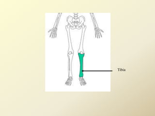
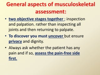
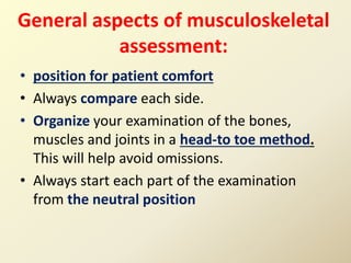
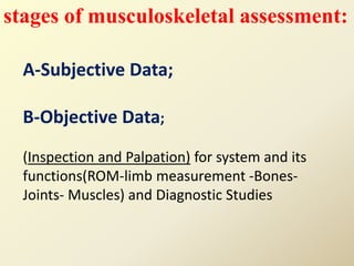
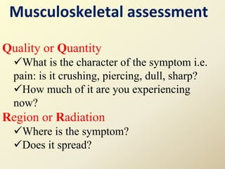
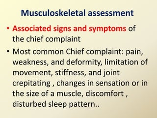
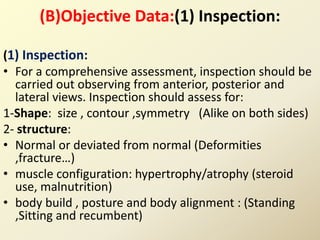
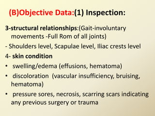
Musculoskeletal assessment involves a thorough subjective and objective examination of the muscles, bones, and joints. The subjective examination includes gathering information on the patient's chief complaint, pain characteristics, functional limitations, and relevant medical history. The objective examination consists of inspection and palpation techniques to evaluate the musculoskeletal system, range of motion, limb measurements, and diagnostic tests. Together, the subjective and objective data aim to determine the degree to which the patient's daily activities are affected by any musculoskeletal problems.
this topic is on bed sores. discusses the definition, etiology , pathophysiology of bed sore development as well as prevention and managemene of pressure sores
this topic explains the nature of pain, signs and symptoms of pain, different types of pain, factors influencing pain, assessment of pain and pharmacological and non pharmacological management of pain.
nursing intervention for patients with musculoskeletal system disorders by Mulugeta Emiru (MSc in Adult health Nursing): Mizan Tepi university. 2017/2018.
Disorders of maxillary sinus / dental implant courses by Indian dental academy Indian dental academy
The Indian Dental Academy is the Leader in continuing dental education , training dentists in all aspects of dentistry and
offering a wide range of dental certified courses in different formats.for more details please visit
www.indiandentalacademy.com
Skeletal system
Divisions of skeletal system,
types of bone,
salient features and functions
of bones of axial and appendicular skeletal system Organization of skeletal muscle,
physiology of muscle contraction,
neuromuscular junction.
The Skeletal System
Skeletal System
Skeletal System Essay
Skeletal System
The Skeletal System Essay
The Skeletal System
Skeletal System Support Body
Skeletal System
The Skeletal System Essay
Chapter 5: the Skeletal System Essay
Healthy Skeletal System
These lecture slides, by Dr Sidra Arshad, offer a quick overview of physiological basis of a normal electrocardiogram.
Learning objectives:
1. Define an electrocardiogram (ECG) and electrocardiography
2. Describe how dipoles generated by the heart produce the waveforms of the ECG
3. Describe the components of a normal electrocardiogram of a typical bipolar leads (limb II)
4. Differentiate between intervals and segments
5. Enlist some common indications for obtaining an ECG
Study Resources:
1. Chapter 11, Guyton and Hall Textbook of Medical Physiology, 14th edition
2. Chapter 9, Human Physiology - From Cells to Systems, Lauralee Sherwood, 9th edition
3. Chapter 29, Ganong’s Review of Medical Physiology, 26th edition
4. Electrocardiogram, StatPearls - https://www.ncbi.nlm.nih.gov/books/NBK549803/
5. ECG in Medical Practice by ABM Abdullah, 4th edition
6. ECG Basics, http://www.nataliescasebook.com/tag/e-c-g-basics
The prostate is an exocrine gland of the male mammalian reproductive system
It is a walnut-sized gland that forms part of the male reproductive system and is located in front of the rectum and just below the urinary bladder
Function is to store and secrete a clear, slightly alkaline fluid that constitutes 10-30% of the volume of the seminal fluid that along with the spermatozoa, constitutes semen
A healthy human prostate measures (4cm-vertical, by 3cm-horizontal, 2cm ant-post ).
It surrounds the urethra just below the urinary bladder. It has anterior, median, posterior and two lateral lobes
It’s work is regulated by androgens which are responsible for male sex characteristics
Generalised disease of the prostate due to hormonal derangement which leads to non malignant enlargement of the gland (increase in the number of epithelial cells and stromal tissue)to cause compression of the urethra leading to symptoms (LUTS
These simplified slides by Dr. Sidra Arshad present an overview of the non-respiratory functions of the respiratory tract.
Learning objectives:
1. Enlist the non-respiratory functions of the respiratory tract
2. Briefly explain how these functions are carried out
3. Discuss the significance of dead space
4. Differentiate between minute ventilation and alveolar ventilation
5. Describe the cough and sneeze reflexes
Study Resources:
1. Chapter 39, Guyton and Hall Textbook of Medical Physiology, 14th edition
2. Chapter 34, Ganong’s Review of Medical Physiology, 26th edition
3. Chapter 17, Human Physiology by Lauralee Sherwood, 9th edition
4. Non-respiratory functions of the lungs https://academic.oup.com/bjaed/article/13/3/98/278874
Ethanol (CH3CH2OH), or beverage alcohol, is a two-carbon alcohol
that is rapidly distributed in the body and brain. Ethanol alters many
neurochemical systems and has rewarding and addictive properties. It
is the oldest recreational drug and likely contributes to more morbidity,
mortality, and public health costs than all illicit drugs combined. The
5th edition of the Diagnostic and Statistical Manual of Mental Disorders
(DSM-5) integrates alcohol abuse and alcohol dependence into a single
disorder called alcohol use disorder (AUD), with mild, moderate,
and severe subclassifications (American Psychiatric Association, 2013).
In the DSM-5, all types of substance abuse and dependence have been
combined into a single substance use disorder (SUD) on a continuum
from mild to severe. A diagnosis of AUD requires that at least two of
the 11 DSM-5 behaviors be present within a 12-month period (mild
AUD: 2–3 criteria; moderate AUD: 4–5 criteria; severe AUD: 6–11 criteria).
The four main behavioral effects of AUD are impaired control over
drinking, negative social consequences, risky use, and altered physiological
effects (tolerance, withdrawal). This chapter presents an overview
of the prevalence and harmful consequences of AUD in the U.S.,
the systemic nature of the disease, neurocircuitry and stages of AUD,
comorbidities, fetal alcohol spectrum disorders, genetic risk factors, and
pharmacotherapies for AUD.
Couples presenting to the infertility clinic- Do they really have infertility...Sujoy Dasgupta
Dr Sujoy Dasgupta presented the study on "Couples presenting to the infertility clinic- Do they really have infertility? – The unexplored stories of non-consummation" in the 13th Congress of the Asia Pacific Initiative on Reproduction (ASPIRE 2024) at Manila on 24 May, 2024.
Title: Sense of Taste
Presenter: Dr. Faiza, Assistant Professor of Physiology
Qualifications:
MBBS (Best Graduate, AIMC Lahore)
FCPS Physiology
ICMT, CHPE, DHPE (STMU)
MPH (GC University, Faisalabad)
MBA (Virtual University of Pakistan)
Learning Objectives:
Describe the structure and function of taste buds.
Describe the relationship between the taste threshold and taste index of common substances.
Explain the chemical basis and signal transduction of taste perception for each type of primary taste sensation.
Recognize different abnormalities of taste perception and their causes.
Key Topics:
Significance of Taste Sensation:
Differentiation between pleasant and harmful food
Influence on behavior
Selection of food based on metabolic needs
Receptors of Taste:
Taste buds on the tongue
Influence of sense of smell, texture of food, and pain stimulation (e.g., by pepper)
Primary and Secondary Taste Sensations:
Primary taste sensations: Sweet, Sour, Salty, Bitter, Umami
Chemical basis and signal transduction mechanisms for each taste
Taste Threshold and Index:
Taste threshold values for Sweet (sucrose), Salty (NaCl), Sour (HCl), and Bitter (Quinine)
Taste index relationship: Inversely proportional to taste threshold
Taste Blindness:
Inability to taste certain substances, particularly thiourea compounds
Example: Phenylthiocarbamide
Structure and Function of Taste Buds:
Composition: Epithelial cells, Sustentacular/Supporting cells, Taste cells, Basal cells
Features: Taste pores, Taste hairs/microvilli, and Taste nerve fibers
Location of Taste Buds:
Found in papillae of the tongue (Fungiform, Circumvallate, Foliate)
Also present on the palate, tonsillar pillars, epiglottis, and proximal esophagus
Mechanism of Taste Stimulation:
Interaction of taste substances with receptors on microvilli
Signal transduction pathways for Umami, Sweet, Bitter, Sour, and Salty tastes
Taste Sensitivity and Adaptation:
Decrease in sensitivity with age
Rapid adaptation of taste sensation
Role of Saliva in Taste:
Dissolution of tastants to reach receptors
Washing away the stimulus
Taste Preferences and Aversions:
Mechanisms behind taste preference and aversion
Influence of receptors and neural pathways
Impact of Sensory Nerve Damage:
Degeneration of taste buds if the sensory nerve fiber is cut
Abnormalities of Taste Detection:
Conditions: Ageusia, Hypogeusia, Dysgeusia (parageusia)
Causes: Nerve damage, neurological disorders, infections, poor oral hygiene, adverse drug effects, deficiencies, aging, tobacco use, altered neurotransmitter levels
Neurotransmitters and Taste Threshold:
Effects of serotonin (5-HT) and norepinephrine (NE) on taste sensitivity
Supertasters:
25% of the population with heightened sensitivity to taste, especially bitterness
Increased number of fungiform papillae
263778731218 Abortion Clinic /Pills In Harare ,sisternakatoto
263778731218 Abortion Clinic /Pills In Harare ,ABORTION WOMEN’S CLINIC +27730423979 IN women clinic we believe that every woman should be able to make choices in her pregnancy. Our job is to provide compassionate care, safety,affordable and confidential services. That’s why we have won the trust from all generations of women all over the world. we use non surgical method(Abortion pills) to terminate…Dr.LISA +27730423979women Clinic is committed to providing the highest quality of obstetrical and gynecological care to women of all ages. Our dedicated staff aim to treat each patient and her health concerns with compassion and respect.Our dedicated group ABORTION WOMEN’S CLINIC +27730423979 IN women clinic we believe that every woman should be able to make choices in her pregnancy. Our job is to provide compassionate care, safety,affordable and confidential services. That’s why we have won the trust from all generations of women all over the world. we use non surgical method(Abortion pills) to terminate…Dr.LISA +27730423979women Clinic is committed to providing the highest quality of obstetrical and gynecological care to women of all ages. Our dedicated staff aim to treat each patient and her health concerns with compassion and respect.Our dedicated group of receptionists, nurses, and physicians have worked together as a teamof receptionists, nurses, and physicians have worked together as a team wwww.lisywomensclinic.co.za/
HOT NEW PRODUCT! BIG SALES FAST SHIPPING NOW FROM CHINA!! EU KU DB BK substit...GL Anaacs
Contact us if you are interested:
Email / Skype : kefaya1771@gmail.com
Threema: PXHY5PDH
New BATCH Ku !!! MUCH IN DEMAND FAST SALE EVERY BATCH HAPPY GOOD EFFECT BIG BATCH !
Contact me on Threema or skype to start big business!!
Hot-sale products:
NEW HOT EUTYLONE WHITE CRYSTAL!!
5cl-adba precursor (semi finished )
5cl-adba raw materials
ADBB precursor (semi finished )
ADBB raw materials
APVP powder
5fadb/4f-adb
Jwh018 / Jwh210
Eutylone crystal
Protonitazene (hydrochloride) CAS: 119276-01-6
Flubrotizolam CAS: 57801-95-3
Metonitazene CAS: 14680-51-4
Payment terms: Western Union,MoneyGram,Bitcoin or USDT.
Deliver Time: Usually 7-15days
Shipping method: FedEx, TNT, DHL,UPS etc.Our deliveries are 100% safe, fast, reliable and discreet.
Samples will be sent for your evaluation!If you are interested in, please contact me, let's talk details.
We specializes in exporting high quality Research chemical, medical intermediate, Pharmaceutical chemicals and so on. Products are exported to USA, Canada, France, Korea, Japan,Russia, Southeast Asia and other countries.
NVBDCP.pptx Nation vector borne disease control programSapna Thakur
NVBDCP was launched in 2003-2004 . Vector-Borne Disease: Disease that results from an infection transmitted to humans and other animals by blood-feeding arthropods, such as mosquitoes, ticks, and fleas. Examples of vector-borne diseases include Dengue fever, West Nile Virus, Lyme disease, and malaria.
Pulmonary Thromboembolism - etilogy, types, medical- Surgical and nursing man...VarunMahajani
Disruption of blood supply to lung alveoli due to blockage of one or more pulmonary blood vessels is called as Pulmonary thromboembolism. In this presentation we will discuss its causes, types and its management in depth.
New Drug Discovery and Development .....NEHA GUPTA
The "New Drug Discovery and Development" process involves the identification, design, testing, and manufacturing of novel pharmaceutical compounds with the aim of introducing new and improved treatments for various medical conditions. This comprehensive endeavor encompasses various stages, including target identification, preclinical studies, clinical trials, regulatory approval, and post-market surveillance. It involves multidisciplinary collaboration among scientists, researchers, clinicians, regulatory experts, and pharmaceutical companies to bring innovative therapies to market and address unmet medical needs.
micro teaching on communication m.sc nursing.pdfAnurag Sharma
Microteaching is a unique model of practice teaching. It is a viable instrument for the. desired change in the teaching behavior or the behavior potential which, in specified types of real. classroom situations, tends to facilitate the achievement of specified types of objectives.
3. Introduction
The musculoskeletal system is the
supporting framework and collectively
the largest system in the body.
It is word of 2 syllables
Muscle + Skeletal
4. So it Consists of:
• A. Muscles (accounts for approximately
50% of the body weight):
• B. Bony structures and connective tissue
(accounts for approximately 25% of the
body weight):
– 1-The Skeleton
– 2-Supportive connective tissues
– 3-Articular system(Joints)
5.
6. 1-Types
1. Skeletal muscles (voluntary
and striated),
2. Cardiac muscles
(involuntary and striated)
3. Smooth/visceral muscles
(involuntary and non-striated)
8. Types of Muscle Contractions:
• 1-isometric contraction, the length of the
muscles remains constant but the force
generated by the muscles is increased; an
example of this is when one pushes against an
immovable wall.
• 2- Isotonic contraction, is characterized by
shortening of the muscle with no increase in
tension within the muscle; an example of this
is flexion of the forearm.
9. .• NB: Many muscle movements are a
combination of isometric and isotonic
contraction. For example, during
walking, isotonic contraction results in
shortening of the leg, and isometric
contraction causes the stiff leg to push
against the floor
10. The function of muscles is
• Movement of body parts: by isotonic &
isometric contractions
• Maintenance of posture
• Production of body heat
11.
12. SKELETAL FUNCTION
Movement
Support: protects the internal body organs
factory which produces red blood cells from the
bone marrow of certain bones and white cells from
the marrow of other bones
a storehouse for minerals - calcium, for
example - which can be supplied to other parts of
the body
13. Consists of:
1. The Skeleton (bones)
2. Articular system (Joints)
3. Supportive connective tissues
(Cartilage, ligaments, tendons)
14. 1-The Skeleton(Bones):
Mobility and weight-bearing capacity are
directly related to the bone’s size and shape.
Bones: composed of : cells, protein matrix,
and mineral deposits.
15. Typs of bones cells:
• 1-Osteoblasts :function in bone
formation by secreting bone matrix.
• 2-Osteocytes are mature bone cells
involved in bone-maintenance functions.
• 3-Osteoclasts: involved in destroying,
resorbing, and remolding bone.
17. Osteogenesis:
• Ossification is the process by which the
bone matrix (collagen fibers and ground
substance) is formed and hardening
minerals (eg, calcium salts) are deposited
on the collagen fibers. The collagen fibers
give tensile strength to the bone, and the
calcium provides compressional strength.
18. regulating factors for Bone
Maintenance:
• 1-Local stress (weight bearing) acts to simulate
bone formation and remodeling. prolonged bed rest?
• 2- vitamin D: promoting absorption of calcium from
the gastrointestinal tract. It also facilitates
mineralization of osteoid tissue.
• 3-Blood supply: With diminished blood supply or
hyperemia (congestion), osteogenesis (bone
formation) and bone density decrease
19. .
• 4-Parathyroid hormone and calcitonin :
calcium homeostasis. demineralization of
bone, and the formation of bone cysts.
Calcitonin, secreted by the thyroid gland in
response to elevated blood calcium levels,
inhibits bone resorption and increases the
deposit of calcium in bone.
• 5-Gerontologic Considerations:
aged person become prone to fracture(
vertebrae, hip, wrist), Weakness,Fatigue and
Falls.
20. Anatomy of the Skeletal
System
Part I: Bones of the Cranium
Part II: Bones of the Appendicular
Skeleton
80. 2- Joints
Joint: the point at which two or more
bones meet. The synotide membrane lines
the joints. It secretes synovial fluid that
acts as a lubricant so the joint can move
smoothly
Components: (Synovial fluid-Cartilage-
Tendons-Ligaments-Bursa)
81. .
Bursa: disc shaped, fluid-filled synovial sacs that
develop at points of friction around joints, between
tendons, cartilage & bonedecrease friction & promote
ease of motion
82. Classification of Joints
• (1) Synarthroses or fibrous
• (2) amphiarthroses or cartilaginous
• (3) diarthroses or synovial = movable joints
85. 3- Supportive connective tissue
(A)Cartilage : cushioning tissue within a joint
so that the bone ends do not rub together
-Hyaline cartilages (trachea, nose and articular
surface of bones)
Elastic cartilage ( ear, epiglottis, and larynx)
Fibrous cartilage (between the vertebral disks,
between bones of the pelvic girdle, knee, and
shoulder).
86. (2)Ligaments
Is a small band of dense, white,
fibrous elastic tissue , connect
bones to each other at the joint
level to limit dislocation and
provide stability while permitting
controlled movement at the joint.
Also support many internal
organs; including the uterus, the
bladder, the liver, and the
diaphragm and supporting the
breasts.
87. (3)Tendons:
connect muscles to bones, When muscles
contract (shorten), tendons at each end of
the muscle cause the bone to move
89. Aims of musculoskeletal assessment:
For the patient presenting with a musculoskeletal
problem his primary complaint is likely to be
that of pain or a decrease in functional ability.
Thus, the aim of the musculoskeletal assessment
is to determine the degree to which
the patient’s activities of daily living
are affected, through a systematic
assessment.
90. General aspects of musculoskeletal
assessment:
• two objective stages together ; inspection
and palpation. rather than inspecting all
joints and then returning to palpate.
• To discover you must uncover but ensure
privacy and dignity.
• Always ask whether the patient has any
pain and if so, assess the pain-free side
first.
91. General aspects of musculoskeletal
assessment:
• position for patient comfort
• Always compare each side.
• Organize your examination of the bones,
muscles and joints in a head-to toe method.
This will help avoid omissions.
• Always start each part of the examination
from the neutral position
93. stages of musculoskeletal assessment:
A-Subjective Data;
B-Objective Data;
(Inspection and Palpation) for system and its
functions(ROM-limb measurement -Bones-
Joints- Muscles) and Diagnostic Studies
95. A-Subjective Data;
)1)Demographic data: Age, sex, Weight gain/loss
and Work………etc
(2) Present history musculoskeletal complaint:
what’s functional limitation?
-Symptoms in single vs multiple joints?
-Acute vs slowly progressive?
-If injurymechanism?
-Prior problems w/area?
-Systemic symptoms?
96. A-Subjective Data;
(3)PQRSTA: useful in gathering data about
any complaint/problem/symptom.
Provocative or Palliative
What causes the symptom?
What makes is better or worse?
What have you done to get relief?
97. Musculoskeletal assessment
Quality or Quantity
What is the character of the symptom i.e.
pain: is it crushing, piercing, dull, sharp?
How much of it are you experiencing
now?
Region or Radiation
Where is the symptom?
Does it spread?
98. Musculoskeletal assessment
• Severity
How does the symptom rate on a severity scale of
1 to 10 with 10 being the most intense?
• Timing &Time
Timing:
When did the symptom begin?
How long does it last (Identify 24 hour pattern of
presenting complaint?
• Time: How often does it occur? Is it sudden or
gradual?
99. • Associated signs and symptoms of
the chief complaint
• Most common Chief complaint: pain,
weakness, and deformity, limitation of
movement, stiffness, and joint
crepitating , changes in sensation or in
the size of a muscle, discomfort ,
disturbed sleep pattern..
Musculoskeletal assessment
100. (4) Effects of presenting
musculoskeletal complaint on:
• activities of daily living: able to care for himself
(independently-with assistance -complete
dependence)
• Activity-Exercise Pattern ,Nutritional-Metabolic
Pattern, Elimination Pattern, Sleep Pattern,
Role-Relationship Pattern
101. (5) Past Health History and
Concurrent health conditions:
(1)Certain illnesses can affect the musculoskeletal
system either directly or indirectly :-
• Tuberculosis, poliomyelitis, diabetes mellitus
parathyroid problems, hemophilia, rickets, soft
tissue infection, and neuromuscular disabilities.
• Arthritic and connective tissue diseases (e.g.,
gout, psoriatic arthritis, systemic lupus
erythematosus)
102. • 2-History of trauma, surgery, period of
prolonged immobilization , Alcohol use
,Smoking, Family history of osteoporosis
• 3- Diet: Adequate amounts of vitamins C
and D, calcium, and protein are essential
for a healthy, intact musculoskeletal
system.
103. 4- Medications:
• for any possible side effects include antiseizure
drugs(osteomalacia),corticosteroids( vascular
necrosis, decrease bone and muscle mass) and
potassium depleting diuretics( muscle cramps
and weakness)
• A history of medication use and response to
pain medication aids in designing medication
management regimens
104. Musculoskeletal Side Effects of
Medications/Substances
-Amphetamines : Muscle hyperactivity
-Anticoagulants: Bleeding into the joints
-Antipsychotics: Dystonic movements, altered
gait
-Caffeine: Muscle hyperactivity
-Corticosteroids: Necrosis of femur head
-Diuretics: Muscle weakness and cramping
-Phenothiazines: Gait disturbances
106. (B)Objective Data:(1) Inspection:
(1) Inspection:
• For a comprehensive assessment, inspection should be
carried out observing from anterior, posterior and
lateral views. Inspection should assess for:
1-Shape: size , contour ,symmetry (Alike on both sides)
2- structure:
• Normal or deviated from normal (Deformities
,fracture…)
• muscle configuration: hypertrophy/atrophy (steroid
use, malnutrition)
• body build , posture and body alignment : (Standing
,Sitting and recumbent)
107.
108. (B)Objective Data:(1) Inspection:
3-structural relationships:(Gait-involuntary
movements -Full Rom of all joints)
- Shoulders level, Scapulae level, Iliac crests level
4- skin condition
• swelling/edema (effusions, hematoma)
• discoloration (vascular insufficiency, bruising,
hematoma)
• pressure sores, necrosis, scarring scars indicating
any previous surgery or trauma
109. In Gait…
Note ( joint and muscle symmetry - extremity
length and muscle deformity-Body alignment-
Use of Assistive devices-Shoes) (type of
gait:Unsteady–Shuffling–Limp–Steady)
111. (2) Palpation
• • Palpate joints, bursal sites, bones and
surrounding muscles.
• • During Palpation: Assess the patient for both
verbal and non-verbal cues of pain, Ask the
patient, ‘Does the pain radiate elsewhere from
the initial region?’
112. Palpation should assess for the
following:( TEC)2
• T: increased temperature (use the back of the hand
above, below and on the joint and compare with
the other side)
• T:tenderness
• E: edema/ swelling
• E: enlargement (bone tumor)
• C: crepitus (osteoarthritis, listen for crepitus as well
as feeling)
• C:Consistency and tone of muscle
113. During assessment
The part may has :
1. Muscles
2. Bones
3. Joints
4. Limb to be measured
so, those must be assessed
116. B-Muscle Measurement:
Muscle mass is measured
circumferentially at the largest
area of the muscle. When
recording measurements,
document the exact location at
which the measurements were
obtained (e.g., the quadriceps
muscle is measured 15 cm above
the patella). This informs the next
examiner of the exact area to
measure and ensures consistency
during reassessment
117. 3-Muscle Strength:
• 1. Assess each group :Strong & Equal
2. Compare each side
3. Scale - 0-5
- It is considered a disability is
muscle strength is less than grade 3.
118. Muscle strength scale
No detection of muscular contraction.
0
A barely detectable flicker or trace of contraction with observation
1
Active movement of body part with elimination of gravity
2
Active movement against gravity only and not against resistance
3
Active movement against gravity and some resistance
4
Active movements against full resistance without evident fatigue
(normal muscle strength)
5
120. 2-Bones
• Examine for:
1- Deformity 2- Tumors
3- Pain: is the pain focal (fracture/trauma,
infection, malignancy, Paget’s disease,
osteoid osteoma), or diffuse (malignancy,
Paget’s disease, osteomalacia, osteoporosis,
metabolic bone disease)?
121. 3-Joint
1- Signs of inflammation, injury (swelling, redness, warmth)?
Deformity? Compare w/opposite side
2- activity and Range of motion–what can’t they do?
Specific limitations? Discrete event (e.g. trauma)?
Mechanism of injury?
3-Palpate joint warmth? Point tenderness? Over what
structure(s)?
4-Strength, neuro-vascular assessment.
5-If acute injury& pain difficult to assess as patient
122. Range of movement (ROM)
• Assess (Type: Active, Passive, Full, Limited,
Stiffness, contractures)
• If ROM is limited – determine the cause (excess
fluid or any loose bodies in the joint e.g. cartilage,
joint surface irregularity e.g. osteoarthritis,
contracture of muscle, ligaments or capsule)
• Range of motion assessed by:
1-goniometer, most accurate which measures the
angle of the joint.
2- Symmetry
123.
124.
125. 4-Limb measurement
• limbs are in the neutral
position.
• the patient is lying straight
• Full length upper limb –
measure from the acromion
process to the end of the
middle finger.
• Full length lower limb – lower
edge of the ileum to tibial
malleolus.
126. • Special Tests
• Phalen’s Test –Ask the
patient to hold the wrist in
acute flexion for 60 seconds.
Numbness or burning
indicate carpel tunnel
syndrome.
127. • “Bulge sign” –assess
for small amount of
fluid on the knee.
Milk upward on the
medial side of the
knee then tap lateral
side of the patella. It
indicated joint
swelling
128. Diagnostic Studies Of
Musculoskeletal System
1-Radiological studies
2- Bone mineral density (BMD) measurements
3-NUCLEAR STUDIES
4-Endoscopic Studies –arthrocentesis,
arthroscopy
5-SYNOVIAL FLUID ANALYSIS
6-Muscle Biopsy
7-Laboratory
130. -X-rays provide information about bone
deformity, joint congruity, bone density,
and calcification in soft tissue.
-Fracture diagnosis and management are
the primary indications for x-ray.
-but it is also useful in the evaluation of
hereditary, developmental, infectious,
inflammatory, neo-plastic, metabolic,
and degenerative disorders
131. RELATED NURSING CARE
• Maintain privacy of patient
• patient is asked to remove some or all of his
clothes and to wear a gown during the exam.
• may also be asked to remove jewelry, removable
dental appliances, eye glasses and any metal
objects or clothing that might interfere with the
x-ray images.
• If contrast medium is used, assess for allergy to
shellfish, iodine, or contrast medium used in
previous tests. If allergy is present, test will not
be performed.
132. A-Fluoroscopy
Real-time x-ray images with digital detectors
X-ray source is underneath table and detector
above, thus shield needs to be placed
underneath patient
133. B-Diskogram
• X-ray of cervical or lumbar intenvertebral disk
is done after injection of contrast media into
nucleus pulpous. Permits visualization of
intenvertebral disk abnormalities
134. C-Computed Tomography
CT uses x-rays to produce cross
sectional images
• X-ray beam is used with a computer
to provide a three-dimensional
picture.
138. D-Magnetic Resonance
Imaging (MRI)
• Radio waves and magnetic field are used to
view soft tissue. Especially useful in the
diagnosis of a vascular necrosis, disk disease,
tumors,; ligament tears, land cartilage tears.
• Patient is placed inside scanning chamber.
• Gadolinium may be injected IV to enhance
visualization of structures
139. MRI
• RELATED NURSING CARE : Assess for metallic
implants or metal on clothing (metallic implants,
such as clips on aneurysms, pacemakers, or
shrapnel, will prohibit having an MRI)
• Contraindicated in patient with eneurysm clips,
metallic implants, pacemakers electronic
devices, hearing aids, and shrapnel.
140. E-Myelogram with or without CT
• Involves injecting a radiographic
contrast medium: Into sac around
nerve roots. CT scan may follow to
show how the bone is affecting the
nerve foots. Very sensitive test for
nerve impingement and can detect
very subtle lesions and injuries.
142. A-Quantitative ultrasound (QUS)
• Evaluates density,
elasticity, and strength
of bone using
ultrasound rather than
radiation. Common
area assessed is
calcaneus's (heel).
143. B-Dual energy x-ray Absorptlomatry
(DEXA)
• Assesses bone density to diagnose
osteoporosis
• -Uses LOW dose radiation to measure bone
density
• - Painless procedure, non-invasive, no special
preparation
• -Advise to remove jewelry
145. Imaging study with the use of a contrast radioactive
material
-Pre-test: Painless procedure, IV radioisotope is used,
no special preparation, pregnancy is
contraindicated
-Intra-test: IV injection, Waiting period of 2 hours
before X-ray, Fluids allowed, Supine position for
scanning
-Post-test: Increase fluid intake to flush out
radioactive material
BONE SCAN:
147. Arthroscopy
A direct visualization of the joint cavity
-Pre-test: consent, explanation of procedure, NPO
-Intra-test: Sedative, Anesthesia, incision will be made
-Post-test: maintain dressing, ambulation as soon as
awake, mild soreness of joint for 2 days, joint rest
for a few days, ice application to relieve discomfort
-NB: If general anesthesia is used, client is NPO after
midnight. Following the procedure, assess for
bleeding and swelling, apply ice to the area if
prescribed, and teach client to avoid excessive use
of the joint for 2 to 3 days.
148. Arthrocentesis
-Done to obtain synovial fluid from a joint for
diagnosis (such as infections /hemorrhage)
or to remove excess fluid. A needle is
inserted through the joint capsule and fluid
is aspirated
151. Urine Tests
• 24 hour creatine-creatinine ratio:- (Creatine
phosphate is the most important storage form of
high-energy phosphate; together with some
other smaller sources, this energy reserve is
sometimes called the creatine phosphate pool).
• Urine Uric acid –24 hr specimen
• Cancer of the bone has increased calcium levels
• Urine deoxypyridino-line
152. Blood tests:
1-Rheumatoid Factor:
Importantly, RF is not a 'test for rheumatoid
arthritis'. It is therefore neither sufficient nor
necessary for the diagnosis. Its principal use is as a
prognostic marker; a high titre at presentation
associates with a poorer prognosis. IgG RF has
greater specificity for major rheumatic disease but
the above caveats still remain.
153. Blood tests:
2-Antinuclear antibodies:
Mainly for diagnoses of SLE
- a negative ANA virtually excludes the diagnosis
- a positive ANA :lupus is suspected. For lupus,
ANA has high sensitivity (virtually 100%)
However, the specificity is low (10-40%) so a
positive result does not make the diagnosis
-ANA directed against double-stranded DNA
(anti-dsDNA) is highly specific for lupus.
157. SYNOVIAL FLUID ANALYSIS
• For: septic arthritis, crystal-associated arthritis and
intra-articular bleeding, and it should be
performed in all patients with acute monoarthritis,
especially with overlying erythema.
• From: sample from most peripheral joints and for
diagnostic purposes only a small volume is
required
• Normal SF: is present in small volume, contains
very few cells, is clear and either colourless or pale
yellow, and has high viscosity
158. • Turbid: joint inflammation, volume increases, the
total cell count and proportion of neutrophils
rise, and the viscosity lowers (due to enzymatic
degradation of hyaluronan and aggrecan).
However, because of considerable variation and
overlap between arthropathies these features
have little diagnostic value.
• Frank pus or 'pyarthrosis‘: results from very high
neutrophil counts and is not specific for sepsis.
Continue..
159. Continue ..
• High concentrations of crystals: mainly urate or
cholesterol, can make SF appear white Non-
uniform blood-staining of SF is common.
• Florid: Uniform blood-staining-haemarthrosis-
commonly accompanies florid synovitis but
may also result from a bleeding diathesis,
trauma or pigmented villonodular synovitis.
160. Continue..
• A lipid layer floating: above blood-stained fluid is
diagnostic of intra-articular fracture with release of
lipid from the bone marrow.
• If sepsis is suspected, SF should be sent for urgent
Gram stain and culture in a sterile universal
container. If gonococcal sepsis or uncommon
organisms are suspected, especially in
immunocompromised patients, the microbiologist
should be consulted to ensure that optimal cultures
are established and that molecular techniques of
antigen detection are used if appropriate
161. BONE BIOPSIES
• BONE BIOPSY:
in metabolic bone diseases
With patients who are
suspected of having
osteomalacia
162. Muscle biopsy
• For myopathy and myositis.
• Needle muscle biopsy of the
quadriceps or deltoid
• is preferred to open surgical
biopsy because it is a simple
procedure which can be
repeated for serial
monitoring of treatment
response