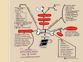
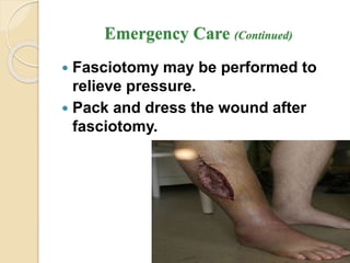
Musculoskeletal System
Assessment & Disorders
Dr Ibraheem Bashayreh, RN, PhD
Skeletal System
 Bone types
 Bone structure
 Bone function
 Bone growth and metabolism affected
by calcium and phosphorous,
calcitonin, vitamin D, parathyroid,
growth hormone, glucocorticoids,
estrogens and androgens, thyroxine,
and insulin.
Bones
 Human skeleton has 206 bones
 Provide structure and support for soft
tissue
 Protect vital organs
Figure 41-1 Bones of the human skeleton.
Figure 41-2 Classification of bones by shape.
Bones
 Compact bone
◦ Smooth and dense
◦ Forms shaft of long bones and outside
layer of other bones
 Spongy bone
◦ Contains spaces
◦ Spongy sections contain bone marrow
Bone Marrow
 Red bone marrow
◦ Found in flat bones of sternum, ribs, and
ileum
◦ Produces blood cells and hemoglobin
 Yellow bone marrow
◦ Found in shaft of long bones
◦ Contains fat and connective tissue
Joints (Articulations)
 Area where two or more bones meet
 Holds skeleton together while allowing
body to move
Joints
 Synarthrosis
◦ Immovable (e.g., skull)
 Amphiarthrosis
◦ Slightly movable (e.g., vertebral joints)
 Diarthrosis or synovial
◦ Freely movable (e.g., shoulders, hips)
Synovial Joints
 Found at all limb articulations
 Surface covered with cartilage
 Joint cavity covered with tough fibrous
capsule
 Cavity lined with synovial membrane
and filled with synovial fluid
Ligaments
 Bands of connective tissue that
connect bone to bone
 Either limit or enhance movement
 Provide joint stability
 Enhance joint strength
Tendons
 Fibrous connective tissue bands that
connect bone to muscles
 Enable bones to move when muscles
contract
Muscles
 Skeletal (voluntary)
◦ Allows voluntary movement
 Smooth (involuntary)
◦ Muscle movement controlled by internal
mechanism
◦ e.g., muscles in bladder wall and GI
system
 Cardiac (involuntary)
◦ Found in heart
Skeletal Muscle
 600 skeletal muscles
 Made up of thick bundles of parallel
fibers
 Each muscle fiber made up of smaller
structure myofibrils
 Myofibrils are strands of repeating
units called sarcomeres
Skeletal Muscle
 Skeletal muscle contracts with the
release of acetylcholine
 The more fibers that contract, the
stronger the muscle contraction
Changes in Older Adult
 Musculoskeletal changes can be due
to:
◦ Aging process
◦ Decreased activity
◦ Lifestyle factors
Changes in Older Adult
 Loss of bone mass in older women
 Joint and disk cartilage dehydrates
causing loss of flexibility contributes to
degenerative joint disease
(osteoarthritis); joints stiffen, lose
range of motion
Changes in Older Adult
 Cause stooped posture, changing
center of gravity
 Elderly at greater risk for falls
 Endocrine changes cause skeletal
muscle atrophy
 Muscle tone decreases
Assessment
 Health history
 Chief complaint
 Onset of problem
 Effect on ADLs
 Precipitating events, e.g., trauma
Assessment
 Examine complaints of pain for
location, duration, radiation character
(sharp dull), aggravating, or alleviating
factors
 Inquire about fever, fatigue, weight
changes, rash, or swelling
Physical Examination
 Posture
 Gait
 Ability to walk with or without assistive
devices
 Ability to feed, toilet, and dress self
 Muscle mass and symmetry
Physical Examination
 Inspect and palpate bone, joints for
visible deformities, tenderness or pain,
swelling, warmth, and ROM
 Assess and compare corresponding
joints
 Palpate joints knees and shoulder for
crepitus
Physical Examination
 Never attempt to move a joint past
normal ROM or past point where
patient experiences pain
 Bulge sign and ballottement sign used
to assess for fluid in the knee joint
 Thomas test performed when hip
flexion contracture suspected
Figure 41-4 Checking for the bulge sign.
Figure 41-5 Checking for ballottement.
Diagnostic Tests
 Blood tests
 Arthrocentesis
 X-rays
 Bone density scan
 CT scan
 MRI
 Ultrasound
 Bone scan
Diagnostic Evaluation
 Imaging Procedures – CT, Bone Scan, MRI
 Nuclear Studies - radioisotope bone density,
 Endoscopic Studies –arthrocentesis,
arthroscopy
 Other Studies –biopsy, synovial fluid,
Arthrogram, venogram,
 Electromyography
 Myelography*
 Laboratory Studies
Musculoskeletal
Assessment – Diagnostic Test
 Laboratory
◦ Urine Tests
 24 hour creatine-
creatinine ratio
 Urine Uric acid –24
hr specimen
 Urine deoxypyridino-
line
 Laboratory
◦ Blood Tests
 Serum muscle
enzymes
 Rheumatoid Factor
 LE Prep/Antinuclear
Antibodies(ANA)
 Erythrocyte
Sedimentation Rate
 Calcium,
Phosphorous, Alkaline
phosphatase
Muscoluloskeletal
Assessment – Diagnostic
 Blood Tests
◦ CBC – Hgb, Hct
◦ Acid phosphatase
◦ Metabolic/Endocrine
◦ Enzymes
Increase creatine kinase,
serum increase
glutamin-oxaloacetic
due to muscle damage,
aldolase, SGOT
Musculoskeletal - Radiographic
 Standard radiography, tomography
and xeroradiography, myelography,
arthrography and CT
 Other diagnostic tests: bone and
muscle biopsy
Arthroscopy
 Fiberoptic tube is inserted into a joint
for direct visualization.
 Client must be able to flex the knee;
exercises are prescribed for ROM.
 Evaluate the neurovascular status of
the affected limb frequently.
 Analgesics are prescribed.
 Monitor for complications.
Bone Scan
 Nuclear medicine procedure in which
amount of radioactive isotope taken
up by bones is evaluated
 Abnormal bone scans show hot spots
due to malignancies or infection
 Cold spot uptakes show areas of bone
that are ischemic
Arthroscopy
 Flexible fiberoptic endoscope used to
view joint structures and tissues
 Used to identify:
◦ Torn tendon and ligaments
◦ Injured meniscus
◦ Inflammatory joint changes
◦ Damaged cartilage
Interventions for Clients with
Musculoskeletal Trauma
 Tissue is subjected to more force than
it can absorb
 Severity depends on:
◦ Amount of force
◦ Location of impact
Musculoskeletal Trauma
 Mild to severe
 Soft tissue
 Fractures
◦ Affect function of muscle, tendons, and
ligaments
 Complete amputation
Preventing Trauma
 Teach importance of using safety
equipment
◦ Seat belts
◦ Bicycle helmets
◦ Football pads
◦ Proper footwear
◦ Protective eyewear
◦ Hard hats
Soft Tissue Trauma
 Contusion
◦ Bleeding into soft tissue
◦ Significant bleeding can cause a
hematoma
◦ Swelling and discoloration (bruise)
Soft Tissue Trauma - Sprain
 Ligament injury (Excessive stretching
of a ligament)
 Twisting motion
 Overstretching or tear
◦ Grade I—mild bleeding and inflammation
◦ Grade II—severe stretching and some
tearing and inflammation and hematoma
◦ Grade III—complete tearing of ligament
◦ Grade IV—bony attachment of ligament
broken away
Sprains
 Treatment of sprains:
◦ first-degree: rest, ice for 24 to 48 hr,
compression bandage, and elevation
◦ second-degree: immobilization, partial
weight bearing as tear heals
◦ third-degree: immobilization for 4 to 6
weeks, possible surgery
Soft Tissue Trauma - Strain
 Microscopic tear in the muscle
 May cause bleeding
 “Pulled muscle”
 Inappropriate lifting or sudden
acceleration-deceleration
Soft Tissue Trauma
 To decrease swelling and pain, and
encourage rest
◦ Ice for first 48 hours
◦ Splint to support extremities and limit
movement
◦ Compression dressing
◦ Elevation to increase venous return and
decrease swelling
◦ NSAIDs
Soft Tissue Trauma
 Diagnosis
◦ X-ray to rule out fracture
◦ MRI
Fractures
 Break in the continuity of bone
◦ Direct blow
◦ Crushing force (compression)
◦ Sudden twisting motions (torsion)
◦ Severe muscle contraction
◦ Disease (pathologic fracture)
Fractures
Classification of Fractures
 Closed or simple
 Open or compound
 Complete or incomplete
 Stable or unstable
 Direction of the fracture line
◦ Oblique
◦ Spiral
◦ Lengthwise plane (greenstick)
Stages of Bone Healing
 Hematoma formation within 48 to 72 hr
after injury
 Hematoma to granulation tissue
 Callus formation
 Osteoblastic proliferation
 Bone remodeling
 Bone healing completed within about 6
weeks; up to 6 months in the older
person
Fractures – Emergency Care
 Immobilize before moving client
 Joint above and below
 Check pulse, color, movement,
sensation before splinting
 Sterile dressing for open wounds
Fractures – Emergency Care
 Fracture reduction
◦ Closed—external manipulation
◦ Open—surgery
Acute Compartment Syndrome
 Serious condition in which
increased pressure within one or
more compartments causes
massive compromise of circulation
to the area
 Prevention of pressure buildup of
blood or fluid accumulation
 Pathophysiologic changes
sometimes referred to as ischemia-
edema cycle
Emergency Care - Acute
Compartment Syndrome
 Within 4 to 6 hr after the onset of
acute compartment syndrome,
neuromuscular damage is
irreversible; the limb can become
useless within 24 to 48 hr.
 Monitor compartment pressures.
(Continued)
Emergency Care (Continued)
 Fasciotomy may be performed to
relieve pressure.
 Pack and dress the wound after
fasciotomy.
Possible Results of Acute Compartment
Syndrome
 Infection
 Motor weakness
 Volkmann’s contractures: (a deformity of
the hand, fingers, and wrist caused by a lack of blood
flow (ischemia) to the muscles of the forearm)
Other Complications of Fractures
 Shock
 Fat embolism syndrome: serious
complication resulting from a
fracture; fat globules are released
from yellow bone marrow into
bloodstream
 Venous thromboembolism
(Continued)
Other Complications of Fractures
(Continued)
 Infection
 Ischemic necrosis
 Fracture blisters, delayed union,
nonunion, and malunion
Musculoskeletal
Complications (continued)
 Muscle Atrophy, loss of muscle strength
range of motion, pressure ulcers, and other
problems associated with immobility
 Embolism/Pneumonia/ARDS
◦ TREATMENT – hydration, albumin,
corticosteroids
 Constipation/Anorexia
 UTI
 DVT
Musculoskeletal Assessment - Fracture
 Change in bone alignment
 Alteration in length of extremity
 Change in shape of bone
 Pain upon movement
 Decreased ROM
 Crepitation
 Ecchymotic skin
(Continued)
Musculoskeletal Assessment – Fracture
(Continued)
 Subcutaneous emphysema with
bubbles under the skin
 Swelling at the fracture site
Special Assessment Considerations
 For fractures of the shoulder and upper
arm, assess client in sitting or standing
position.
 Support the affected arm to promote
comfort.
 For distal areas of the arm, assess
client in a supine position.
 For fracture of lower extremities and
pelvis, client is in supine position.
CAST
CAST
Casts
 Rigid device that immobilizes the
affected body part while allowing other
body parts to move
 Cast materials: plaster, fiberglass,
polyester-cotton
 Types of casts for various parts of the
body: arm, leg, brace, body
(Continued)
Casts (Continued)
 Cast care and client education
 Cast complications: infection,
circulation impairment, peripheral
nerve damage, complications of
immobility
Managing Care of the Patient in a Cast
 Casting Materials
 Relieving Pain
 Improving Mobility
 Promoting Healing
 Neurovascular Function
 Potential Complications
Cast, Splint, Braces, and Traction
Management Considerations
 Arm Casts
 Leg Casts
 Body or Spica Casts
 Splints and Braces
 External Fixator
 Traction
POLYESTER/FIBERGLASS
UPPER EXTREMITY CAST
LOWER EXTREMITY CAST
Musculoskeletal
Nursing Care - Casts
◦ Neurovascular
 Check
color/capillary refill
 Temperature
 Pulse
 Movement
 Sensation
 Traction Nursing Care
◦ Pin Site care
◦ Skin and
neurovascular check
Cast Care (continued)
 Elevate Extremity
 Exercises – to unaffected side; isometric
exercises to affected extremity
 Keep heel off mattress
 Handle with palms of hands if cast wet
 Turn every two hours till dry
 Notify MD at once of wound drainage
 Do not place items under cast.
Traction
 Application of a pulling force to the
body to provide reduction,
alignment, and rest at that site
 Types of traction: skin, skeletal,
plaster, brace, circumferential
(Continued)
Traction (Continued)
 Traction care:
◦ Maintain correct balance between
traction pull and counter traction force
◦ Care of weights
◦ Skin inspection
◦ Pin care
◦ Assessment of neurovascular status
Musculoskeletal – Fractures
Treatment
 Primary Goal – reduce fracture-
◦ Realign and immobilize
 Medications
◦ Analgesics, antibiotics, tetanus toxoid
 Closed Reduction – Manual and Cast;
External Fixation Device
 Traction; Splints; Braces
 Surgery
◦ Open reduction with internal fixation
◦ Reconstructive surgery
◦ Endoprosthetic replacement
Figure 42-5 In external fixation, pins placed through the bone above and below the fracture are attached to external
fixation rods that hold the pins and bone in place.
Nursing Management
 Positioning
 Strengthening Exercises
 Potential Complications
Musculoskeletal
Nursing Care
 Promote comfort
 Assess infection
 Promote mobility
 Teach safety
 Vital Signs
 Flotation, sheep skin
 Nutrition
 Vital Signs
 Monitor elimination
 Elevate extremity
to decrease
swelling/ ice pack
 Teach skin care,
cast care, diet,
complications
Operative Procedures
 Open reduction with internal
fixation
 External fixation
 Postoperative care: similar to that
for any surgery; certain
complications specific to fractures
and musculoskeletal surgery
include fat embolism and venous
thromboembolism
Managing the Patient Undergoing
Orthopedic Surgery
 Joint Replacement
 Total Hip Replacement
 Total Knee Replacement
Risk for Infection
 Interventions include:
◦ Apply strict aseptic technique for
dressing changes and wound
irrigations.
◦ Assess for local inflammation
◦ Report purulent drainage immediately
to health care provider.
(Continued)
Risk for Infection (Continued)
◦ Assess for pneumonia and urinary
tract infection.
◦ Administer broad-spectrum antibiotics
prophylactically.
Imbalanced Nutrition: Less Than Body
Requirements
 Interventions include:
◦ Diet high in protein, calories, and
calcium, supplemental vitamins B and
C
◦ Frequent small feedings and
supplements of high-protein liquids
◦ Intake of foods high in iron
Upper Extremity Fractures
 Fractures include those of the:
◦ Clavicle
◦ Scapula
◦ Humerus
◦ Olecranon
◦ Radius and ulna
◦ Wrist and hand
Lower Extremity Fractures
 Fractures include those of the:
◦ Femur
◦ Patella
◦ Tibia and fibula
◦ Ankle and foot
Fractures of the Hip
 Intracapsular or extracapsular
 Treatment of choice: surgical
repair, when possible, to allow the
older client to get out of bed
 Open reduction with internal
fixation
 Intramedullary rod, pins, a
prosthesis, or a fixed sliding plate
 Prosthetic device
Fractures of the Pelvis
 Associated internal damage the
chief concern in fracture
management of pelvic fractures
 Non–weight-bearing fracture of the
pelvis
 Weight-bearing fracture of the
pelvis
Compression Fractures of the Spine
 Most are associated with
osteoporosis rather than acute
spinal injury.
 Multiple hairline fractures result
when bone mass diminishes.
(Continued)
Compression Fractures of the Spine
(Continued)
 Nonsurgical management includes
bedrest, analgesics, and physical
therapy.
 Minimally invasive surgeries are
vertebroplasty and kyphoplasty, in
which bone cement is injected.
(Continued)
Amputations
 Surgical amputation
 Traumatic amputation
 Levels of amputation
 Complications of amputations:
hemorrhage, infection, phantom
limb pain, problems associated
with immobility, neuroma (a growth or
tumour of nerve tissue), flexion contracture
Amputation
Nursing Management
◦ relieving pain
◦ minimizing altered sensory
perception
◦ promoting wound healing
◦ enhancing body image
◦ self-care
Phantom Limb Pain
 Phantom limb pain is a frequent
complication of amputation.
 Client complains of pain at the site
of the removed body part, most
often shortly after surgery.
 Pain is intense burning feeling,
crushing sensation or cramping.
 Some clients feel that the removed
body part is in a distorted position.
Management of Phantom Pain
 Phantom limb pain must be
distinguished from stump pain
because they are managed
differently.
 Recognize that this pain is real and
interferes with the amputee’s
activities of daily living.
(Continued)
Management of Phantom Pain
(Continued)
 Some studies have shown that
opioids are not as effective for
phantom limb pain as they are for
residual limb pain.
 Other drugs include intravenous
infusion calcitonin, beta blockers,
anticonvulsants, and
antispasmodics.
Exercise After Amputation
 ROM to prevent flexion
contractures, particularly of the hip
and knee
 Trapeze and overhead frame
 Firm mattress
 Prone position every 3 to 4 hours
 Elevation of lower-leg residual limb
controversial
Prostheses
 Devices to help shape and shrink
the residual limb and help client
readapt
 Wrapping of elastic bandages
 Individual fitting of the prosthesis;
special care
Crush Syndrome
 Can occur when leg or arm injury
includes multiple compartments
 Characterized by acute compartment
syndrome, hypovolemia, hyperkalemia,
rhabdomyolysis, and acute tubular
necrosis
 Treatment: adequate intravenous fluids,
low-dose dopamine, sodium
bicarbonate, kayexalate, and
hemodialysis
Metabolic Bone Disorders
Osteoporosis
Osteomalcia
Paget’s Disease
Osteoporosis
 A disease in which loss of bone exceeds
rate of bone formation; usually increase in
older women, white race, nulliparity.
 Clinical Manifestations – bone pain,
decrease movement.
 Treatment – Calcium, Vit. D, estrogen
replacement, Calcitonin, fluoride, estrogen
with progestin, SERM (Selective Estrogen
Receptor Modulator) with anti-estrogens,
exercise.
 Pathologic fracture-safety.
Classification of Osteoporosis
 Generalized osteoporosis occurs most
commonly in postmenopausal women
and men in their 60s and 70s.
 Secondary osteoporosis results from
an associated medical condition such
as hyperparathyroidism, long-term
drug therapy, long-term immobility.
 Regional osteoporosis occurs when a
limb is immobilized.
Health Promotion/Illness Prevention -
Osteoporosis
 Ensure adequate calcium intake.
 Avoid sedentary life style (a type of
lifestyle with a lack of physical
exercise) .
 Continue program of weight-
bearing exercises.
Osteoporosis - Assessment
 Physical assessment
 Psychosocial assessment
 Laboratory assessment
 Radiographic assessment
Os
O
s
t
e
o
p
r
o
s
i
s
O
s
t
e
o
p
r
o
r
s
i
Drug Therapy
Osteoporosis
 Hormone replacement therapy
 Parathyroid hormone
 Calcium and vitamin D
 Bisphosphonates
 Selective estrogen receptor
modulators
 Calcitonin
 Other agents used with varying
results
Diet Therapy - Osteoporosis
 Protein
 Magnesium
 Vitamin K
 Trace minerals
 Calcium and vitamin D
 Avoid alcohol and caffeine
Fall Prevention - Osteoporosis
 Hazard-free environment
 High-risk assessment through
programs such as Falling Star
protocol
 Hip protectors that prevent hip
fracture in case of a fall
Others - Osteoporosis
 Exercise
 Pain management
 Orthotic devices
Osteomalacia
 Softening of the bone tissue
characterized by inadequate
mineralization of osteoid
 Vitamin D deficiency, lack of
sunlight exposure
 Similar, but not the same as
osteoporosis
 Major treatment: vitamin D from
exposure to sun and certain foods
Paget’s Disease of the Bone
 Metabolic disorder of bone remodeling,
or turnover; increased resorption (the
process by which osteoclasts break down bone and
release the minerals, resulting in a transfer of calcium
from bone fluid to the blood) of loss results in
bone deposits that are weak, enlarged,
and disorganized
 Nonsurgical management: calcitonin,
selected bisphosphonates, mithramycin
 Surgical management: tibial osteotomy
or partial or total joint replacement
Paget’s Disease
 An imbalance of increase osteoblast and
osteoclast cells; thickening and
hypertrophy.
 Bone pain most common symptom;
bony enlargement and deformities
usually bilateral, kyphosis, long bone.
 Analgesics, meds bisphosphonates and
calcitonin, NSAID, assistance devices,
and hot/cold treatment.
Osteomyelitis
 A condition caused by the invasion
by one or more pathogenic
microorganisms that stimulates the
inflammatory response in bone
tissue
 Exogenous, endogenous,
hematogenous, contiguous
Osteomyelitis
 Infection of bone; causative agent – Staph/Strept
 Typical signs and symptoms : Acute osteomyelitis
include:
 Fever that may be abrupt
 Irritability or lethargy in young children
 Pain in the area of the infection
 Swelling, warmth and redness over the area of the
infection
 Chronic osteomyelitis include:
 Warmth, swelling and redness over the area of the
infection
 Pain or tenderness in the affected area
 Chronic fatigue
 Drainage from an open wound near the area of the
infection
 Fever, sometimes
 Treatment – IV antibiotic; long term for 4-6 months
Surgical Management
Osteomyelitis
 Sequestrectomy (Surgical removal of a
sequestrum), a detached piece of necrotic bone
that often migrates to a wound, abscess, etc.
 Bone grafts
 Bone segment transfers
 Muscle flaps
 Amputation
Bone Tumors
Benign Bone Tumors
Malignant Bone Tumors
Metastatic Bone Disease
Bone Tumors
 Benign bone tumors
(noncancerous):
◦ Chrondrogenic tumors:
osteochondroma, chondroma
◦ Osteogenic tumors: osteoid osteoma,
osteoblastoma, giant cell tumor
◦ Fibrogenic tumors
Interventions
 Nondrug pain relief measures
 Drug therapy: analgesics, NSAIDs
 Surgical therapy: curettage (simple
excision of the tumor tissue), joint
replacement, or arthrodesis
Malignant Bone Tumors
 Primary tumors, those tumors that
originate in the bone
◦ Osteosarcoma
◦ Ewing’s sarcoma
◦ Chondrosarcoma
◦ Fibrosarcoma
◦ Metastatic bone disease
Osteosarcoma
 Cancer of the bone – metastasis to the
lung is common. Most in long bones.
 Clinical manifestations – dull pain,
swelling, intermittent but increases per
time; night pain common.
 Treatment – radiation, chemotherapy,
hormonal therapy, surgical excision with
prosthetics, assistance devices,
palliative measures.
Treatment Cancer of Bone
 Interventions include:
◦ Treatment aimed at reducing the size or
removing the tumor
◦ Drug therapy; chemotherapy
◦ Radiation therapy
◦ Surgical management
◦ Promotion of physical mobility with ROM
exercises
Cancer of Bone
Anticipatory Grieving
 Interventions include:
◦ Active listening
◦ Encouraging client and family to
verbalize feelings
◦ Making appropriate referrals
◦ Helping client and others to cope with
the loss and grieving
◦ Promoting the physician-client
relationship
Cancer of Bone
Disturbed Body Image
 Interventions include:
◦ Recognize and accept the client’s view
of body image alteration.
◦ Establish and maintain a trusting
nurse-client relationship.
◦ Emphasize the client’s strengths and
remaining capabilities.
◦ Establish realistic mutual goals.
Potential for Fractures
Bone Cancer
 Interventions
◦ Nonsurgical management: radiation
therapy and strengthening exercises.
◦ Surgical management: replace as much of
the defective bone as possible, avoid a
second procedure, and return client to a
functioning state with a minimum of
hospitalization and immobilization.
Carpal Tunnel Syndrome
 Common condition; the median
nerve in the wrist becomes
compressed, causing pain and
numbness
 Common repetitive strain injury via
occupational or sports motions
 Nonsurgical management: drug
therapy and immobilization
 Possible surgical management
Scoliosis
 Abnormal spinal curvature of various
degrees or severity involving
shortening of muscles and
ligaments.
 Milwaukee brace (a back brace used in the
treatment of spinal curvatures) , internal
fixative devices.
Scoliosis
 Changes in muscles and ligaments
on the concave side of the spinal
column
 Congenital, neuromuscular, or
idiopathic in type
 Assessment: complete history, pain
assessment, observation of posture
 Interventions: exercise, weight
reduction, bracing, casting, surgery
Musculoskeletal System lecture 3.ppt

Musculoskeletal System lecture 3.ppt

  • 1.
    Musculoskeletal System Assessment &Disorders Dr Ibraheem Bashayreh, RN, PhD
  • 2.
    Skeletal System  Bonetypes  Bone structure  Bone function  Bone growth and metabolism affected by calcium and phosphorous, calcitonin, vitamin D, parathyroid, growth hormone, glucocorticoids, estrogens and androgens, thyroxine, and insulin.
  • 3.
    Bones  Human skeletonhas 206 bones  Provide structure and support for soft tissue  Protect vital organs
  • 4.
    Figure 41-1 Bonesof the human skeleton.
  • 5.
    Figure 41-2 Classificationof bones by shape.
  • 6.
    Bones  Compact bone ◦Smooth and dense ◦ Forms shaft of long bones and outside layer of other bones  Spongy bone ◦ Contains spaces ◦ Spongy sections contain bone marrow
  • 7.
    Bone Marrow  Redbone marrow ◦ Found in flat bones of sternum, ribs, and ileum ◦ Produces blood cells and hemoglobin  Yellow bone marrow ◦ Found in shaft of long bones ◦ Contains fat and connective tissue
  • 8.
    Joints (Articulations)  Areawhere two or more bones meet  Holds skeleton together while allowing body to move
  • 9.
    Joints  Synarthrosis ◦ Immovable(e.g., skull)  Amphiarthrosis ◦ Slightly movable (e.g., vertebral joints)  Diarthrosis or synovial ◦ Freely movable (e.g., shoulders, hips)
  • 10.
    Synovial Joints  Foundat all limb articulations  Surface covered with cartilage  Joint cavity covered with tough fibrous capsule  Cavity lined with synovial membrane and filled with synovial fluid
  • 11.
    Ligaments  Bands ofconnective tissue that connect bone to bone  Either limit or enhance movement  Provide joint stability  Enhance joint strength
  • 12.
    Tendons  Fibrous connectivetissue bands that connect bone to muscles  Enable bones to move when muscles contract
  • 13.
    Muscles  Skeletal (voluntary) ◦Allows voluntary movement  Smooth (involuntary) ◦ Muscle movement controlled by internal mechanism ◦ e.g., muscles in bladder wall and GI system  Cardiac (involuntary) ◦ Found in heart
  • 14.
    Skeletal Muscle  600skeletal muscles  Made up of thick bundles of parallel fibers  Each muscle fiber made up of smaller structure myofibrils  Myofibrils are strands of repeating units called sarcomeres
  • 15.
    Skeletal Muscle  Skeletalmuscle contracts with the release of acetylcholine  The more fibers that contract, the stronger the muscle contraction
  • 16.
    Changes in OlderAdult  Musculoskeletal changes can be due to: ◦ Aging process ◦ Decreased activity ◦ Lifestyle factors
  • 17.
    Changes in OlderAdult  Loss of bone mass in older women  Joint and disk cartilage dehydrates causing loss of flexibility contributes to degenerative joint disease (osteoarthritis); joints stiffen, lose range of motion
  • 18.
    Changes in OlderAdult  Cause stooped posture, changing center of gravity  Elderly at greater risk for falls  Endocrine changes cause skeletal muscle atrophy  Muscle tone decreases
  • 19.
    Assessment  Health history Chief complaint  Onset of problem  Effect on ADLs  Precipitating events, e.g., trauma
  • 20.
    Assessment  Examine complaintsof pain for location, duration, radiation character (sharp dull), aggravating, or alleviating factors  Inquire about fever, fatigue, weight changes, rash, or swelling
  • 21.
    Physical Examination  Posture Gait  Ability to walk with or without assistive devices  Ability to feed, toilet, and dress self  Muscle mass and symmetry
  • 25.
    Physical Examination  Inspectand palpate bone, joints for visible deformities, tenderness or pain, swelling, warmth, and ROM  Assess and compare corresponding joints  Palpate joints knees and shoulder for crepitus
  • 26.
    Physical Examination  Neverattempt to move a joint past normal ROM or past point where patient experiences pain  Bulge sign and ballottement sign used to assess for fluid in the knee joint  Thomas test performed when hip flexion contracture suspected
  • 27.
    Figure 41-4 Checkingfor the bulge sign.
  • 28.
    Figure 41-5 Checkingfor ballottement.
  • 29.
    Diagnostic Tests  Bloodtests  Arthrocentesis  X-rays  Bone density scan  CT scan  MRI  Ultrasound  Bone scan
  • 30.
    Diagnostic Evaluation  ImagingProcedures – CT, Bone Scan, MRI  Nuclear Studies - radioisotope bone density,  Endoscopic Studies –arthrocentesis, arthroscopy  Other Studies –biopsy, synovial fluid, Arthrogram, venogram,  Electromyography  Myelography*  Laboratory Studies
  • 31.
    Musculoskeletal Assessment – DiagnosticTest  Laboratory ◦ Urine Tests  24 hour creatine- creatinine ratio  Urine Uric acid –24 hr specimen  Urine deoxypyridino- line  Laboratory ◦ Blood Tests  Serum muscle enzymes  Rheumatoid Factor  LE Prep/Antinuclear Antibodies(ANA)  Erythrocyte Sedimentation Rate  Calcium, Phosphorous, Alkaline phosphatase
  • 32.
    Muscoluloskeletal Assessment – Diagnostic Blood Tests ◦ CBC – Hgb, Hct ◦ Acid phosphatase ◦ Metabolic/Endocrine ◦ Enzymes Increase creatine kinase, serum increase glutamin-oxaloacetic due to muscle damage, aldolase, SGOT
  • 33.
    Musculoskeletal - Radiographic Standard radiography, tomography and xeroradiography, myelography, arthrography and CT  Other diagnostic tests: bone and muscle biopsy
  • 34.
    Arthroscopy  Fiberoptic tubeis inserted into a joint for direct visualization.  Client must be able to flex the knee; exercises are prescribed for ROM.  Evaluate the neurovascular status of the affected limb frequently.  Analgesics are prescribed.  Monitor for complications.
  • 36.
    Bone Scan  Nuclearmedicine procedure in which amount of radioactive isotope taken up by bones is evaluated  Abnormal bone scans show hot spots due to malignancies or infection  Cold spot uptakes show areas of bone that are ischemic
  • 37.
    Arthroscopy  Flexible fiberopticendoscope used to view joint structures and tissues  Used to identify: ◦ Torn tendon and ligaments ◦ Injured meniscus ◦ Inflammatory joint changes ◦ Damaged cartilage
  • 38.
    Interventions for Clientswith Musculoskeletal Trauma
  • 39.
    Musculoskeletal Trauma  Tissueis subjected to more force than it can absorb  Severity depends on: ◦ Amount of force ◦ Location of impact
  • 40.
    Musculoskeletal Trauma  Mildto severe  Soft tissue  Fractures ◦ Affect function of muscle, tendons, and ligaments  Complete amputation
  • 41.
    Preventing Trauma  Teachimportance of using safety equipment ◦ Seat belts ◦ Bicycle helmets ◦ Football pads ◦ Proper footwear ◦ Protective eyewear ◦ Hard hats
  • 42.
    Soft Tissue Trauma Contusion ◦ Bleeding into soft tissue ◦ Significant bleeding can cause a hematoma ◦ Swelling and discoloration (bruise)
  • 43.
    Soft Tissue Trauma- Sprain  Ligament injury (Excessive stretching of a ligament)  Twisting motion  Overstretching or tear ◦ Grade I—mild bleeding and inflammation ◦ Grade II—severe stretching and some tearing and inflammation and hematoma ◦ Grade III—complete tearing of ligament ◦ Grade IV—bony attachment of ligament broken away
  • 44.
    Sprains  Treatment ofsprains: ◦ first-degree: rest, ice for 24 to 48 hr, compression bandage, and elevation ◦ second-degree: immobilization, partial weight bearing as tear heals ◦ third-degree: immobilization for 4 to 6 weeks, possible surgery
  • 45.
    Soft Tissue Trauma- Strain  Microscopic tear in the muscle  May cause bleeding  “Pulled muscle”  Inappropriate lifting or sudden acceleration-deceleration
  • 46.
    Soft Tissue Trauma To decrease swelling and pain, and encourage rest ◦ Ice for first 48 hours ◦ Splint to support extremities and limit movement ◦ Compression dressing ◦ Elevation to increase venous return and decrease swelling ◦ NSAIDs
  • 47.
    Soft Tissue Trauma Diagnosis ◦ X-ray to rule out fracture ◦ MRI
  • 49.
    Fractures  Break inthe continuity of bone ◦ Direct blow ◦ Crushing force (compression) ◦ Sudden twisting motions (torsion) ◦ Severe muscle contraction ◦ Disease (pathologic fracture)
  • 50.
    Fractures Classification of Fractures Closed or simple  Open or compound  Complete or incomplete  Stable or unstable  Direction of the fracture line ◦ Oblique ◦ Spiral ◦ Lengthwise plane (greenstick)
  • 51.
    Stages of BoneHealing  Hematoma formation within 48 to 72 hr after injury  Hematoma to granulation tissue  Callus formation  Osteoblastic proliferation  Bone remodeling  Bone healing completed within about 6 weeks; up to 6 months in the older person
  • 52.
    Fractures – EmergencyCare  Immobilize before moving client  Joint above and below  Check pulse, color, movement, sensation before splinting  Sterile dressing for open wounds
  • 53.
    Fractures – EmergencyCare  Fracture reduction ◦ Closed—external manipulation ◦ Open—surgery
  • 55.
    Acute Compartment Syndrome Serious condition in which increased pressure within one or more compartments causes massive compromise of circulation to the area  Prevention of pressure buildup of blood or fluid accumulation  Pathophysiologic changes sometimes referred to as ischemia- edema cycle
  • 56.
    Emergency Care -Acute Compartment Syndrome  Within 4 to 6 hr after the onset of acute compartment syndrome, neuromuscular damage is irreversible; the limb can become useless within 24 to 48 hr.  Monitor compartment pressures. (Continued)
  • 57.
    Emergency Care (Continued) Fasciotomy may be performed to relieve pressure.  Pack and dress the wound after fasciotomy.
  • 58.
    Possible Results ofAcute Compartment Syndrome  Infection  Motor weakness  Volkmann’s contractures: (a deformity of the hand, fingers, and wrist caused by a lack of blood flow (ischemia) to the muscles of the forearm)
  • 59.
    Other Complications ofFractures  Shock  Fat embolism syndrome: serious complication resulting from a fracture; fat globules are released from yellow bone marrow into bloodstream  Venous thromboembolism (Continued)
  • 60.
    Other Complications ofFractures (Continued)  Infection  Ischemic necrosis  Fracture blisters, delayed union, nonunion, and malunion
  • 61.
    Musculoskeletal Complications (continued)  MuscleAtrophy, loss of muscle strength range of motion, pressure ulcers, and other problems associated with immobility  Embolism/Pneumonia/ARDS ◦ TREATMENT – hydration, albumin, corticosteroids  Constipation/Anorexia  UTI  DVT
  • 62.
    Musculoskeletal Assessment -Fracture  Change in bone alignment  Alteration in length of extremity  Change in shape of bone  Pain upon movement  Decreased ROM  Crepitation  Ecchymotic skin (Continued)
  • 63.
    Musculoskeletal Assessment –Fracture (Continued)  Subcutaneous emphysema with bubbles under the skin  Swelling at the fracture site
  • 64.
    Special Assessment Considerations For fractures of the shoulder and upper arm, assess client in sitting or standing position.  Support the affected arm to promote comfort.  For distal areas of the arm, assess client in a supine position.  For fracture of lower extremities and pelvis, client is in supine position.
  • 65.
  • 66.
    Casts  Rigid devicethat immobilizes the affected body part while allowing other body parts to move  Cast materials: plaster, fiberglass, polyester-cotton  Types of casts for various parts of the body: arm, leg, brace, body (Continued)
  • 67.
    Casts (Continued)  Castcare and client education  Cast complications: infection, circulation impairment, peripheral nerve damage, complications of immobility
  • 68.
    Managing Care ofthe Patient in a Cast  Casting Materials  Relieving Pain  Improving Mobility  Promoting Healing  Neurovascular Function  Potential Complications
  • 69.
    Cast, Splint, Braces,and Traction Management Considerations  Arm Casts  Leg Casts  Body or Spica Casts  Splints and Braces  External Fixator  Traction
  • 71.
  • 72.
  • 74.
  • 78.
    Musculoskeletal Nursing Care -Casts ◦ Neurovascular  Check color/capillary refill  Temperature  Pulse  Movement  Sensation  Traction Nursing Care ◦ Pin Site care ◦ Skin and neurovascular check
  • 79.
    Cast Care (continued) Elevate Extremity  Exercises – to unaffected side; isometric exercises to affected extremity  Keep heel off mattress  Handle with palms of hands if cast wet  Turn every two hours till dry  Notify MD at once of wound drainage  Do not place items under cast.
  • 80.
    Traction  Application ofa pulling force to the body to provide reduction, alignment, and rest at that site  Types of traction: skin, skeletal, plaster, brace, circumferential (Continued)
  • 81.
    Traction (Continued)  Tractioncare: ◦ Maintain correct balance between traction pull and counter traction force ◦ Care of weights ◦ Skin inspection ◦ Pin care ◦ Assessment of neurovascular status
  • 84.
    Musculoskeletal – Fractures Treatment Primary Goal – reduce fracture- ◦ Realign and immobilize  Medications ◦ Analgesics, antibiotics, tetanus toxoid  Closed Reduction – Manual and Cast; External Fixation Device  Traction; Splints; Braces  Surgery ◦ Open reduction with internal fixation ◦ Reconstructive surgery ◦ Endoprosthetic replacement
  • 86.
    Figure 42-5 Inexternal fixation, pins placed through the bone above and below the fracture are attached to external fixation rods that hold the pins and bone in place.
  • 88.
    Nursing Management  Positioning Strengthening Exercises  Potential Complications
  • 89.
    Musculoskeletal Nursing Care  Promotecomfort  Assess infection  Promote mobility  Teach safety  Vital Signs  Flotation, sheep skin  Nutrition  Vital Signs  Monitor elimination  Elevate extremity to decrease swelling/ ice pack  Teach skin care, cast care, diet, complications
  • 90.
    Operative Procedures  Openreduction with internal fixation  External fixation  Postoperative care: similar to that for any surgery; certain complications specific to fractures and musculoskeletal surgery include fat embolism and venous thromboembolism
  • 91.
    Managing the PatientUndergoing Orthopedic Surgery  Joint Replacement  Total Hip Replacement  Total Knee Replacement
  • 93.
    Risk for Infection Interventions include: ◦ Apply strict aseptic technique for dressing changes and wound irrigations. ◦ Assess for local inflammation ◦ Report purulent drainage immediately to health care provider. (Continued)
  • 94.
    Risk for Infection(Continued) ◦ Assess for pneumonia and urinary tract infection. ◦ Administer broad-spectrum antibiotics prophylactically.
  • 95.
    Imbalanced Nutrition: LessThan Body Requirements  Interventions include: ◦ Diet high in protein, calories, and calcium, supplemental vitamins B and C ◦ Frequent small feedings and supplements of high-protein liquids ◦ Intake of foods high in iron
  • 96.
    Upper Extremity Fractures Fractures include those of the: ◦ Clavicle ◦ Scapula ◦ Humerus ◦ Olecranon ◦ Radius and ulna ◦ Wrist and hand
  • 97.
    Lower Extremity Fractures Fractures include those of the: ◦ Femur ◦ Patella ◦ Tibia and fibula ◦ Ankle and foot
  • 99.
    Fractures of theHip  Intracapsular or extracapsular  Treatment of choice: surgical repair, when possible, to allow the older client to get out of bed  Open reduction with internal fixation  Intramedullary rod, pins, a prosthesis, or a fixed sliding plate  Prosthetic device
  • 103.
    Fractures of thePelvis  Associated internal damage the chief concern in fracture management of pelvic fractures  Non–weight-bearing fracture of the pelvis  Weight-bearing fracture of the pelvis
  • 104.
    Compression Fractures ofthe Spine  Most are associated with osteoporosis rather than acute spinal injury.  Multiple hairline fractures result when bone mass diminishes. (Continued)
  • 105.
    Compression Fractures ofthe Spine (Continued)  Nonsurgical management includes bedrest, analgesics, and physical therapy.  Minimally invasive surgeries are vertebroplasty and kyphoplasty, in which bone cement is injected. (Continued)
  • 106.
    Amputations  Surgical amputation Traumatic amputation  Levels of amputation  Complications of amputations: hemorrhage, infection, phantom limb pain, problems associated with immobility, neuroma (a growth or tumour of nerve tissue), flexion contracture
  • 107.
    Amputation Nursing Management ◦ relievingpain ◦ minimizing altered sensory perception ◦ promoting wound healing ◦ enhancing body image ◦ self-care
  • 108.
    Phantom Limb Pain Phantom limb pain is a frequent complication of amputation.  Client complains of pain at the site of the removed body part, most often shortly after surgery.  Pain is intense burning feeling, crushing sensation or cramping.  Some clients feel that the removed body part is in a distorted position.
  • 109.
    Management of PhantomPain  Phantom limb pain must be distinguished from stump pain because they are managed differently.  Recognize that this pain is real and interferes with the amputee’s activities of daily living. (Continued)
  • 110.
    Management of PhantomPain (Continued)  Some studies have shown that opioids are not as effective for phantom limb pain as they are for residual limb pain.  Other drugs include intravenous infusion calcitonin, beta blockers, anticonvulsants, and antispasmodics.
  • 111.
    Exercise After Amputation ROM to prevent flexion contractures, particularly of the hip and knee  Trapeze and overhead frame  Firm mattress  Prone position every 3 to 4 hours  Elevation of lower-leg residual limb controversial
  • 114.
    Prostheses  Devices tohelp shape and shrink the residual limb and help client readapt  Wrapping of elastic bandages  Individual fitting of the prosthesis; special care
  • 115.
    Crush Syndrome  Canoccur when leg or arm injury includes multiple compartments  Characterized by acute compartment syndrome, hypovolemia, hyperkalemia, rhabdomyolysis, and acute tubular necrosis  Treatment: adequate intravenous fluids, low-dose dopamine, sodium bicarbonate, kayexalate, and hemodialysis
  • 116.
  • 117.
    Osteoporosis  A diseasein which loss of bone exceeds rate of bone formation; usually increase in older women, white race, nulliparity.  Clinical Manifestations – bone pain, decrease movement.  Treatment – Calcium, Vit. D, estrogen replacement, Calcitonin, fluoride, estrogen with progestin, SERM (Selective Estrogen Receptor Modulator) with anti-estrogens, exercise.  Pathologic fracture-safety.
  • 118.
    Classification of Osteoporosis Generalized osteoporosis occurs most commonly in postmenopausal women and men in their 60s and 70s.  Secondary osteoporosis results from an associated medical condition such as hyperparathyroidism, long-term drug therapy, long-term immobility.  Regional osteoporosis occurs when a limb is immobilized.
  • 119.
    Health Promotion/Illness Prevention- Osteoporosis  Ensure adequate calcium intake.  Avoid sedentary life style (a type of lifestyle with a lack of physical exercise) .  Continue program of weight- bearing exercises.
  • 120.
    Osteoporosis - Assessment Physical assessment  Psychosocial assessment  Laboratory assessment  Radiographic assessment
  • 122.
  • 123.
  • 124.
    Drug Therapy Osteoporosis  Hormonereplacement therapy  Parathyroid hormone  Calcium and vitamin D  Bisphosphonates  Selective estrogen receptor modulators  Calcitonin  Other agents used with varying results
  • 125.
    Diet Therapy -Osteoporosis  Protein  Magnesium  Vitamin K  Trace minerals  Calcium and vitamin D  Avoid alcohol and caffeine
  • 126.
    Fall Prevention -Osteoporosis  Hazard-free environment  High-risk assessment through programs such as Falling Star protocol  Hip protectors that prevent hip fracture in case of a fall
  • 127.
    Others - Osteoporosis Exercise  Pain management  Orthotic devices
  • 128.
    Osteomalacia  Softening ofthe bone tissue characterized by inadequate mineralization of osteoid  Vitamin D deficiency, lack of sunlight exposure  Similar, but not the same as osteoporosis  Major treatment: vitamin D from exposure to sun and certain foods
  • 129.
    Paget’s Disease ofthe Bone  Metabolic disorder of bone remodeling, or turnover; increased resorption (the process by which osteoclasts break down bone and release the minerals, resulting in a transfer of calcium from bone fluid to the blood) of loss results in bone deposits that are weak, enlarged, and disorganized  Nonsurgical management: calcitonin, selected bisphosphonates, mithramycin  Surgical management: tibial osteotomy or partial or total joint replacement
  • 130.
    Paget’s Disease  Animbalance of increase osteoblast and osteoclast cells; thickening and hypertrophy.  Bone pain most common symptom; bony enlargement and deformities usually bilateral, kyphosis, long bone.  Analgesics, meds bisphosphonates and calcitonin, NSAID, assistance devices, and hot/cold treatment.
  • 131.
    Osteomyelitis  A conditioncaused by the invasion by one or more pathogenic microorganisms that stimulates the inflammatory response in bone tissue  Exogenous, endogenous, hematogenous, contiguous
  • 132.
    Osteomyelitis  Infection ofbone; causative agent – Staph/Strept  Typical signs and symptoms : Acute osteomyelitis include:  Fever that may be abrupt  Irritability or lethargy in young children  Pain in the area of the infection  Swelling, warmth and redness over the area of the infection  Chronic osteomyelitis include:  Warmth, swelling and redness over the area of the infection  Pain or tenderness in the affected area  Chronic fatigue  Drainage from an open wound near the area of the infection  Fever, sometimes  Treatment – IV antibiotic; long term for 4-6 months
  • 133.
    Surgical Management Osteomyelitis  Sequestrectomy(Surgical removal of a sequestrum), a detached piece of necrotic bone that often migrates to a wound, abscess, etc.  Bone grafts  Bone segment transfers  Muscle flaps  Amputation
  • 134.
    Bone Tumors Benign BoneTumors Malignant Bone Tumors Metastatic Bone Disease
  • 135.
    Bone Tumors  Benignbone tumors (noncancerous): ◦ Chrondrogenic tumors: osteochondroma, chondroma ◦ Osteogenic tumors: osteoid osteoma, osteoblastoma, giant cell tumor ◦ Fibrogenic tumors
  • 136.
    Interventions  Nondrug painrelief measures  Drug therapy: analgesics, NSAIDs  Surgical therapy: curettage (simple excision of the tumor tissue), joint replacement, or arthrodesis
  • 137.
    Malignant Bone Tumors Primary tumors, those tumors that originate in the bone ◦ Osteosarcoma ◦ Ewing’s sarcoma ◦ Chondrosarcoma ◦ Fibrosarcoma ◦ Metastatic bone disease
  • 138.
    Osteosarcoma  Cancer ofthe bone – metastasis to the lung is common. Most in long bones.  Clinical manifestations – dull pain, swelling, intermittent but increases per time; night pain common.  Treatment – radiation, chemotherapy, hormonal therapy, surgical excision with prosthetics, assistance devices, palliative measures.
  • 139.
    Treatment Cancer ofBone  Interventions include: ◦ Treatment aimed at reducing the size or removing the tumor ◦ Drug therapy; chemotherapy ◦ Radiation therapy ◦ Surgical management ◦ Promotion of physical mobility with ROM exercises
  • 140.
    Cancer of Bone AnticipatoryGrieving  Interventions include: ◦ Active listening ◦ Encouraging client and family to verbalize feelings ◦ Making appropriate referrals ◦ Helping client and others to cope with the loss and grieving ◦ Promoting the physician-client relationship
  • 141.
    Cancer of Bone DisturbedBody Image  Interventions include: ◦ Recognize and accept the client’s view of body image alteration. ◦ Establish and maintain a trusting nurse-client relationship. ◦ Emphasize the client’s strengths and remaining capabilities. ◦ Establish realistic mutual goals.
  • 142.
    Potential for Fractures BoneCancer  Interventions ◦ Nonsurgical management: radiation therapy and strengthening exercises. ◦ Surgical management: replace as much of the defective bone as possible, avoid a second procedure, and return client to a functioning state with a minimum of hospitalization and immobilization.
  • 143.
    Carpal Tunnel Syndrome Common condition; the median nerve in the wrist becomes compressed, causing pain and numbness  Common repetitive strain injury via occupational or sports motions  Nonsurgical management: drug therapy and immobilization  Possible surgical management
  • 144.
    Scoliosis  Abnormal spinalcurvature of various degrees or severity involving shortening of muscles and ligaments.  Milwaukee brace (a back brace used in the treatment of spinal curvatures) , internal fixative devices.
  • 145.
    Scoliosis  Changes inmuscles and ligaments on the concave side of the spinal column  Congenital, neuromuscular, or idiopathic in type  Assessment: complete history, pain assessment, observation of posture  Interventions: exercise, weight reduction, bracing, casting, surgery