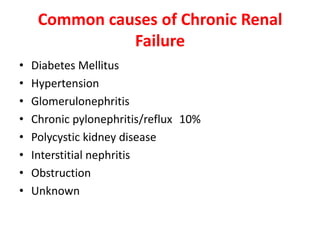
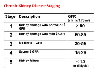
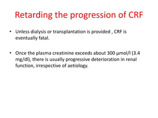
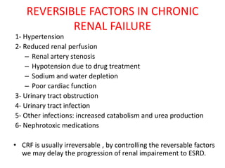
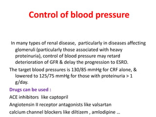
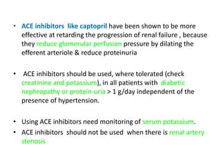
CHRONIC RENAL FAILURE

Chronic renal failure (CRF) refers to an irreversible deterioration in
renal function which classically develops over a period of years .
• Initially, CRF manifest only as a biochemical abnormality.

• Eventually, loss of the excretory, metabolic and endocrine
  functions of the kidney leads to the development of the
  clinical symptoms and signs of renal failure, which are
  referred to as uraemia.

• End stage renal disease (ESRD):renal failure which need renal
  replacement therapy.
• When death is likely without renal replacement therapy, it is
  called end-stage renal failure (ESRF).
Aetiology of CRF
• CRF may be caused by any condition which destroys the
  normal structure and function of the kidney.
Common causes of Chronic Renal
               Failure
•   Diabetes Mellitus
•   Hypertension
•   Glomerulonephritis
•   Chronic pylonephritis/reflux 10%
•   Polycystic kidney disease
•   Interstitial nephritis
•   Obstruction
•   Unknown
Diabetes (most common cause)      Hypertension


                                Chronic Glomerulonephritis
    Polycystic Kidney Disease
Pathogenesis of CRF
• Disturbances in water, electrolyte and acid-base balance
  contribute to the clinical picture in patients with CRF, but the
  exact pathogenesis of the clinical syndrome of uraemia is
  unknown.
• Many substances present in abnormal concentration in the
  plasma have been suspected as being 'uraemic toxins', and
  uraemia is probably caused by the accumulation of various
  intermediary products of metabolism.
Clinical Features of CRF
Major manifestations of Uremic
              Syndrome
The Uremic Syndrome:
Gastrointestinal
  -Anorexia , Nausea , Vomiting , Disturbance of taste , Gastritis
   Peptic ulcer , GI bleeding.
Cardiovascular
  - Cardiomegally , arrhythmias , pericarditis , accelerated
   atherosclerosis.
Pulmonary
  -Pneumonitis , pleuritis, pleural effusion , non-cardiogenic
   pulmonary oedema.
Haematologic
  - Anaemia , bleeding disorders , leukocyte dysfunction.
Skin
   -Pruritus , yellow pigmentation , earthy colour face.
Musculoskeletal
   -Muscle weakness , gout & pseudogout , renal osteodystrophy .
Endocrine / Metabolic
-Hyperparathyroidism , amenorrhea , impotence , hyperlipidemia
   increased insulin resistance.

Acid base / Electrolytes
-Acidosis , hyperkalemia , fluid overload , hypocalcemia
  , hyperphosphatemia , hypermagnesemia.

Nervous system
    -Central : irritability , insomnia , lethargy , seizures , coma.
    -Peripheral : glove-and stocking sensory loss , restless leg
  , foot drop or wrist drop.
Clinical assessment
• Renal failure may present as a raised blood urea and
  creatinine found during routine examination, often
  accompanied by hypertension, proteinuria or anaemia.
• When renal function deteriorates slowly, patients may remain
  asymptomatic until GFR falls below 20 - 30 ml/minute(
  normal range 80 – 120 mL/ min ).
• Nocturia, due to the loss of concentrating ability and
  increased osmotic load per nephron, is often an early
  symptom.
• Thereafter, due to the widespread effects of renal
  failure, symptoms and signs may develop that are related to
  almost every body system .
• Patients may present with complaints which are not
  obviously renal in origin, such as tiredness or
  breathlessness.
• In ESRF (stage 5) patients appear ill and anaemic.

• There may be unusually deep respiration related to
  metabolic acidosis (Kussmaul's respiration), anorexia and
  nausea.
• Later, hiccoughs, pruritus, vomiting, muscular
  twitching, fits, drowsiness and coma ensue.
• Specific manifestations of Uremia
Specific manifestations of Uremia
• Gastrointestinal manifestations

• Are common at low GFRs, including:
• anorexia followed by nausea, and vomiting is commonly seen.

• There is a higher incidence of peptic ulcer disease in uraemic
  patients.
Anaemia
• Anaemia is common in CRF; it usually correlates with the severity
  of renal failure and contributes to many of the non-specific
  symptoms of CRF.

• Several mechanisms are implicated(causes of anaemia inCRF):
 1- Relative deficiency of erythropoietin
 2- Diminished erythropoiesis due to toxic effects of uraemia on
  marrow precursor cells
 3- Reduced red cell survival
 4- Increased blood loss due to capillary fragility and poor platelet
  function
 5- Reduced dietary intake and absorption of iron and other
  haematinics.
• Plasma erythropoietin is usually within the normal range
  and thus inappropriately low for the degree of anaemia.

• In patients with polycystic kidneys, anaemia is often less
  severe or absent, while in some interstitial disorders it
  appears disproportionately severe for the degree of renal
  failure. This is probably because of the effects of these
  disorders on the interstitial fibroblasts that secrete
  erythropoietin
Acidosis
Declining renal function is associated with metabolic
   acidosis , which is often asymptomatic. There may be
   unusually deep respiration related to metabolic acidosis
   (Kussmaul's respiration).
Effects of metabolic acidosis :
• Sustained acidosis results in protons being buffered in
   bone in place of calcium, thus aggravating metabolic
   bone disease.
• Acidosis may also contribute to reduced renal function
   and increased tissue catabolism.
Cardiovascular disease and lipids
• CRF is an independent risk factor for occlusive cardiovascular
  disease.

• Hypertension develops in approximately 80% of patients
  with CRF.
   In part, this is caused by sodium retention.
  Chronically diseased kidneys also tend to hypersecrete
  renin, leading to high circulating concentrations of
  renin, angiotensin II and aldosterone.
  This is exaggerated if there is renal under-perfusion related to
  renal vascular disease.
  Hypertension must be controlled, as it causes further vascular
  and glomerular damage and worsening of renal failure

• Atherosclerosis is common and may be accelerated by
  hypertension.
• Pericarditis is common in untreated or inadequately
   treated ESRF.
  It may lead to pericardial tamponade and, later, constrictive
   pericarditis.

• Hypercholesterolaemia
   is almost universal in patients with significant proteinuria, and
   increased triglyceride levels are also common in patients with
   CRF.
  It has been suggested that as well as influencing the
   development of vascular disease, this may accelerate the
   progression of chronic renal disease.
Renal osteodystrophy
• This metabolic bone disease which accompanies CRF
  consists of a mixture of osteomalacia, hyperparathyroid
  bone disease (osteitis fibrosa), osteoporosis and
  osteosclerosis .
Osteomalacia
• Results from diminished activity of the renal 1α-
  hydroxylase enzyme, with failure to convert
  cholecalciferol to its active metabolite, 1,25-
  dihydroxycholecalciferol.
• A deficiency of the latter leads to diminished intestinal
  absorption of calcium, hypocalcaemia and reduction in
  the calcification of osteoid in bone.
• Osteitis fibrosa results from this secondary hyperparathyroidism
  .
• The parathyroid glands are stimulated by the low plasma
  calcium, and also by hyperphosphataemia.
• In some patients tertiary or autonomous hyperparathyroidism with
  hypercalcaemia develops.


• Osteoporosis occurs in many patients , possibly related to
  malnutrition.


• Osteosclerosis is seen mainly in the sacral area, at the base of the
  skull and in the vertebrae; the cause of this unusual reaction is not known.
Myopathy
• Generalised myopathy is due to a combination of
  poor nutrition, hyperparathyroidism, vitamin D
  deficiency and disorders of electrolyte metabolism.


• Muscle cramps are common, and quinine sulphate may
  be helpful.


• The 'restless leg syndrome', in which the patient's
  legs are jumpy during the night, may be troublesome and is
  often improved by clonazepam
Neuropathy
• Neuropathy results from demyelination of medullated
  fibres, with the longer fibres being involved at an earlier
  stage.

• Sensory neuropathy may cause paraesthesiae. Amitriptyline
  and gabapentin may provide some symptom relief.

• Motor neuropathy may present as foot drop.

• Uraemic autonomic neuropathy may cause delayed gastric
  empty-ing, diarrhoea and postural hypotension.

• Clinical manifestations of neuropathy appear late in the
  course of CRF but may improve or even resolve once dialysis
  is established.
Endocrine Function
    A number of hormonal abnormalities may be present of
    which the most important are hyperprolactinaemia &
    hyperparathyroidism .
•   In both sexes there is loss of libido and sexual
    function, related at least in part to hyperprolactinaemia .
    In women amenorrhoea is common.
•   The half-life of insulin is prolonged in CRF due to reduced
    tubular metabolism of insulin; insulin requirements may
    therefore decline in diabetic patients in end-stage CRF.
•   However, there is also a post-receptor defect in insulin
    action, leading to relative insulin resistance.
•   This latter abnormality is improved by dialysis treatment.
Bleeding
• There is an increased bleeding tendency in renal failure which
  manifests in patients with advanced disease as cutaneous
  ecchymoses and mucosal bleeds.

• Platelet function is impaired and bleeding time prolonged.

• Adequate dialysis treatment partially corrects the bleeding
  tendency.
Infection
• Cellular and humoral immunity are impaired, with increased
  susceptibility to infection.

• Infections are the second most common cause of death in
  dialysis patients, after cardiovascular disease; they must be
  recognised and treated promptly.
Acute or Chronic Renal failure?
•   History
•   Previous renal function test.
•   Small kidneys on u/s
•   Anaemia.
•   Bone changes.
• Investigations and management of CRF
Investigations and management of CRF
• There are several aspects to the management
  of CRF:
• Identify the underlying renal disease.
• Look for reversible factors which are making renal
  function worse .
• Attempt to prevent further renal damage.
• Attempt to limit the adverse effects of the loss of renal
  function.
• Institute renal replacement therapy (dialysis or
  transplantation)
.At presentation the nature of the underlying
 disease should be determined, if possible, by
history, examination, testing of
 biochemistry, immunology, radiology and
 biopsy .

• The degree of renal failure is assessed and
  complications are documented.
Investigations
 -Blood urea & serum creatinine : increased
- Serum electrolytes
        .serum calcium :decreased
        .serum potassium : increased ( risky )
        .serum uric acid : increased(but rarely cause gout )
        .serum phosphate : increased
General urine examination: for: protein , RBC , features of UTI ( may
   need urine culture ) , cast ( waxy broad cast is characteristic for CRF)
Complete blood picture: usually there is anaemia.
PH of blood : metabolic acidosis.
Viral markers : HBsAg , Anti-HCV Ab & HIV test (if the patient need
   dialysis ) (vaccination against hepatitis B if no previous infection;
   isolation of dialysis machine if positive) .
ECG: look for
  -Features of hyperkalemia (hyperacute T- wave ,then prolongation
   of PR- interval & QRS ,then loss of P-wave , & if not managed may
   cause asystole )
  -Features of pericardial effusion( low voltage ECG ).
  -Features of IHD.
Echocardiography: look for any evidence of pericardial effusion
                      or cardiomyopathy ( DCMP).
CXR: look for features of pulmonary oedema , pleural effusion , chest
   infection , enlarged cardiac shadow.
Abdominal Ultrasoud : look for the size & ecchogenecity of the
   kidneys & if there is ascitis.
    In CRF , usually bilateral small kidneys (except polycystic kidney
   disease , amyloidosis , hydronephrosis , diabetic nephropathy )
X- ray of bone : for evidence of renal osteodystrophy.
GFR


Renal biopsy : only indicated in CRF with normal size kidneys
  ( not indicated in CRF with small size kidneys ).

Patients often have bilateral small kidneys at presentation, and
  in such a situation renal biopsy is usually inadvisable because
  of the difficulty in making a histological diagnosis in severely
  damaged kidneys and the fact that treatment is unlikely to
  improve renal function significantly.
• If diagnosis is not known further investigation
  needed as :
•   Immunoglobulins and protein electrophoresis
•   Urinary Bence Jones protein
•   Complement
•   ANA: and dsDNA if ANA is positive
•   Rheumatoid factor
•   ANCA: in all possible inflammatory renal disease
•   Anti-GBM: in all possible inflammatory renal disease
•   Cryoglobulins: if cryoglobulinaemia is clinically suspected
Hyperkalemia & EKG
•   K > 5.5 -6
•   Tall, peaked T’s
•   Wide QRS
•   Prolong PR
•   Diminished P
•   Prolonged QT
•   QRS-T merge – sine
    wave
Chronic Kidney Disease Staging

Stage Description                        GFR
                                         (ml/min/1.73 m2)

   1    Kidney damage with normal or           90
        GFR
        Kidney damage with mild  GFR         60-89
   2
        Moderate  GFR                        30-59
   3
        Severe  GFR                          15-29
   4
        Kidney failure                         < 15
   5
                                            (or dialysis)
Retarding the progression of CRF
• Unless dialysis or transplantation is provided , CRF is
  eventually fatal.

• Once the plasma creatinine exceeds about 300 μmol/l (3.4
  mg/dl), there is usually progressive deterioration in renal
  function, irrespective of aetiology.
REVERSIBLE FACTORS IN CHRONIC
            RENAL FAILURE
1- Hypertension
2- Reduced renal perfusion
   – Renal artery stenosis
   – Hypotension due to drug treatment
   – Sodium and water depletion
   – Poor cardiac function
3- Urinary tract obstruction
4- Urinary tract infection
5- Other infections: increased catabolism and urea production
6- Nephrotoxic medications

• CRF is usually irreversable , by controlling the reversable factors
  we may delay the progression of renal impairement to ESRD.
Control of blood pressure

 In many types of renal disease, particularly in diseases affecting
   glomeruli (particularly those associated with heavy
   proteinuria), control of blood pressure may retard
   deterioration of GFR & delay the progression to ESRD.
The target blood pressures is 130/85 mmHg for CRF alone, &
   lowered to 125/75 mmHg for those with proteinuria > 1
   g/day.
Drugs can be used :
ACE inhibitors like captopril
Angiotensin II receptor antagonists like valsartan
calcium channel blockers like diltizem , amlodipine …
• ACE inhibitors like captopril have been shown to be more
  effective at retarding the progression of renal failure , because
  they reduce glomerular perfusion pressure by dilating the
  efferent arteriole & reduce proteinuria

• ACE inhibitors should be used, where tolerated (check
  creatinine and potassium), in all patients with diabetic
  nephropathy or protein-uria > 1 g/day independent of the
  presence of hypertension.

• Using ACE inhibitors need monitoring of serum potassium.
• ACE inhibitors should not be used when there is renal artery
  stenosis
Diet

Dietary protein
For patients on renal replacement therapy , severe protein
  restriction is not recommended because carry a risk of
  inducing malnutrition.
Moderate restriction (to 60 g protein per day) should be
  accompanied by an adequate intake of calories
  calories(30-35kcal/kg/d) to prevent malnutrition.
Anorexia and muscle loss may indicate a need to
  commence dialysis treatment.
Treatment of Anaemia of CRF
Recombinant human erythropoietin (Eprex ) is effective in
   correcting the anaemia of CRF .
  dose : 50 Iu / kg once or twice wk.ly
  route : subcutaneously (or IV for patients on haemodialysis ).
The target haemoglobin is usually between 10 and 12 g/dl.

Complications of treatment include increased blood pressure, and
  adjustment of antihypertensive medication is often necessary.
  There is also an increase in blood coagulability and an increased
  incidence of thrombosis of the arteriovenous fistulae used for
  haemodialysis.
Erythropoietin is less effective in the presence of :
iron deficiency, active inflammation or malignancy, or in patients with
   aluminium overload which may occur in dialysis.
 Causes of Erythropoietin resistant
• Iron deficiency
• Active inflamation
• Malignancy
• Secondary hyperparathyroid
• Aluminum overload
• Pure red cell aplasia

These factors should be sought and, if possible, corrected before
  treatment.

Iron supplementation should be used to keep ferritin > 100 μg/l and
   transferrin saturation > 20%.
Fluid and electrolyte balance
• Due to the reduced ability of the failing kidney to concentrate the
  urine, a relatively high urine volume is needed to excrete products
  of metabolism and a fluid intake of around 3 litres/day is desirable
   ( when there is no fluid overload “ no oedema “ ) fluid retention
  sometimes lead to episodic pulmonary oedema .
• Fluid intake:urine volume +500ml

.Limitation of potassium intake (e.g. 70 mmol/day) and
   sodium intake (e.g. 100 mmol/day) may be required in late CRF if
   there is evidence of accumulation
.Low phosphate diet(600-1000mg/d)
Hyperkalemia Treatment
•   Calcium gluconate
•   Sodium Bicarbonate
•   Insulin/glucose
•   Sodium bicarbonate
•   Frusemide
•   Salbutamol
•   Hemodialysis
• Some patients with so-called salt-wasting' disease
  may require a high sodium and water intake, including
  supplements of sodium salts, to prevent fluid depletion and
  worsening of renal function.
• This is most often seen in patients with renal cystic
  disease, obstructive uropathy, reflux nephropathy or other
  tubulo-interstitial diseases, and is not seen in patients with
  glomerular disease.
Treatment of Acidosis

• The plasma bicarbonate should be maintained above 22
  mmol/l by giving sodium bicarbonate supplements
  (starting dose of 1 g 8-hourly, increasing as required).

• The increased sodium intake may induce hypertension or
  oedema.
• calcium carbonate (up to 3 g daily) is an alternative that
  is also used to bind dietary phosphate.
Treatment of Hypercholesterolaemia
 • Statin ( as simvastatin ) achieve substantial
   reductions in lipids in chronic renal disease.

Hyperlipidemia is more if the CRF is due to glomerular diseases due
to the increased hepatic lipoprotein synthesis that is triggered by
reduced oncotic pressure .
Treatment of Bleeding
• Adequate dialysis treatment partially corrects the bleeding
  tendency.
Renal osteodystrophy
• To minimise the effects of CRF on bone, plasma calcium and
  phosphate should be kept as near to normal as possible.


• Treatment of Hypocalcaemia
• is corrected by giving 1α-hydroxylated synthetic analogues of
  vitamin D. The dose is adjusted to avoid hypercalcaemia. This
  will usually prevent or control osteomalacia.

•   .
• Treatment of Hyperphosphataemia
• Is controlled by dietary restriction of foods with high
  phosphate content (milk, cheese, eggs)
• The use of phosphate-binding drugs administered with
  food.
• These agents form insoluble complexes with dietary
  phosphate and prevent its absorption (e.g. calcium
  carbonate 500 mg with each meal and aluminium
  hydroxide 300 – 600 mg before each meal).we should
  avoid aluminium toxicity.

• Secondary hyperparathyroidism is usually prevented or
  controlled by these measures but, in severe bone disease
  with autonomous parathyroid
  function, parathyroidectomy may become necessary
Treatment of renal osteodystropy
Vitamin D (0.25ug/d for prophylactic, 0.5ug/d
  for symptomatic)
Low phosphate diet
Calcium carbonate (1-6g/d)
parathyroidectomy
Treatment of Myopathy
• Muscle cramps are common, and quinine sulphate may be
  helpful.

• The 'restless leg syndrome', in which the patient's legs are
  jumpy during the night, may be troublesome and is often
  improved by clonazepam
Treatment of Neuropathy

• Clinical manifestations of neuropathy appear late in the
  course of CRF but may improve or even resolve once dialysis
  is established.

• Sensory neuropathy may cause paraesthesiae. Amitriptyline
  and gabapentin may provide some symptom relief.
Treatment of Endocrine disorders


• Treatment with dopamine agonists is sometimes
  useful for amenorrhoea or galactorrhoea in women

• Insulin requirements usually decline in diabetic
  patients in end-stage CRF ( CRF patients are liable for
  hypoglycemia ).

• insulin resistance is improved by dialysis treatment.
Gastrointestinal manifestations
• Are common at low GFRs, including anorexia followed by
  nausea, and vomiting is commonly seen & improve by
  dialysis.
• There is a higher incidence of peptic ulcer disease in uraemic
  patients and H2-receptor antagonists or proton pump
  inhibitors are commonly used.
Depression
• Depression is common in patients on or
  approaching renal replacement therapy and
  support should be provided for both them and
  their relatives

medicine.CRF2.(dr.kawa)

  • 1.
  • 2.
    CHRONIC RENAL FAILURE Chronicrenal failure (CRF) refers to an irreversible deterioration in renal function which classically develops over a period of years .
  • 3.
    • Initially, CRFmanifest only as a biochemical abnormality. • Eventually, loss of the excretory, metabolic and endocrine functions of the kidney leads to the development of the clinical symptoms and signs of renal failure, which are referred to as uraemia. • End stage renal disease (ESRD):renal failure which need renal replacement therapy. • When death is likely without renal replacement therapy, it is called end-stage renal failure (ESRF).
  • 4.
    Aetiology of CRF •CRF may be caused by any condition which destroys the normal structure and function of the kidney.
  • 5.
    Common causes ofChronic Renal Failure • Diabetes Mellitus • Hypertension • Glomerulonephritis • Chronic pylonephritis/reflux 10% • Polycystic kidney disease • Interstitial nephritis • Obstruction • Unknown
  • 6.
    Diabetes (most commoncause) Hypertension Chronic Glomerulonephritis Polycystic Kidney Disease
  • 7.
    Pathogenesis of CRF •Disturbances in water, electrolyte and acid-base balance contribute to the clinical picture in patients with CRF, but the exact pathogenesis of the clinical syndrome of uraemia is unknown. • Many substances present in abnormal concentration in the plasma have been suspected as being 'uraemic toxins', and uraemia is probably caused by the accumulation of various intermediary products of metabolism.
  • 8.
  • 9.
    Major manifestations ofUremic Syndrome The Uremic Syndrome: Gastrointestinal -Anorexia , Nausea , Vomiting , Disturbance of taste , Gastritis Peptic ulcer , GI bleeding. Cardiovascular - Cardiomegally , arrhythmias , pericarditis , accelerated atherosclerosis. Pulmonary -Pneumonitis , pleuritis, pleural effusion , non-cardiogenic pulmonary oedema. Haematologic - Anaemia , bleeding disorders , leukocyte dysfunction.
  • 10.
    Skin -Pruritus , yellow pigmentation , earthy colour face. Musculoskeletal -Muscle weakness , gout & pseudogout , renal osteodystrophy . Endocrine / Metabolic -Hyperparathyroidism , amenorrhea , impotence , hyperlipidemia increased insulin resistance. Acid base / Electrolytes -Acidosis , hyperkalemia , fluid overload , hypocalcemia , hyperphosphatemia , hypermagnesemia. Nervous system -Central : irritability , insomnia , lethargy , seizures , coma. -Peripheral : glove-and stocking sensory loss , restless leg , foot drop or wrist drop.
  • 11.
    Clinical assessment • Renalfailure may present as a raised blood urea and creatinine found during routine examination, often accompanied by hypertension, proteinuria or anaemia. • When renal function deteriorates slowly, patients may remain asymptomatic until GFR falls below 20 - 30 ml/minute( normal range 80 – 120 mL/ min ). • Nocturia, due to the loss of concentrating ability and increased osmotic load per nephron, is often an early symptom. • Thereafter, due to the widespread effects of renal failure, symptoms and signs may develop that are related to almost every body system .
  • 12.
    • Patients maypresent with complaints which are not obviously renal in origin, such as tiredness or breathlessness. • In ESRF (stage 5) patients appear ill and anaemic. • There may be unusually deep respiration related to metabolic acidosis (Kussmaul's respiration), anorexia and nausea. • Later, hiccoughs, pruritus, vomiting, muscular twitching, fits, drowsiness and coma ensue.
  • 13.
  • 14.
    Specific manifestations ofUremia • Gastrointestinal manifestations • Are common at low GFRs, including: • anorexia followed by nausea, and vomiting is commonly seen. • There is a higher incidence of peptic ulcer disease in uraemic patients.
  • 15.
    Anaemia • Anaemia iscommon in CRF; it usually correlates with the severity of renal failure and contributes to many of the non-specific symptoms of CRF. • Several mechanisms are implicated(causes of anaemia inCRF): 1- Relative deficiency of erythropoietin 2- Diminished erythropoiesis due to toxic effects of uraemia on marrow precursor cells 3- Reduced red cell survival 4- Increased blood loss due to capillary fragility and poor platelet function 5- Reduced dietary intake and absorption of iron and other haematinics.
  • 16.
    • Plasma erythropoietinis usually within the normal range and thus inappropriately low for the degree of anaemia. • In patients with polycystic kidneys, anaemia is often less severe or absent, while in some interstitial disorders it appears disproportionately severe for the degree of renal failure. This is probably because of the effects of these disorders on the interstitial fibroblasts that secrete erythropoietin
  • 17.
    Acidosis Declining renal functionis associated with metabolic acidosis , which is often asymptomatic. There may be unusually deep respiration related to metabolic acidosis (Kussmaul's respiration). Effects of metabolic acidosis : • Sustained acidosis results in protons being buffered in bone in place of calcium, thus aggravating metabolic bone disease. • Acidosis may also contribute to reduced renal function and increased tissue catabolism.
  • 18.
    Cardiovascular disease andlipids • CRF is an independent risk factor for occlusive cardiovascular disease. • Hypertension develops in approximately 80% of patients with CRF. In part, this is caused by sodium retention. Chronically diseased kidneys also tend to hypersecrete renin, leading to high circulating concentrations of renin, angiotensin II and aldosterone. This is exaggerated if there is renal under-perfusion related to renal vascular disease. Hypertension must be controlled, as it causes further vascular and glomerular damage and worsening of renal failure • Atherosclerosis is common and may be accelerated by hypertension.
  • 19.
    • Pericarditis iscommon in untreated or inadequately treated ESRF. It may lead to pericardial tamponade and, later, constrictive pericarditis. • Hypercholesterolaemia is almost universal in patients with significant proteinuria, and increased triglyceride levels are also common in patients with CRF. It has been suggested that as well as influencing the development of vascular disease, this may accelerate the progression of chronic renal disease.
  • 20.
    Renal osteodystrophy • Thismetabolic bone disease which accompanies CRF consists of a mixture of osteomalacia, hyperparathyroid bone disease (osteitis fibrosa), osteoporosis and osteosclerosis . Osteomalacia • Results from diminished activity of the renal 1α- hydroxylase enzyme, with failure to convert cholecalciferol to its active metabolite, 1,25- dihydroxycholecalciferol. • A deficiency of the latter leads to diminished intestinal absorption of calcium, hypocalcaemia and reduction in the calcification of osteoid in bone.
  • 21.
    • Osteitis fibrosaresults from this secondary hyperparathyroidism . • The parathyroid glands are stimulated by the low plasma calcium, and also by hyperphosphataemia. • In some patients tertiary or autonomous hyperparathyroidism with hypercalcaemia develops. • Osteoporosis occurs in many patients , possibly related to malnutrition. • Osteosclerosis is seen mainly in the sacral area, at the base of the skull and in the vertebrae; the cause of this unusual reaction is not known.
  • 22.
    Myopathy • Generalised myopathyis due to a combination of poor nutrition, hyperparathyroidism, vitamin D deficiency and disorders of electrolyte metabolism. • Muscle cramps are common, and quinine sulphate may be helpful. • The 'restless leg syndrome', in which the patient's legs are jumpy during the night, may be troublesome and is often improved by clonazepam
  • 23.
    Neuropathy • Neuropathy resultsfrom demyelination of medullated fibres, with the longer fibres being involved at an earlier stage. • Sensory neuropathy may cause paraesthesiae. Amitriptyline and gabapentin may provide some symptom relief. • Motor neuropathy may present as foot drop. • Uraemic autonomic neuropathy may cause delayed gastric empty-ing, diarrhoea and postural hypotension. • Clinical manifestations of neuropathy appear late in the course of CRF but may improve or even resolve once dialysis is established.
  • 24.
    Endocrine Function A number of hormonal abnormalities may be present of which the most important are hyperprolactinaemia & hyperparathyroidism . • In both sexes there is loss of libido and sexual function, related at least in part to hyperprolactinaemia . In women amenorrhoea is common. • The half-life of insulin is prolonged in CRF due to reduced tubular metabolism of insulin; insulin requirements may therefore decline in diabetic patients in end-stage CRF. • However, there is also a post-receptor defect in insulin action, leading to relative insulin resistance. • This latter abnormality is improved by dialysis treatment.
  • 25.
    Bleeding • There isan increased bleeding tendency in renal failure which manifests in patients with advanced disease as cutaneous ecchymoses and mucosal bleeds. • Platelet function is impaired and bleeding time prolonged. • Adequate dialysis treatment partially corrects the bleeding tendency.
  • 26.
    Infection • Cellular andhumoral immunity are impaired, with increased susceptibility to infection. • Infections are the second most common cause of death in dialysis patients, after cardiovascular disease; they must be recognised and treated promptly.
  • 27.
    Acute or ChronicRenal failure? • History • Previous renal function test. • Small kidneys on u/s • Anaemia. • Bone changes.
  • 28.
    • Investigations andmanagement of CRF
  • 29.
    Investigations and managementof CRF • There are several aspects to the management of CRF: • Identify the underlying renal disease. • Look for reversible factors which are making renal function worse . • Attempt to prevent further renal damage. • Attempt to limit the adverse effects of the loss of renal function. • Institute renal replacement therapy (dialysis or transplantation)
  • 30.
    .At presentation thenature of the underlying disease should be determined, if possible, by history, examination, testing of biochemistry, immunology, radiology and biopsy . • The degree of renal failure is assessed and complications are documented.
  • 31.
    Investigations -Blood urea& serum creatinine : increased - Serum electrolytes .serum calcium :decreased .serum potassium : increased ( risky ) .serum uric acid : increased(but rarely cause gout ) .serum phosphate : increased General urine examination: for: protein , RBC , features of UTI ( may need urine culture ) , cast ( waxy broad cast is characteristic for CRF) Complete blood picture: usually there is anaemia. PH of blood : metabolic acidosis. Viral markers : HBsAg , Anti-HCV Ab & HIV test (if the patient need dialysis ) (vaccination against hepatitis B if no previous infection; isolation of dialysis machine if positive) .
  • 32.
    ECG: look for -Features of hyperkalemia (hyperacute T- wave ,then prolongation of PR- interval & QRS ,then loss of P-wave , & if not managed may cause asystole ) -Features of pericardial effusion( low voltage ECG ). -Features of IHD. Echocardiography: look for any evidence of pericardial effusion or cardiomyopathy ( DCMP). CXR: look for features of pulmonary oedema , pleural effusion , chest infection , enlarged cardiac shadow. Abdominal Ultrasoud : look for the size & ecchogenecity of the kidneys & if there is ascitis. In CRF , usually bilateral small kidneys (except polycystic kidney disease , amyloidosis , hydronephrosis , diabetic nephropathy )
  • 33.
    X- ray ofbone : for evidence of renal osteodystrophy. GFR Renal biopsy : only indicated in CRF with normal size kidneys ( not indicated in CRF with small size kidneys ). Patients often have bilateral small kidneys at presentation, and in such a situation renal biopsy is usually inadvisable because of the difficulty in making a histological diagnosis in severely damaged kidneys and the fact that treatment is unlikely to improve renal function significantly.
  • 34.
    • If diagnosisis not known further investigation needed as : • Immunoglobulins and protein electrophoresis • Urinary Bence Jones protein • Complement • ANA: and dsDNA if ANA is positive • Rheumatoid factor • ANCA: in all possible inflammatory renal disease • Anti-GBM: in all possible inflammatory renal disease • Cryoglobulins: if cryoglobulinaemia is clinically suspected
  • 35.
    Hyperkalemia & EKG • K > 5.5 -6 • Tall, peaked T’s • Wide QRS • Prolong PR • Diminished P • Prolonged QT • QRS-T merge – sine wave
  • 37.
    Chronic Kidney DiseaseStaging Stage Description GFR (ml/min/1.73 m2) 1 Kidney damage with normal or   90 GFR Kidney damage with mild  GFR 60-89 2 Moderate  GFR 30-59 3 Severe  GFR 15-29 4 Kidney failure < 15 5 (or dialysis)
  • 38.
    Retarding the progressionof CRF • Unless dialysis or transplantation is provided , CRF is eventually fatal. • Once the plasma creatinine exceeds about 300 μmol/l (3.4 mg/dl), there is usually progressive deterioration in renal function, irrespective of aetiology.
  • 39.
    REVERSIBLE FACTORS INCHRONIC RENAL FAILURE 1- Hypertension 2- Reduced renal perfusion – Renal artery stenosis – Hypotension due to drug treatment – Sodium and water depletion – Poor cardiac function 3- Urinary tract obstruction 4- Urinary tract infection 5- Other infections: increased catabolism and urea production 6- Nephrotoxic medications • CRF is usually irreversable , by controlling the reversable factors we may delay the progression of renal impairement to ESRD.
  • 40.
    Control of bloodpressure In many types of renal disease, particularly in diseases affecting glomeruli (particularly those associated with heavy proteinuria), control of blood pressure may retard deterioration of GFR & delay the progression to ESRD. The target blood pressures is 130/85 mmHg for CRF alone, & lowered to 125/75 mmHg for those with proteinuria > 1 g/day. Drugs can be used : ACE inhibitors like captopril Angiotensin II receptor antagonists like valsartan calcium channel blockers like diltizem , amlodipine …
  • 41.
    • ACE inhibitorslike captopril have been shown to be more effective at retarding the progression of renal failure , because they reduce glomerular perfusion pressure by dilating the efferent arteriole & reduce proteinuria • ACE inhibitors should be used, where tolerated (check creatinine and potassium), in all patients with diabetic nephropathy or protein-uria > 1 g/day independent of the presence of hypertension. • Using ACE inhibitors need monitoring of serum potassium. • ACE inhibitors should not be used when there is renal artery stenosis
  • 42.
    Diet Dietary protein For patientson renal replacement therapy , severe protein restriction is not recommended because carry a risk of inducing malnutrition. Moderate restriction (to 60 g protein per day) should be accompanied by an adequate intake of calories calories(30-35kcal/kg/d) to prevent malnutrition. Anorexia and muscle loss may indicate a need to commence dialysis treatment.
  • 43.
    Treatment of Anaemiaof CRF Recombinant human erythropoietin (Eprex ) is effective in correcting the anaemia of CRF . dose : 50 Iu / kg once or twice wk.ly route : subcutaneously (or IV for patients on haemodialysis ). The target haemoglobin is usually between 10 and 12 g/dl. Complications of treatment include increased blood pressure, and adjustment of antihypertensive medication is often necessary. There is also an increase in blood coagulability and an increased incidence of thrombosis of the arteriovenous fistulae used for haemodialysis.
  • 44.
    Erythropoietin is lesseffective in the presence of : iron deficiency, active inflammation or malignancy, or in patients with aluminium overload which may occur in dialysis. Causes of Erythropoietin resistant • Iron deficiency • Active inflamation • Malignancy • Secondary hyperparathyroid • Aluminum overload • Pure red cell aplasia These factors should be sought and, if possible, corrected before treatment. Iron supplementation should be used to keep ferritin > 100 μg/l and transferrin saturation > 20%.
  • 45.
    Fluid and electrolytebalance • Due to the reduced ability of the failing kidney to concentrate the urine, a relatively high urine volume is needed to excrete products of metabolism and a fluid intake of around 3 litres/day is desirable ( when there is no fluid overload “ no oedema “ ) fluid retention sometimes lead to episodic pulmonary oedema . • Fluid intake:urine volume +500ml .Limitation of potassium intake (e.g. 70 mmol/day) and sodium intake (e.g. 100 mmol/day) may be required in late CRF if there is evidence of accumulation .Low phosphate diet(600-1000mg/d)
  • 46.
    Hyperkalemia Treatment • Calcium gluconate • Sodium Bicarbonate • Insulin/glucose • Sodium bicarbonate • Frusemide • Salbutamol • Hemodialysis
  • 47.
    • Some patientswith so-called salt-wasting' disease may require a high sodium and water intake, including supplements of sodium salts, to prevent fluid depletion and worsening of renal function. • This is most often seen in patients with renal cystic disease, obstructive uropathy, reflux nephropathy or other tubulo-interstitial diseases, and is not seen in patients with glomerular disease.
  • 48.
    Treatment of Acidosis •The plasma bicarbonate should be maintained above 22 mmol/l by giving sodium bicarbonate supplements (starting dose of 1 g 8-hourly, increasing as required). • The increased sodium intake may induce hypertension or oedema. • calcium carbonate (up to 3 g daily) is an alternative that is also used to bind dietary phosphate.
  • 49.
    Treatment of Hypercholesterolaemia • Statin ( as simvastatin ) achieve substantial reductions in lipids in chronic renal disease. Hyperlipidemia is more if the CRF is due to glomerular diseases due to the increased hepatic lipoprotein synthesis that is triggered by reduced oncotic pressure .
  • 50.
    Treatment of Bleeding •Adequate dialysis treatment partially corrects the bleeding tendency.
  • 51.
    Renal osteodystrophy • Tominimise the effects of CRF on bone, plasma calcium and phosphate should be kept as near to normal as possible. • Treatment of Hypocalcaemia • is corrected by giving 1α-hydroxylated synthetic analogues of vitamin D. The dose is adjusted to avoid hypercalcaemia. This will usually prevent or control osteomalacia. • .
  • 52.
    • Treatment ofHyperphosphataemia • Is controlled by dietary restriction of foods with high phosphate content (milk, cheese, eggs) • The use of phosphate-binding drugs administered with food. • These agents form insoluble complexes with dietary phosphate and prevent its absorption (e.g. calcium carbonate 500 mg with each meal and aluminium hydroxide 300 – 600 mg before each meal).we should avoid aluminium toxicity. • Secondary hyperparathyroidism is usually prevented or controlled by these measures but, in severe bone disease with autonomous parathyroid function, parathyroidectomy may become necessary
  • 53.
    Treatment of renalosteodystropy Vitamin D (0.25ug/d for prophylactic, 0.5ug/d for symptomatic) Low phosphate diet Calcium carbonate (1-6g/d) parathyroidectomy
  • 54.
    Treatment of Myopathy •Muscle cramps are common, and quinine sulphate may be helpful. • The 'restless leg syndrome', in which the patient's legs are jumpy during the night, may be troublesome and is often improved by clonazepam
  • 55.
    Treatment of Neuropathy •Clinical manifestations of neuropathy appear late in the course of CRF but may improve or even resolve once dialysis is established. • Sensory neuropathy may cause paraesthesiae. Amitriptyline and gabapentin may provide some symptom relief.
  • 56.
    Treatment of Endocrinedisorders • Treatment with dopamine agonists is sometimes useful for amenorrhoea or galactorrhoea in women • Insulin requirements usually decline in diabetic patients in end-stage CRF ( CRF patients are liable for hypoglycemia ). • insulin resistance is improved by dialysis treatment.
  • 57.
    Gastrointestinal manifestations • Arecommon at low GFRs, including anorexia followed by nausea, and vomiting is commonly seen & improve by dialysis. • There is a higher incidence of peptic ulcer disease in uraemic patients and H2-receptor antagonists or proton pump inhibitors are commonly used.
  • 58.
    Depression • Depression iscommon in patients on or approaching renal replacement therapy and support should be provided for both them and their relatives