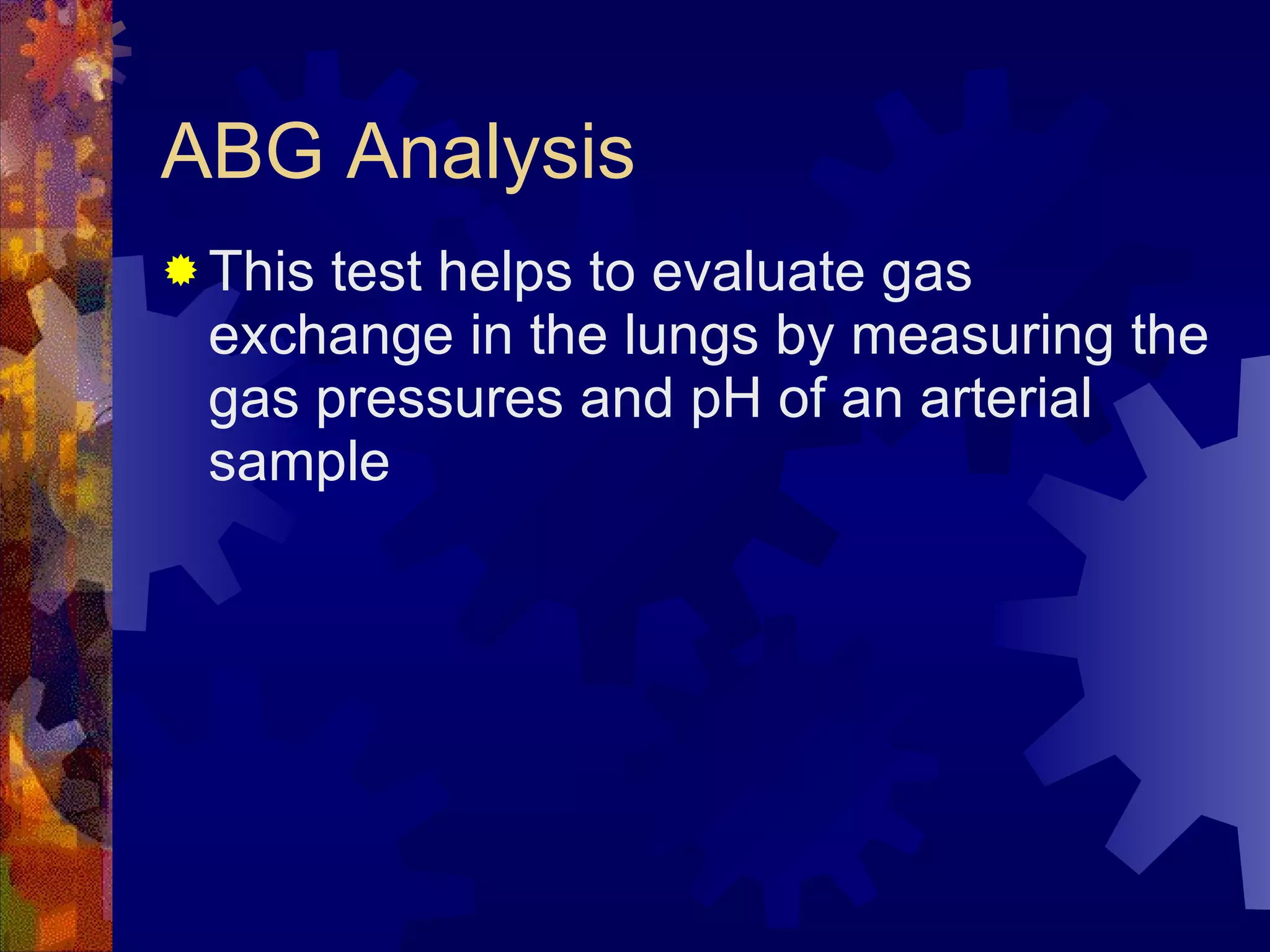
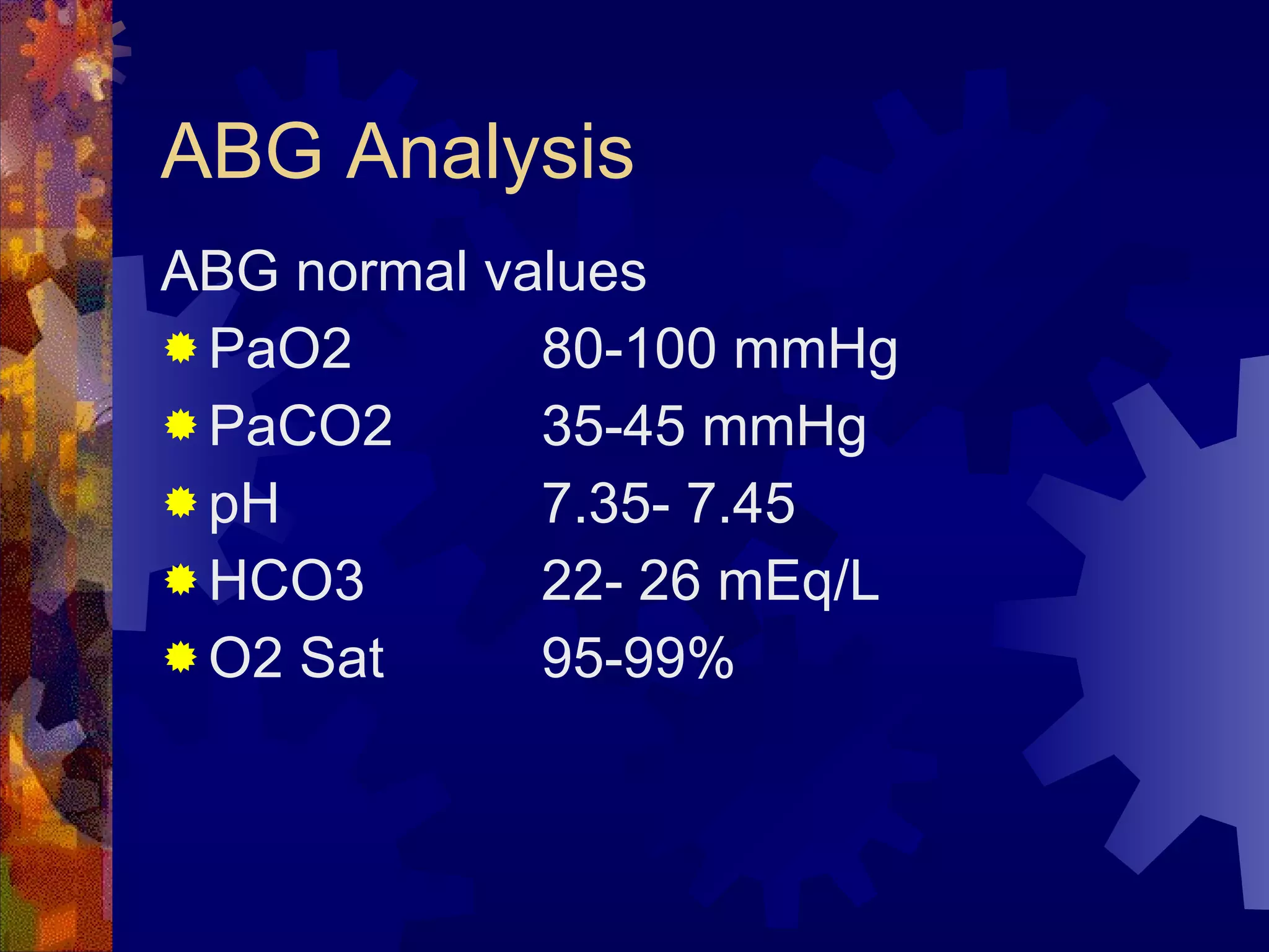
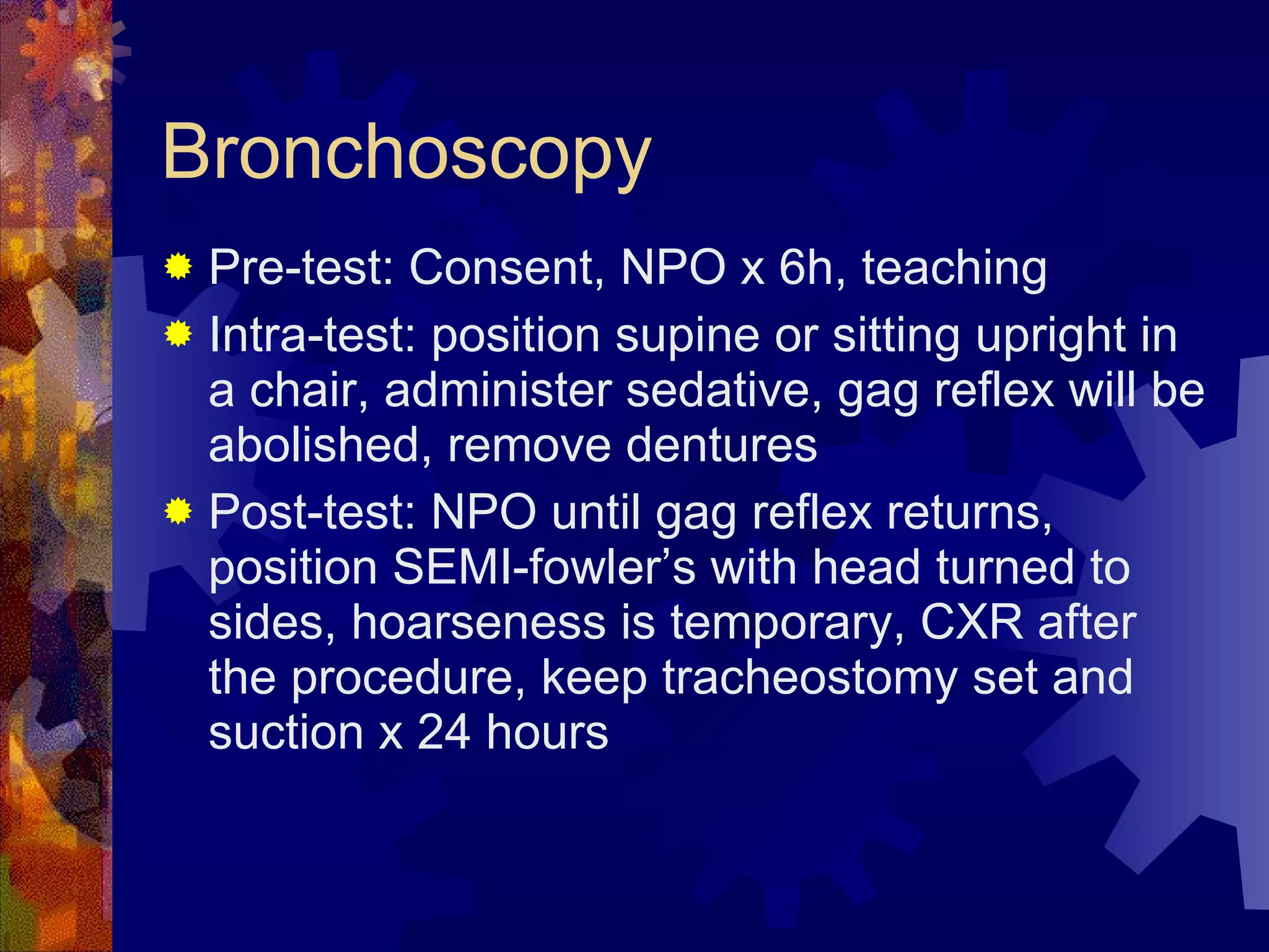
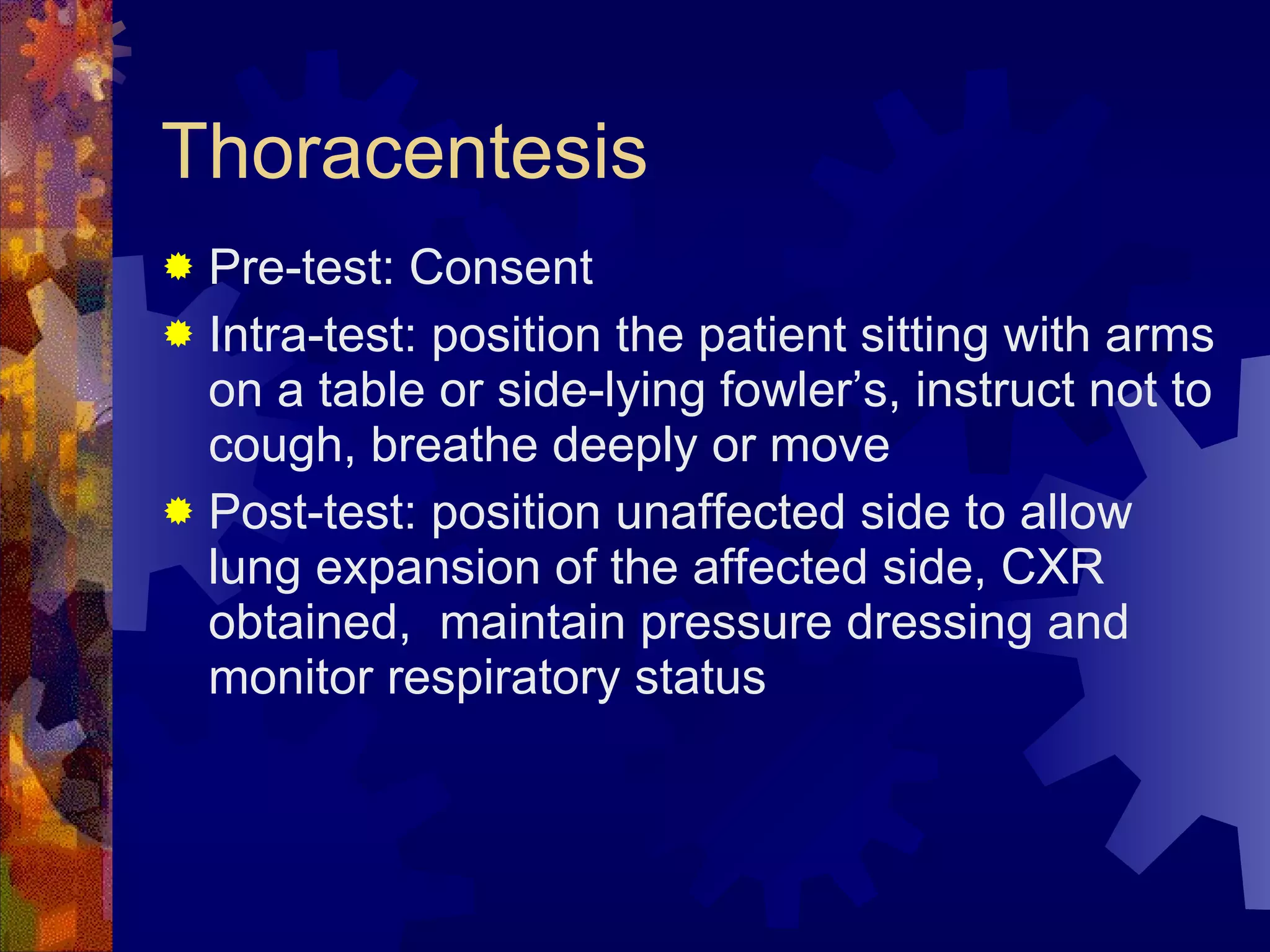
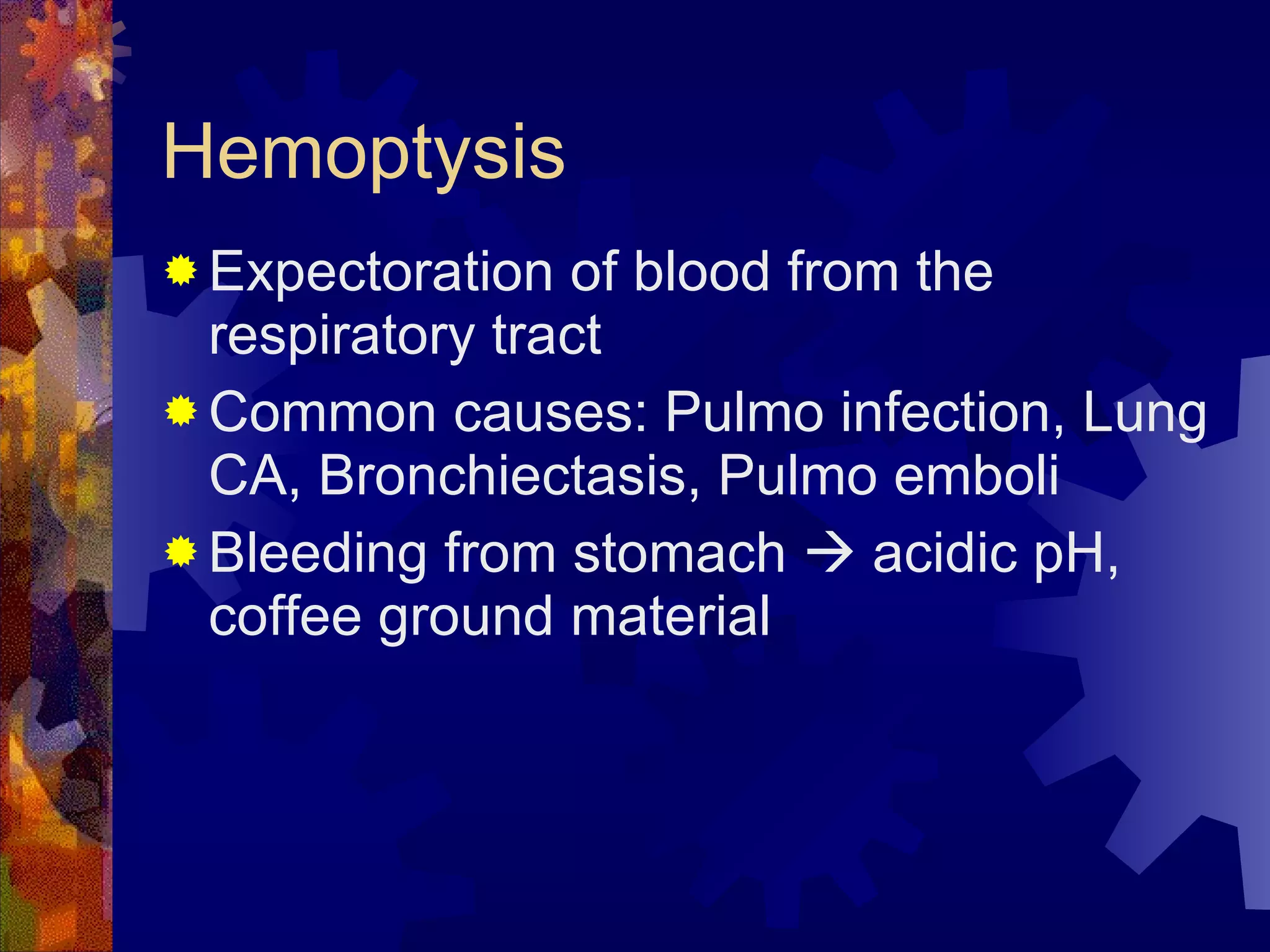
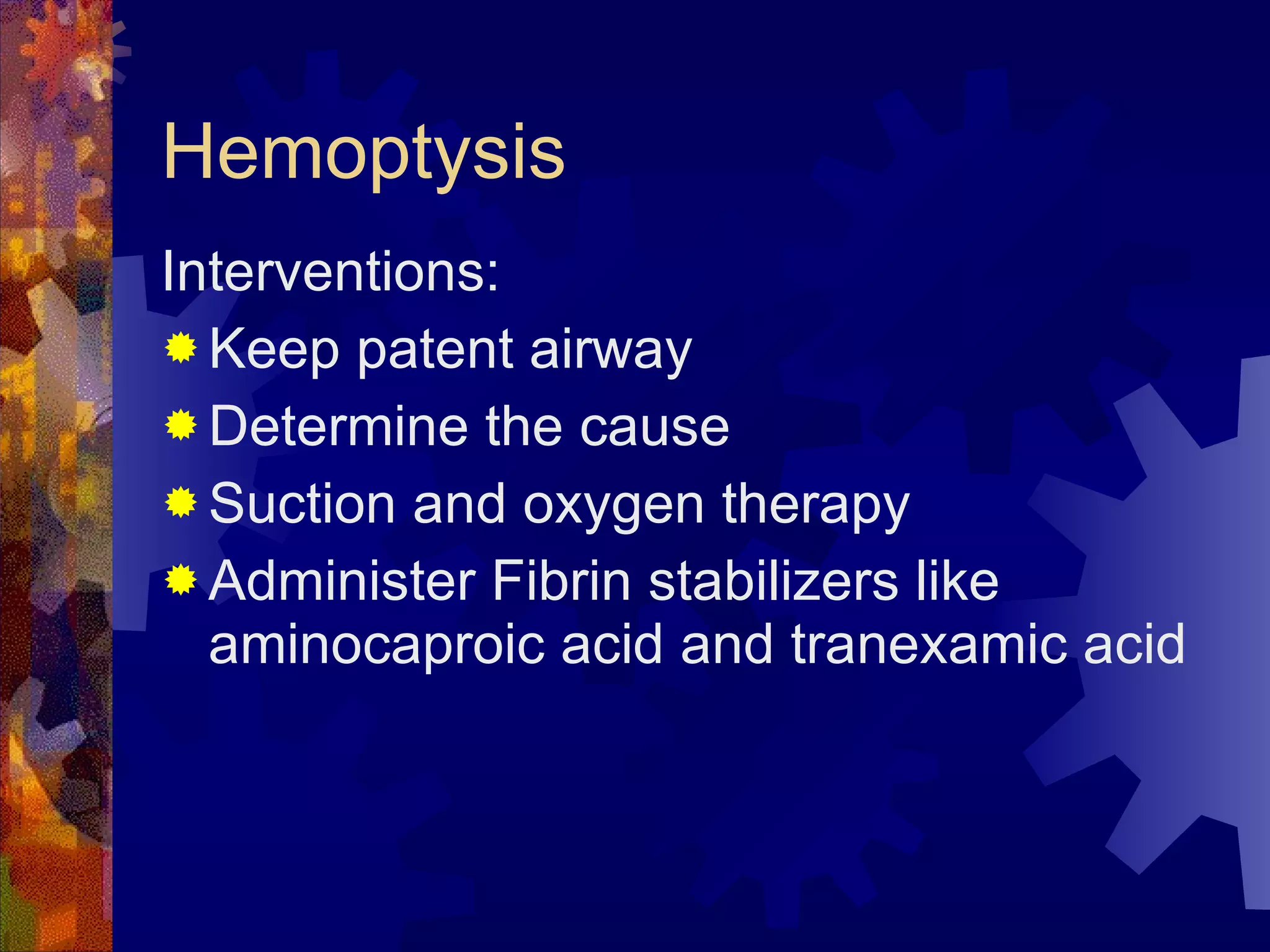
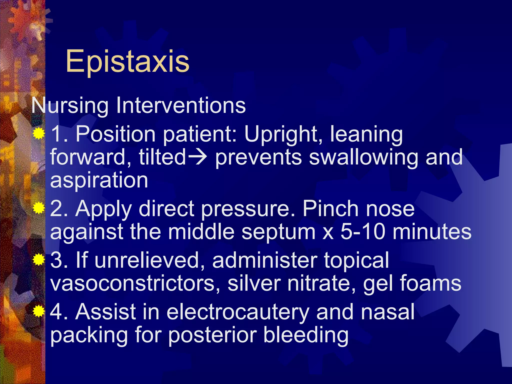
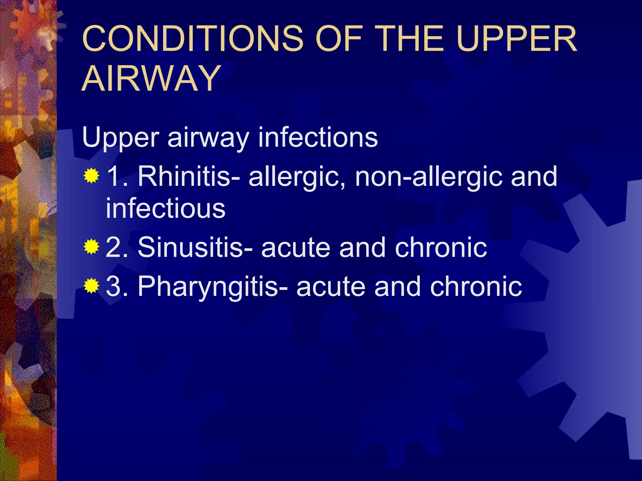
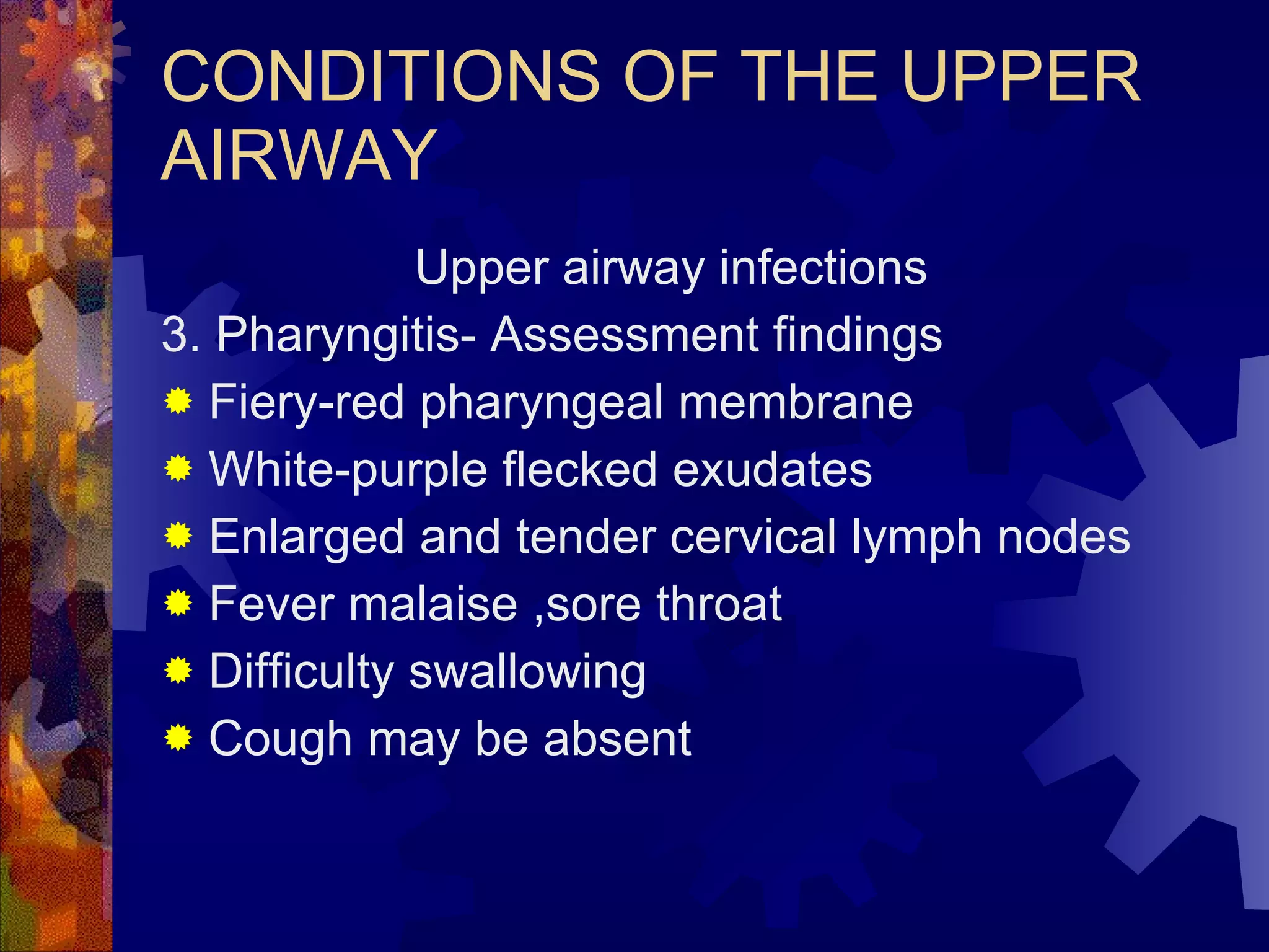
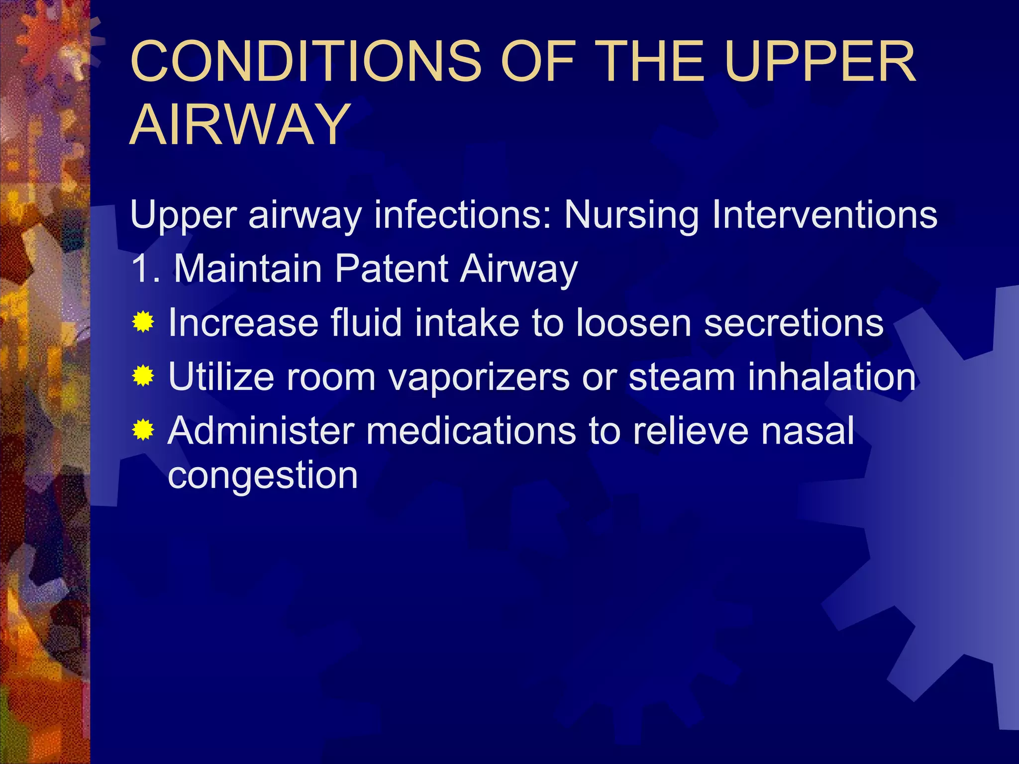
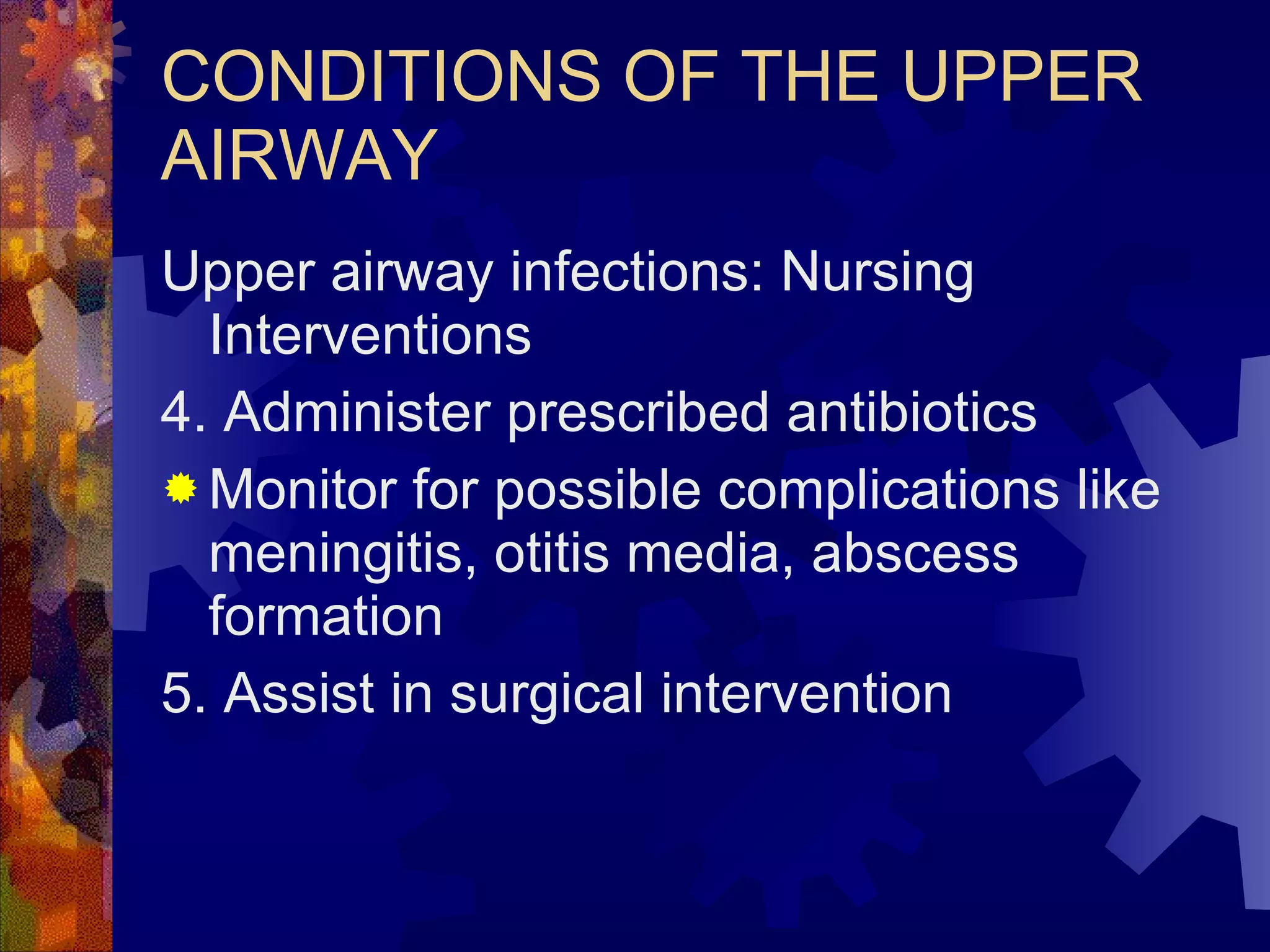
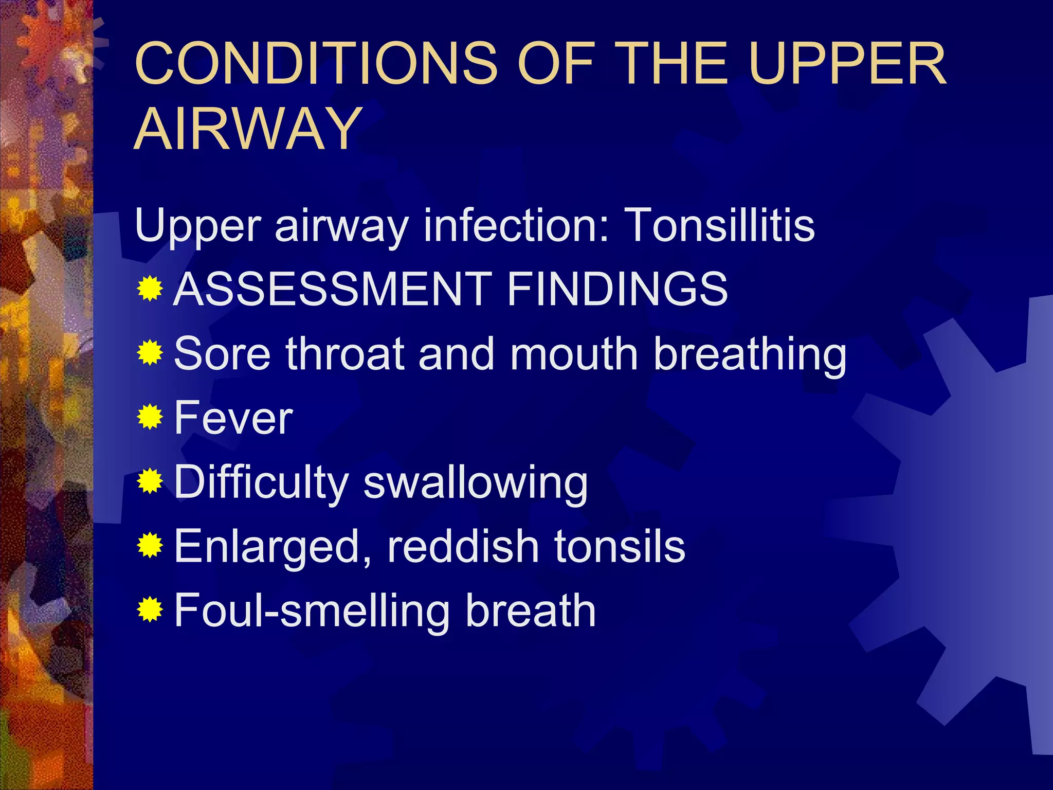
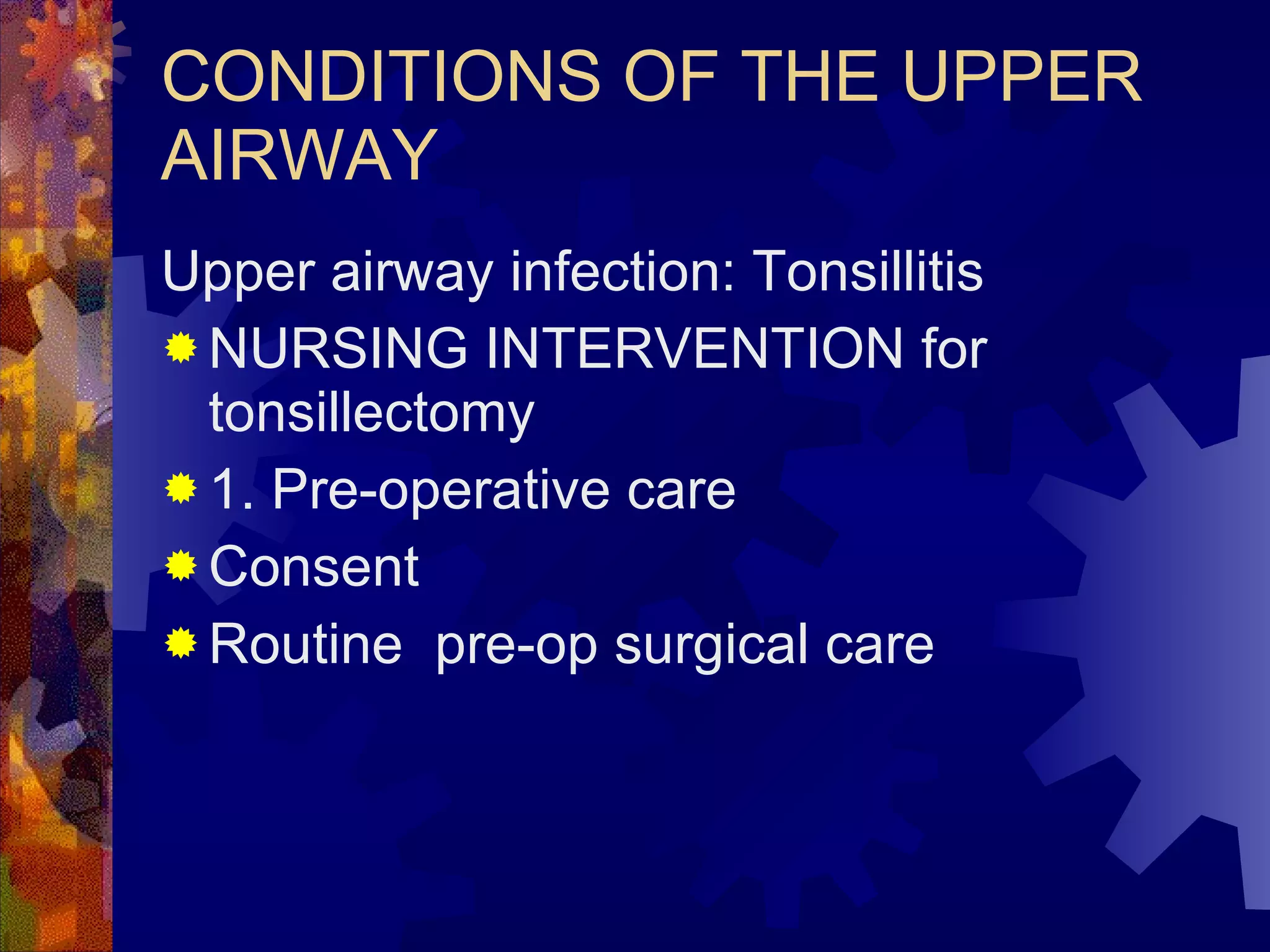
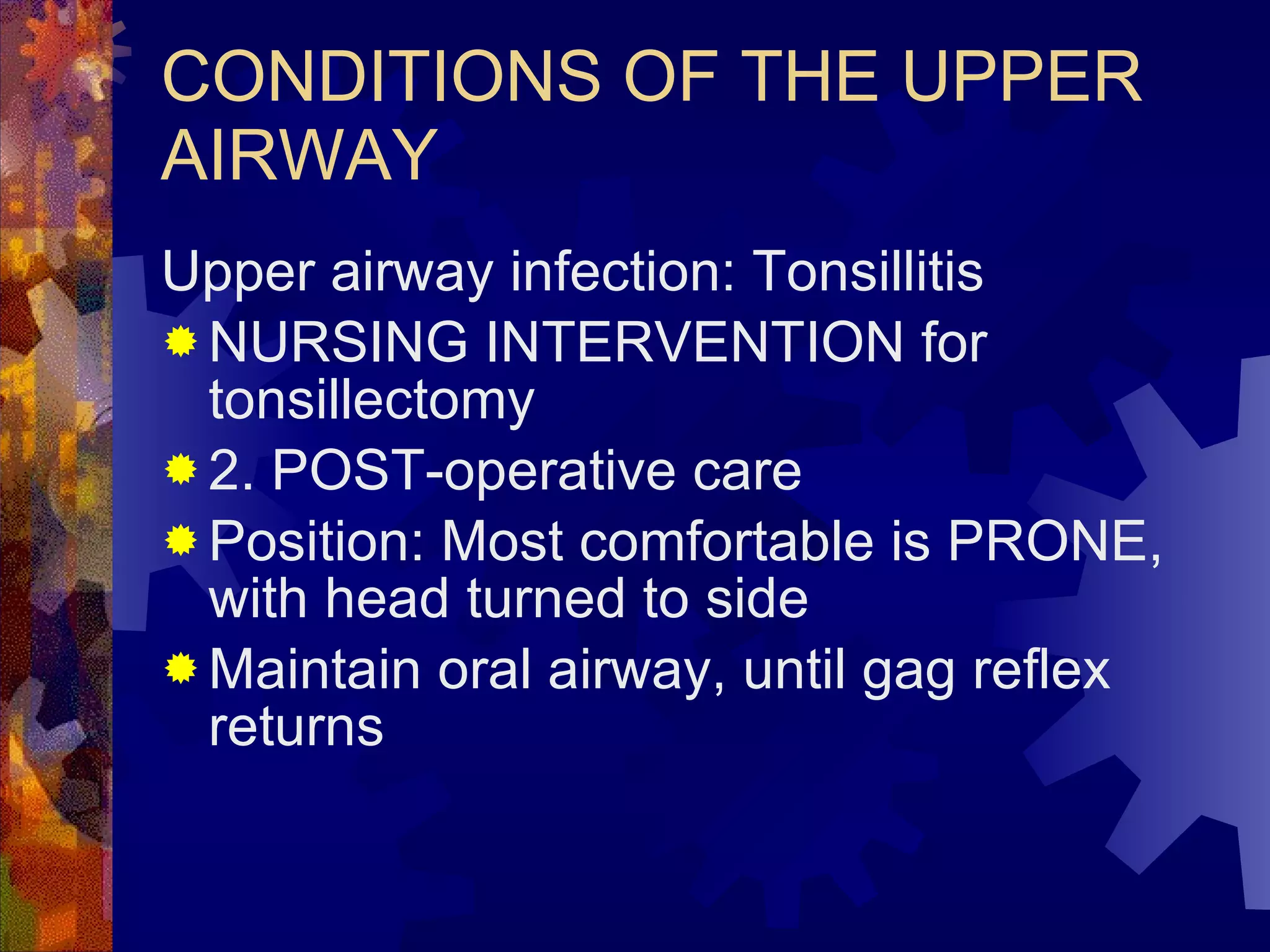
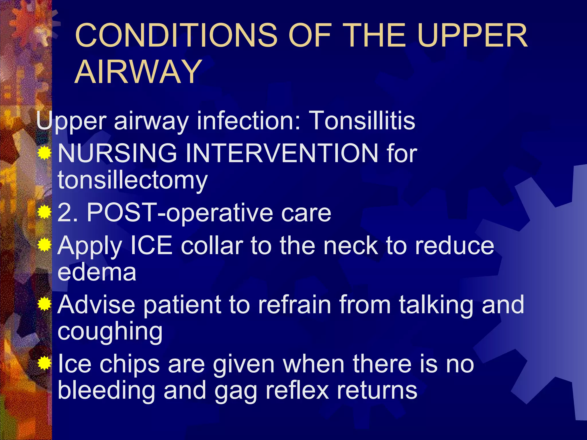
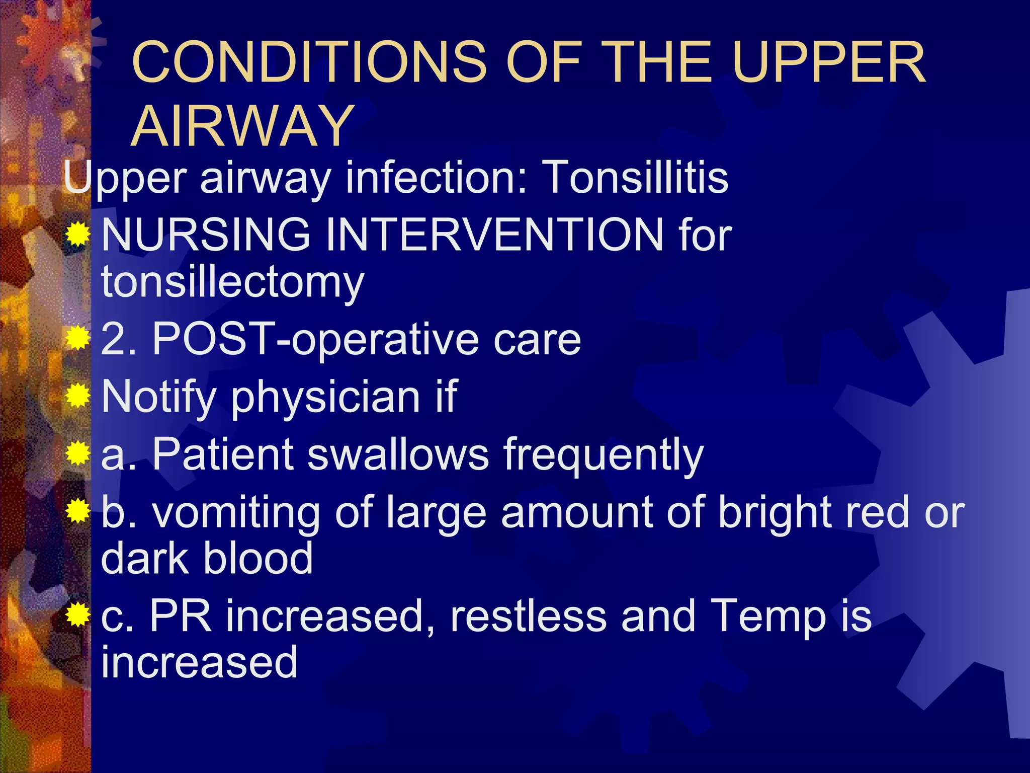
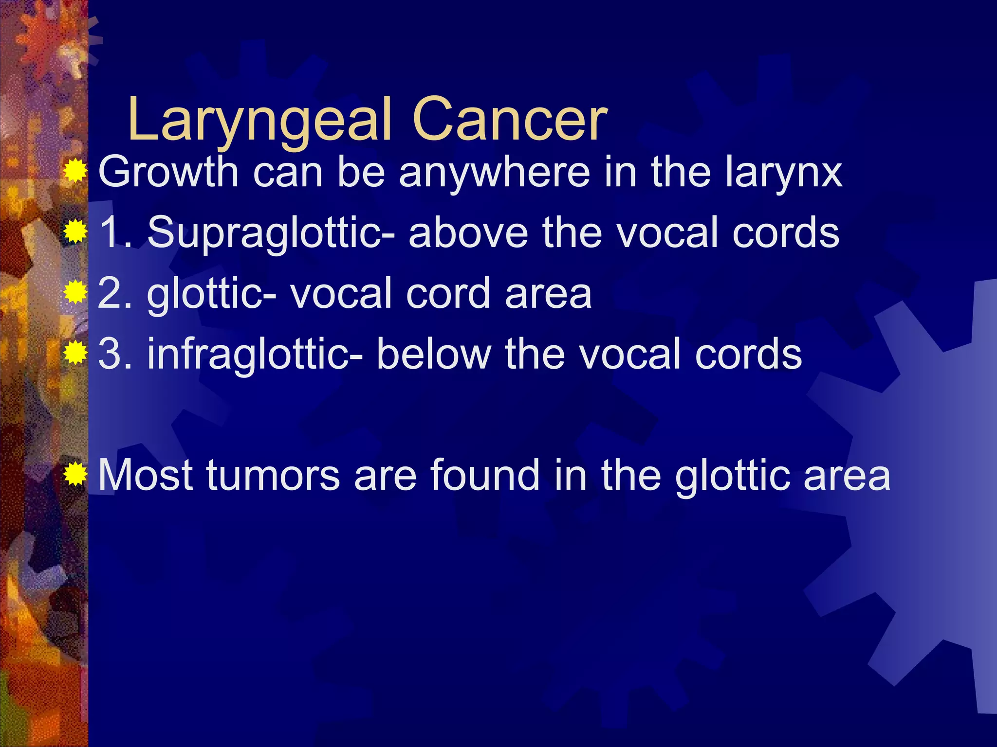
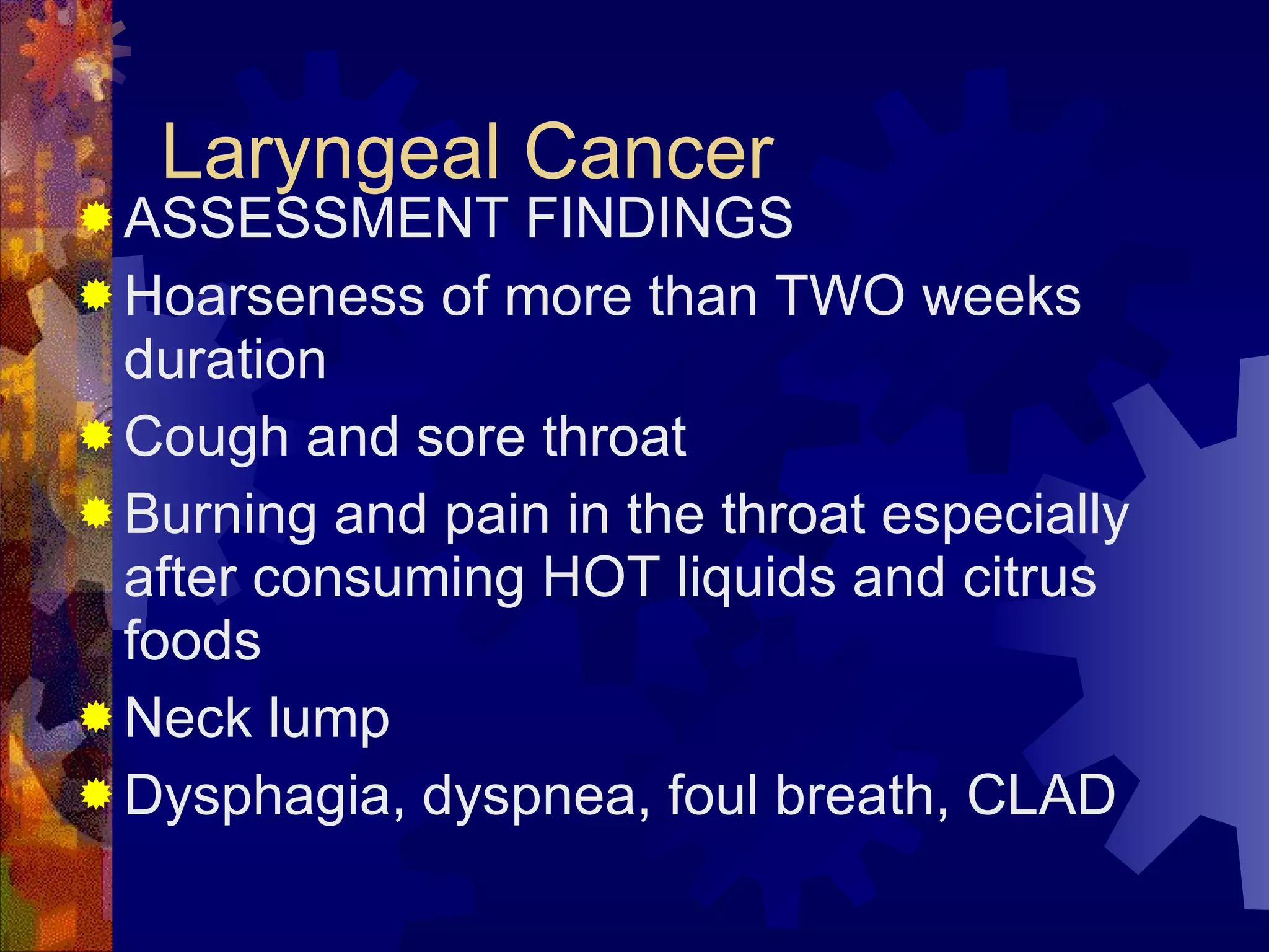
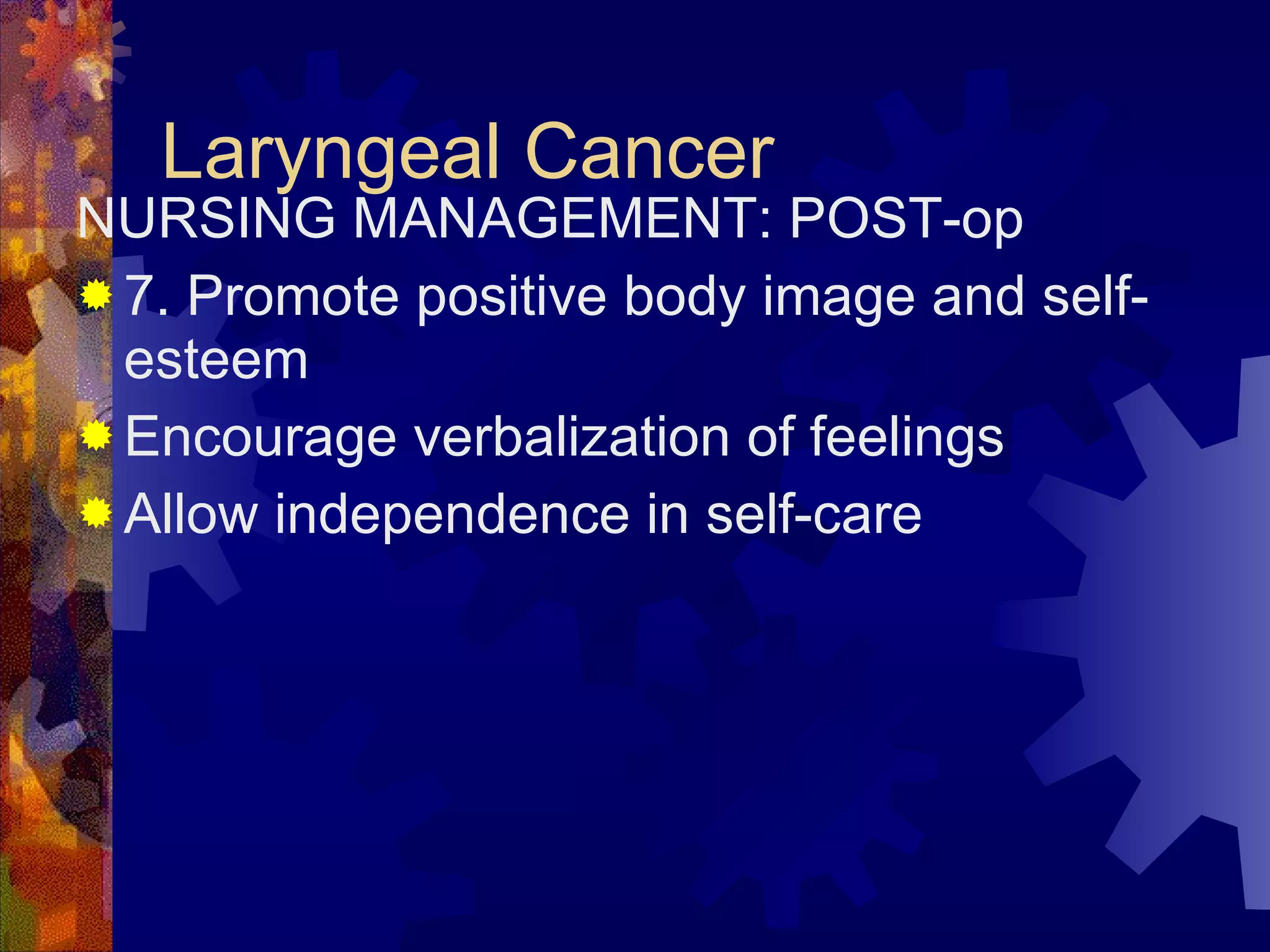
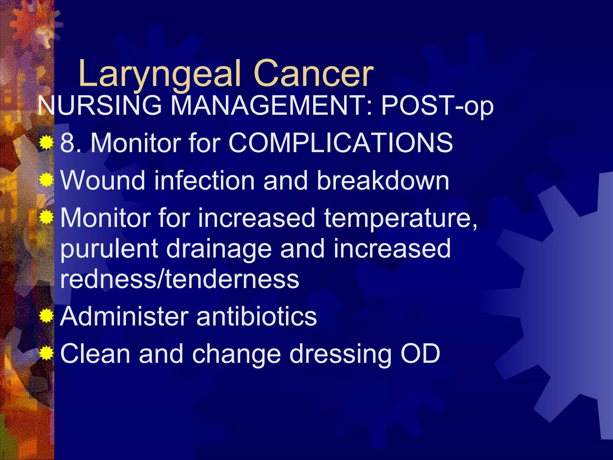
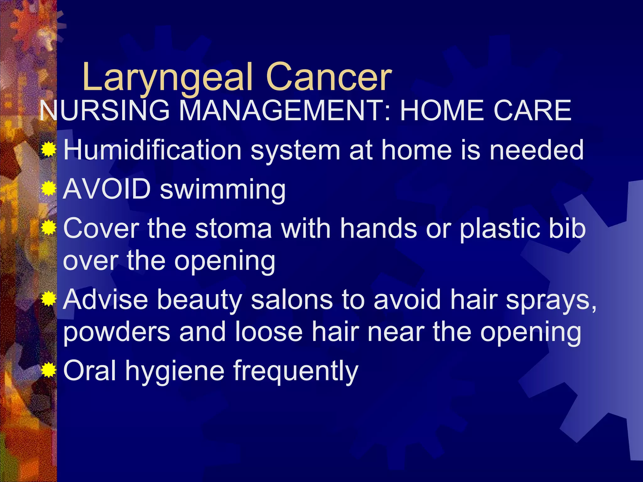
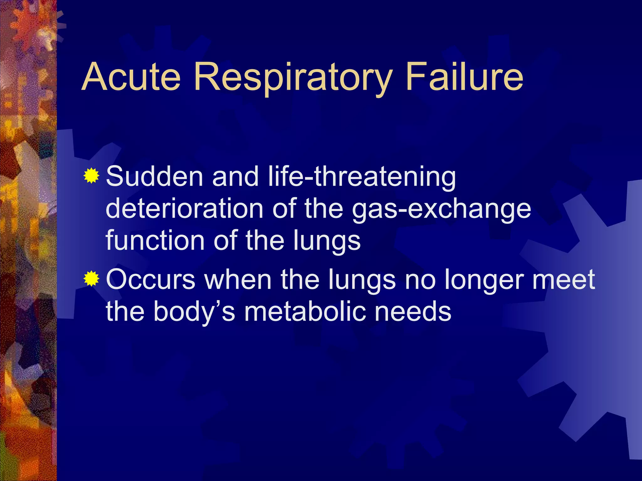
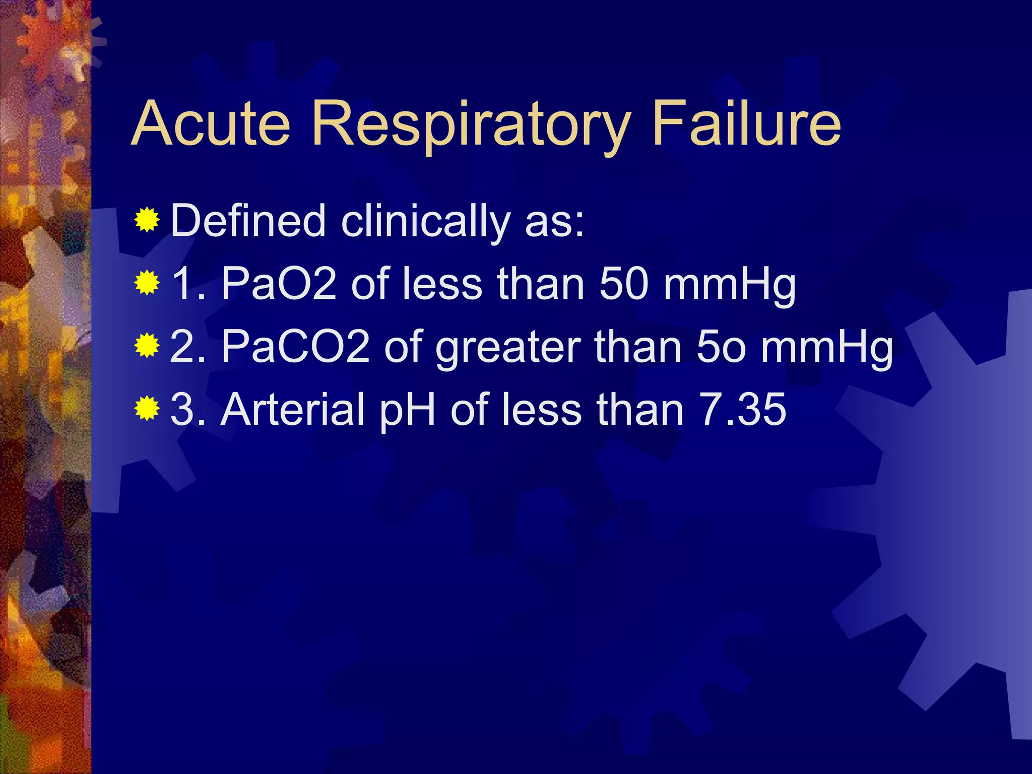
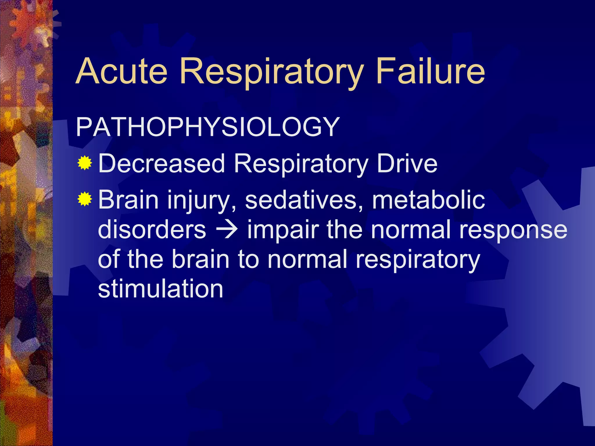
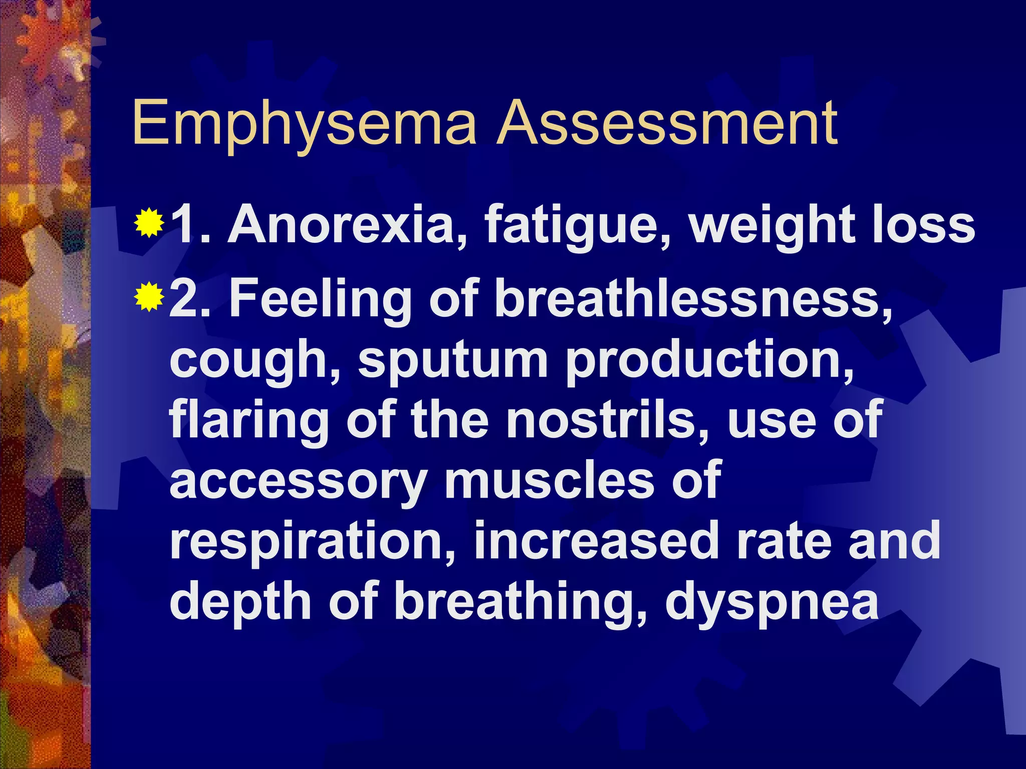
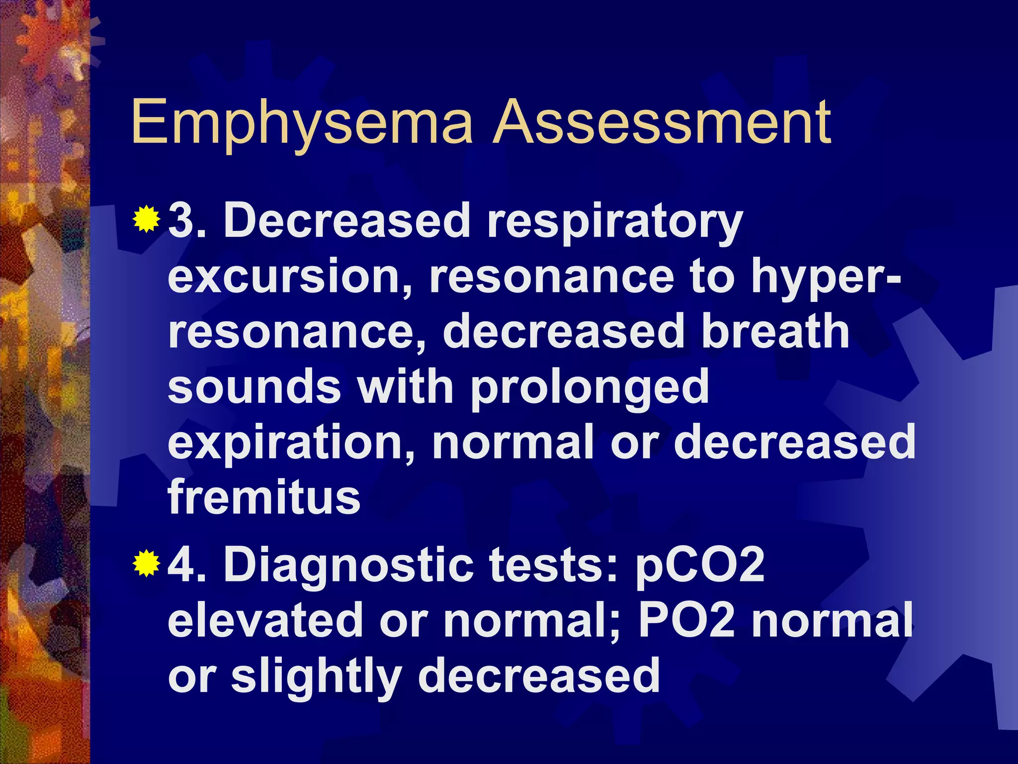
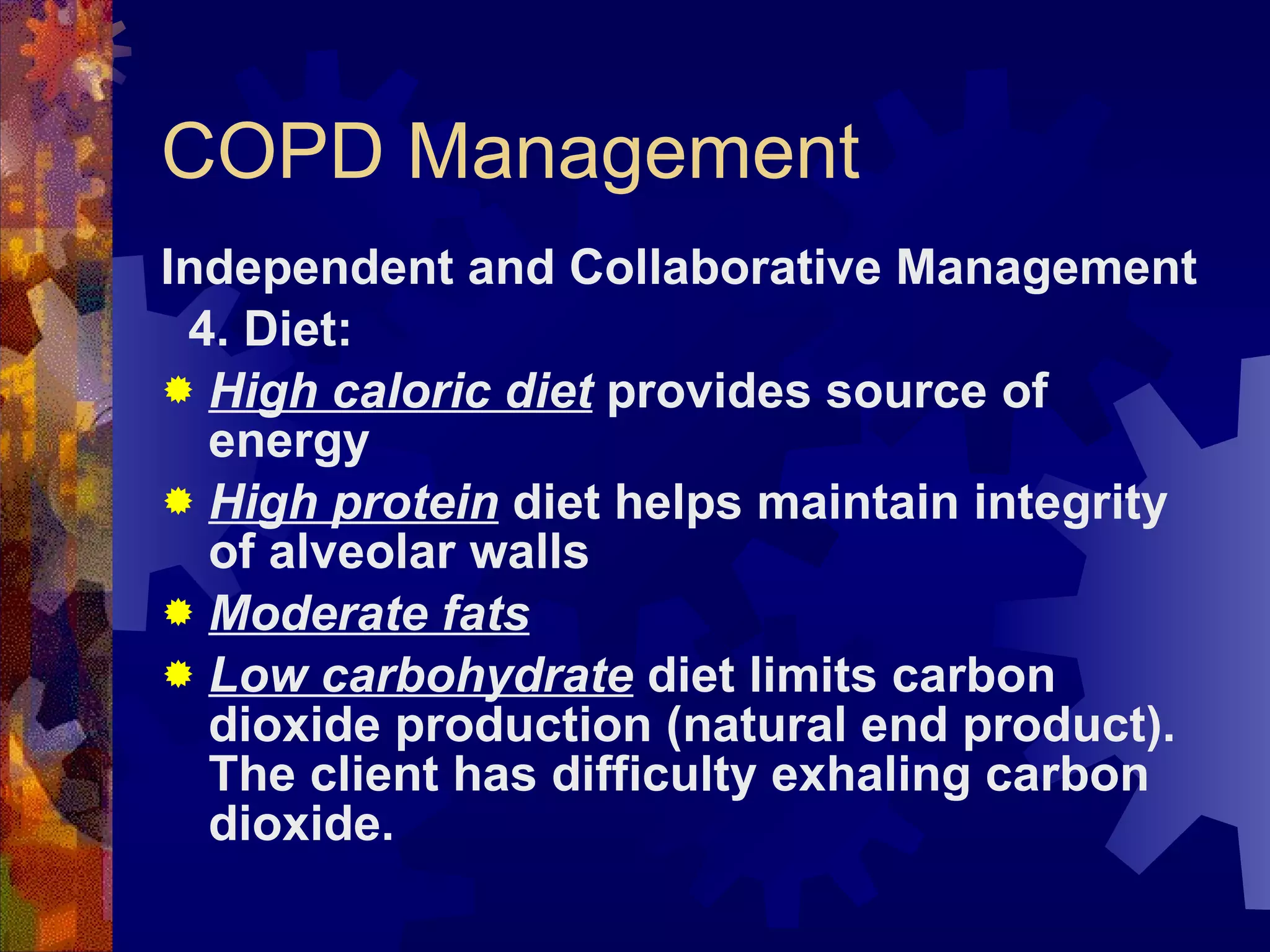
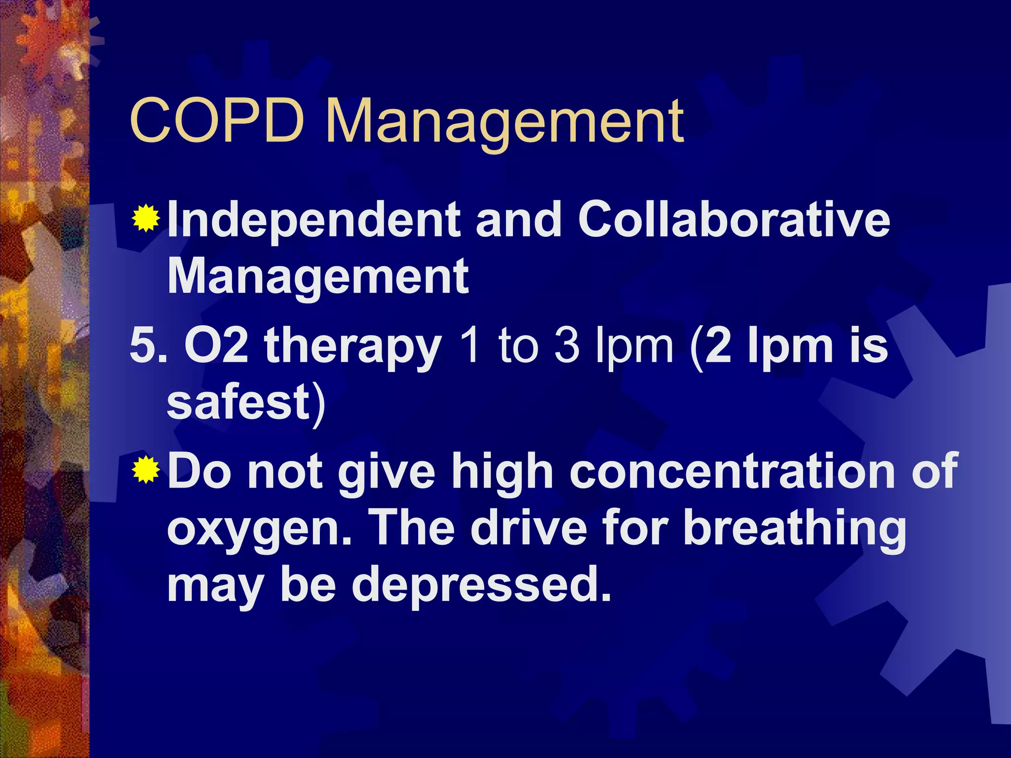
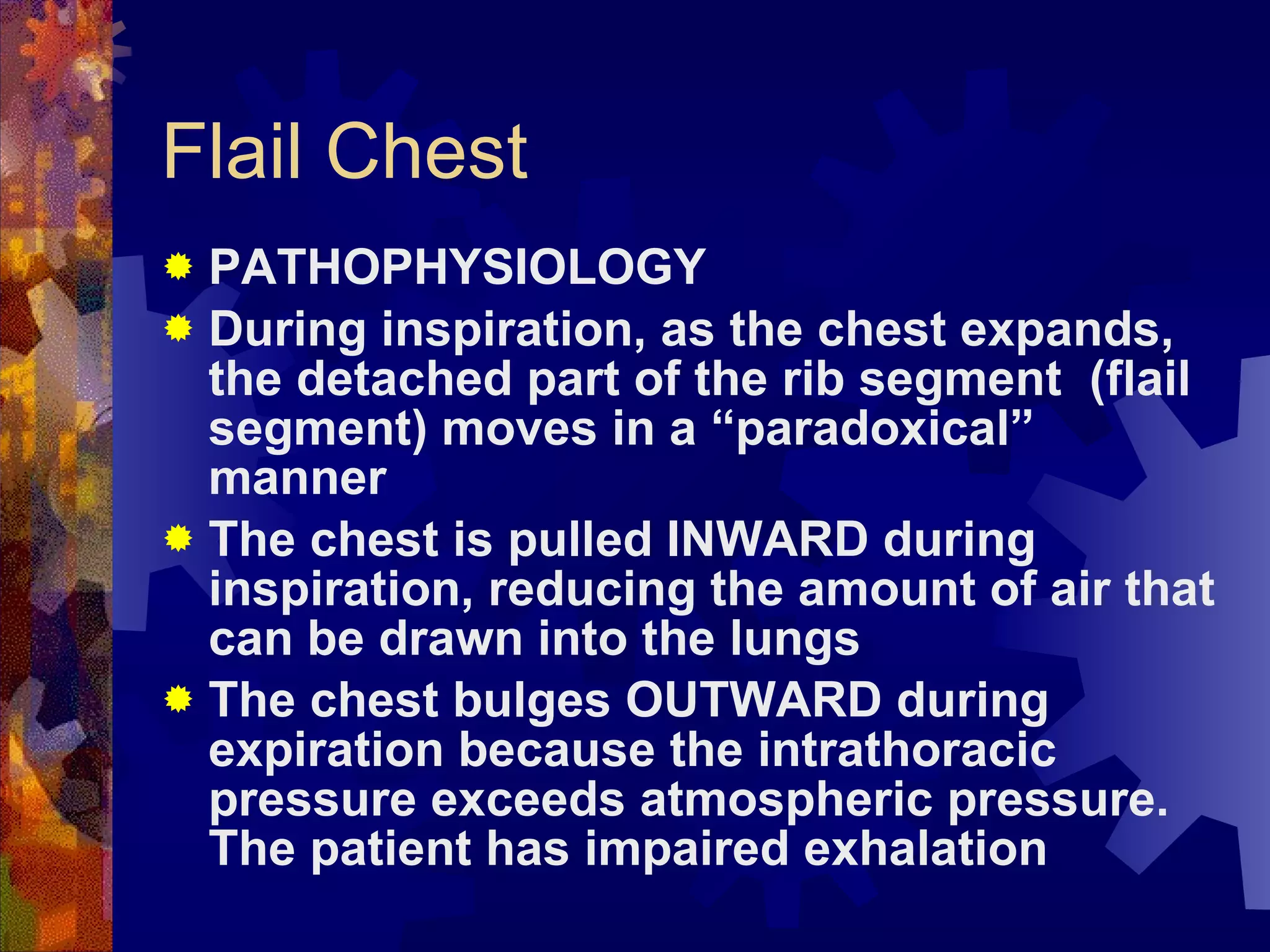
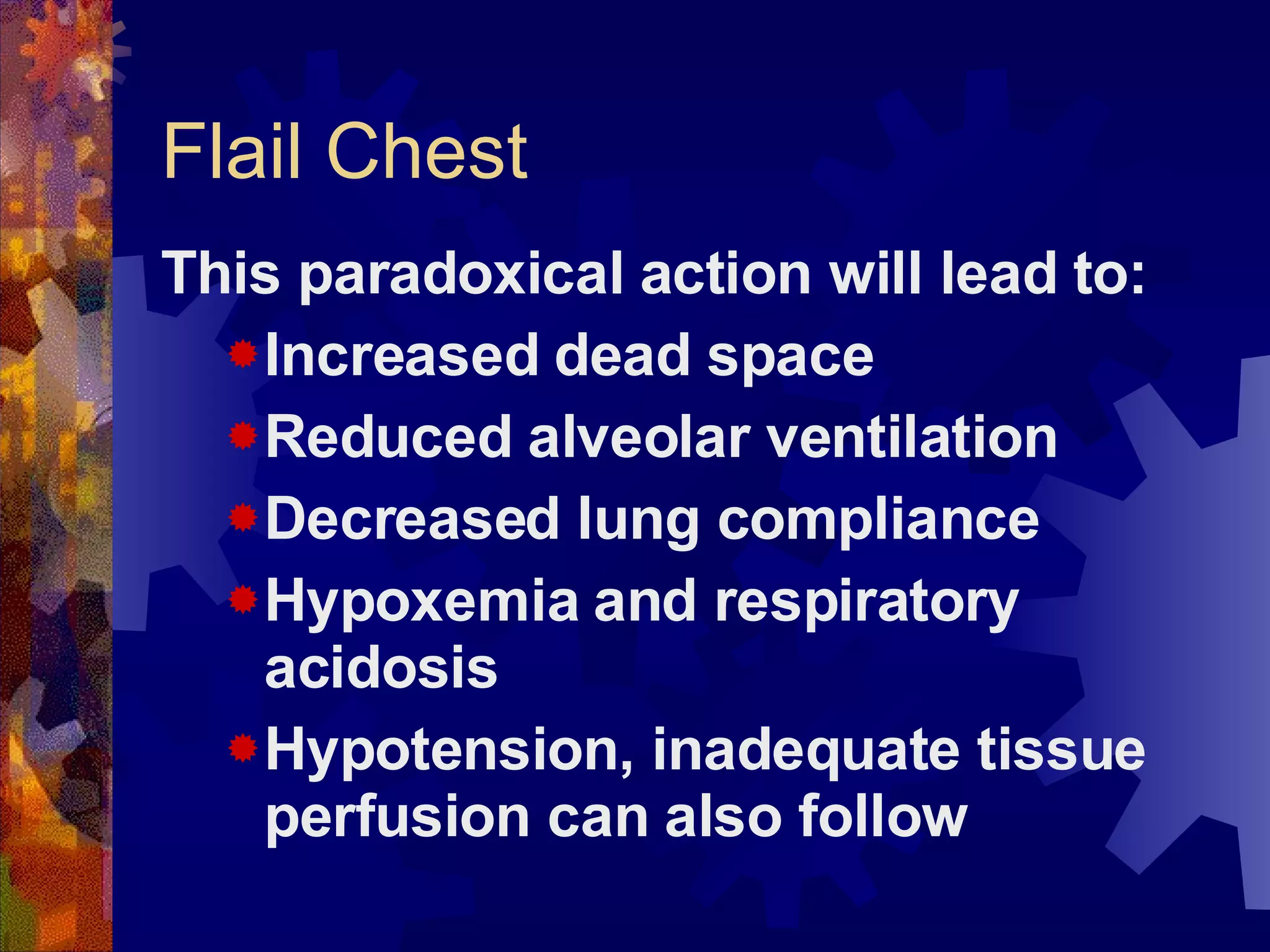
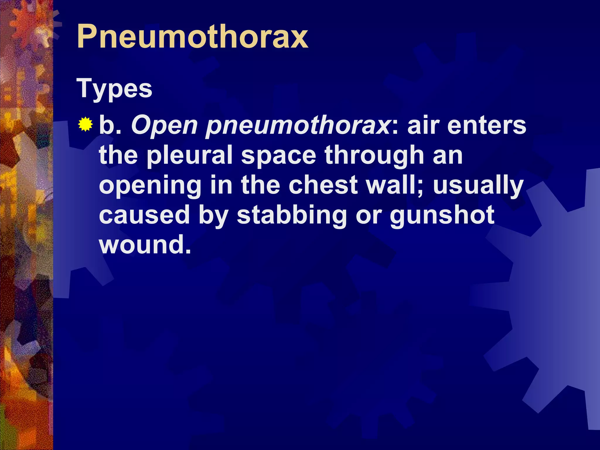
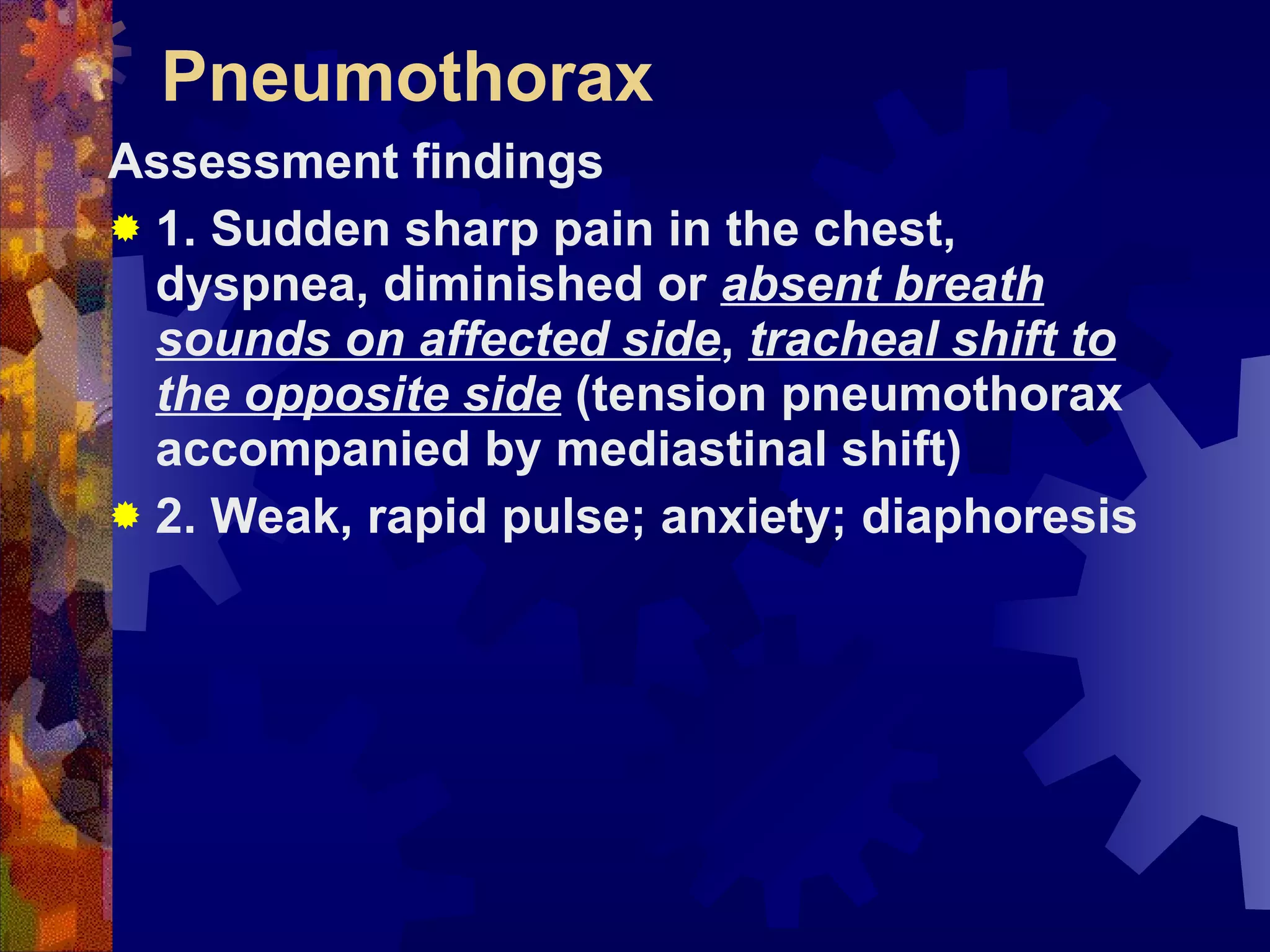
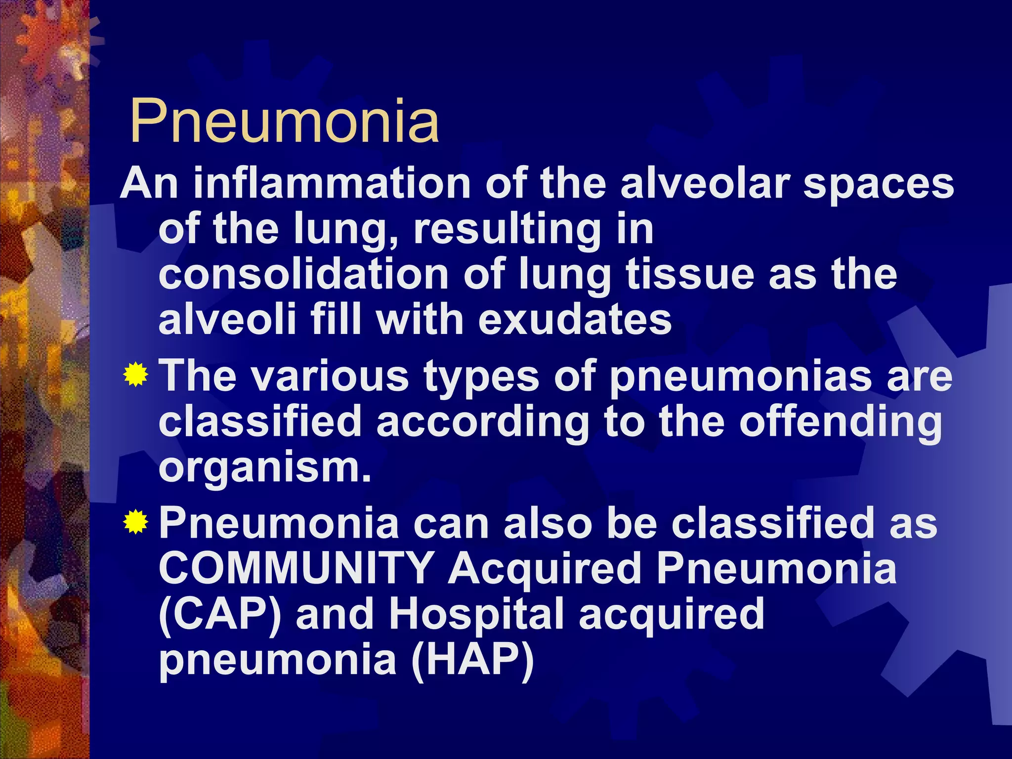
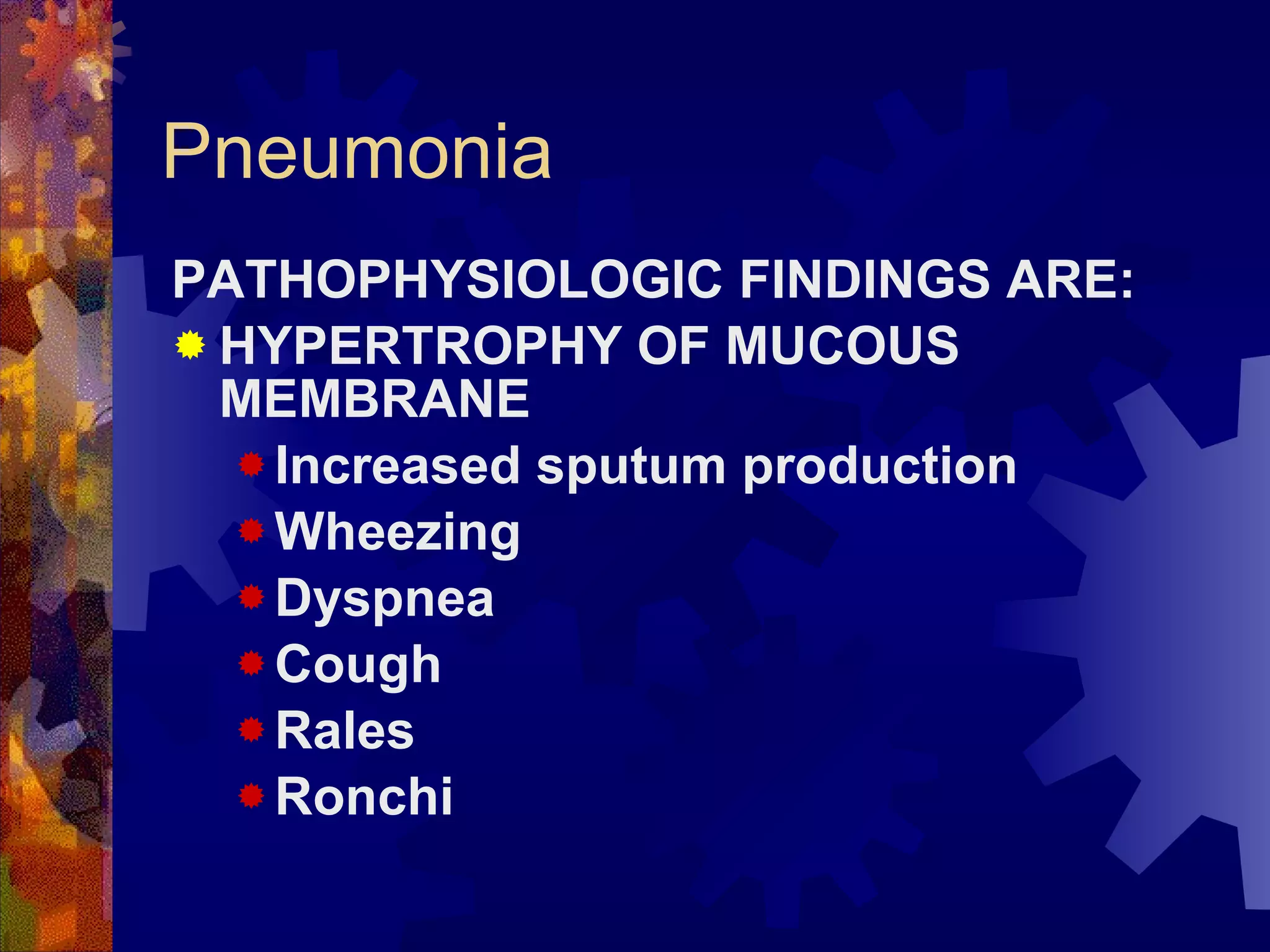
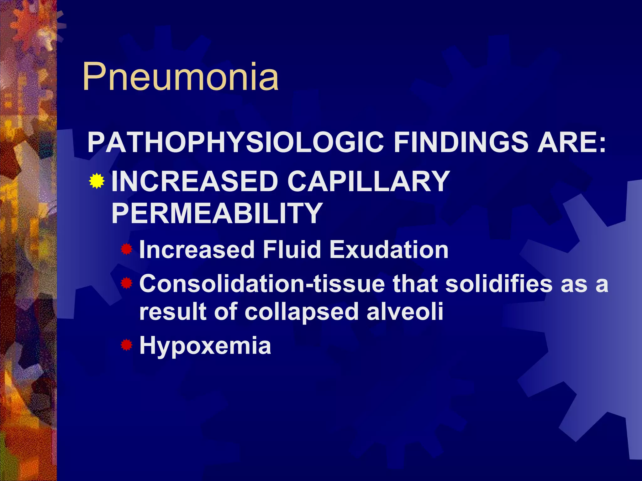
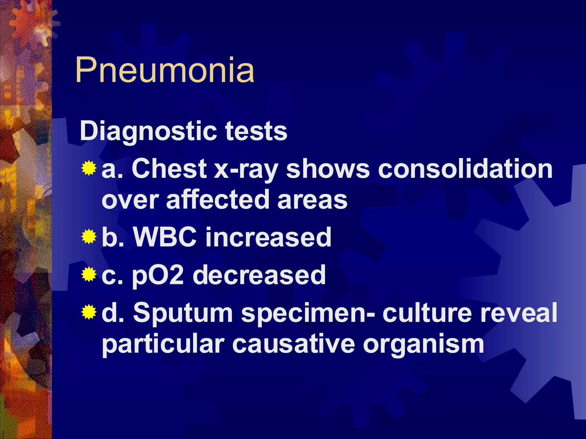
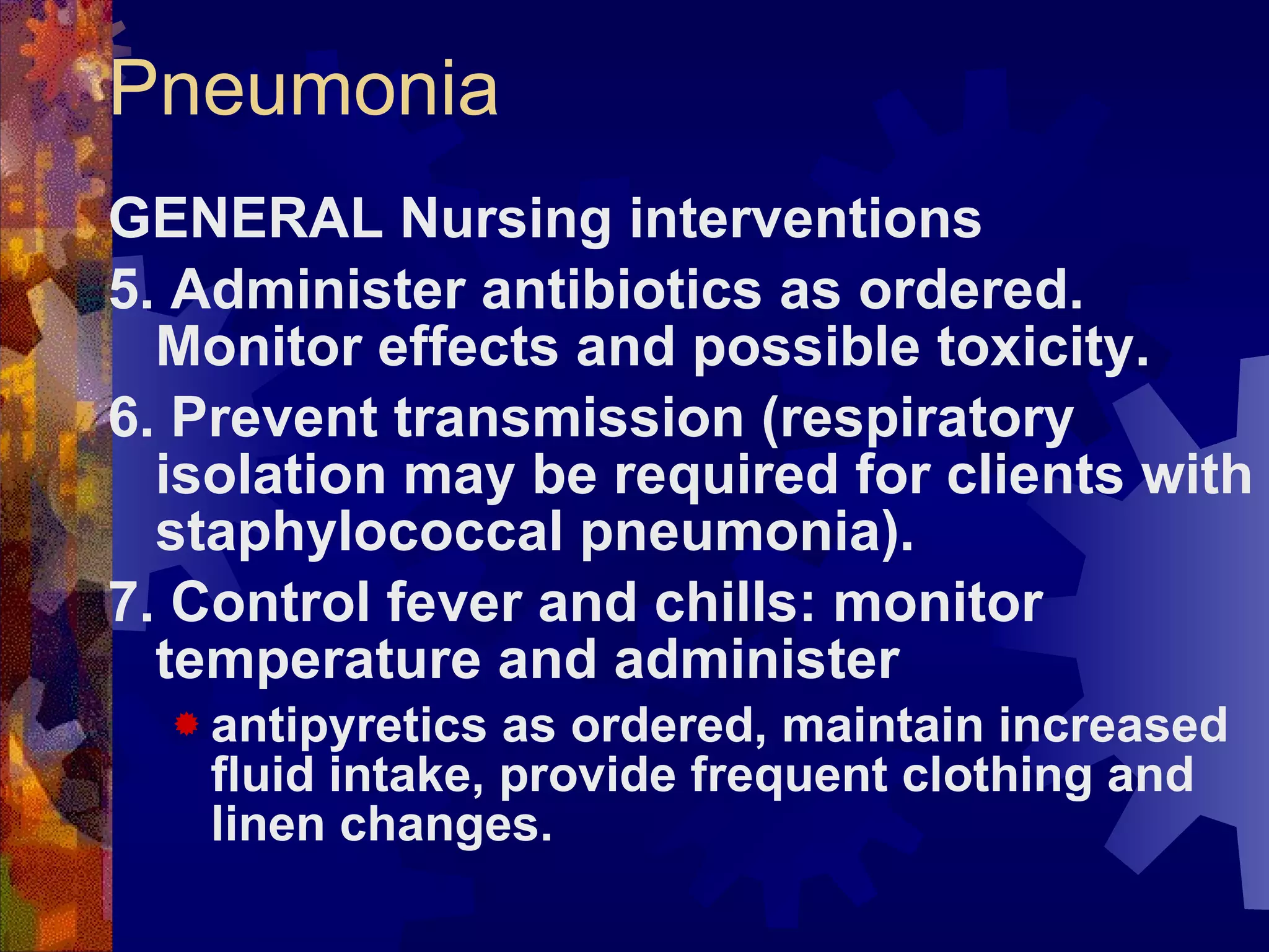
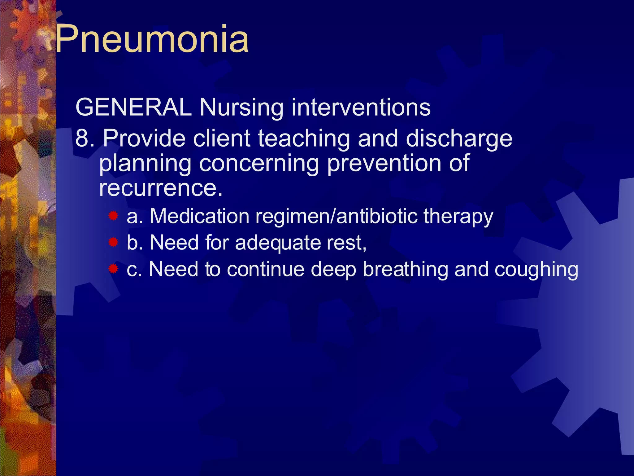
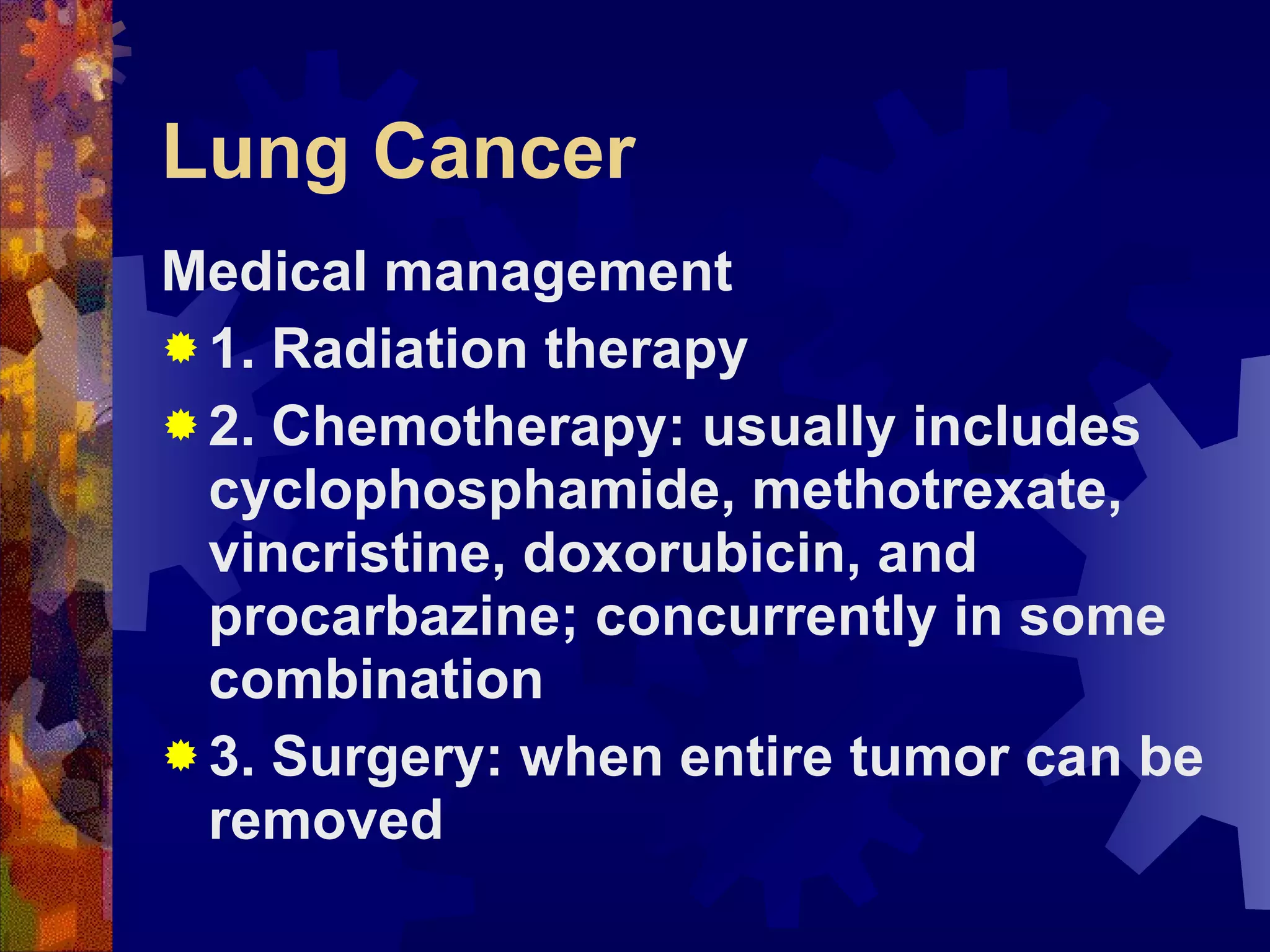
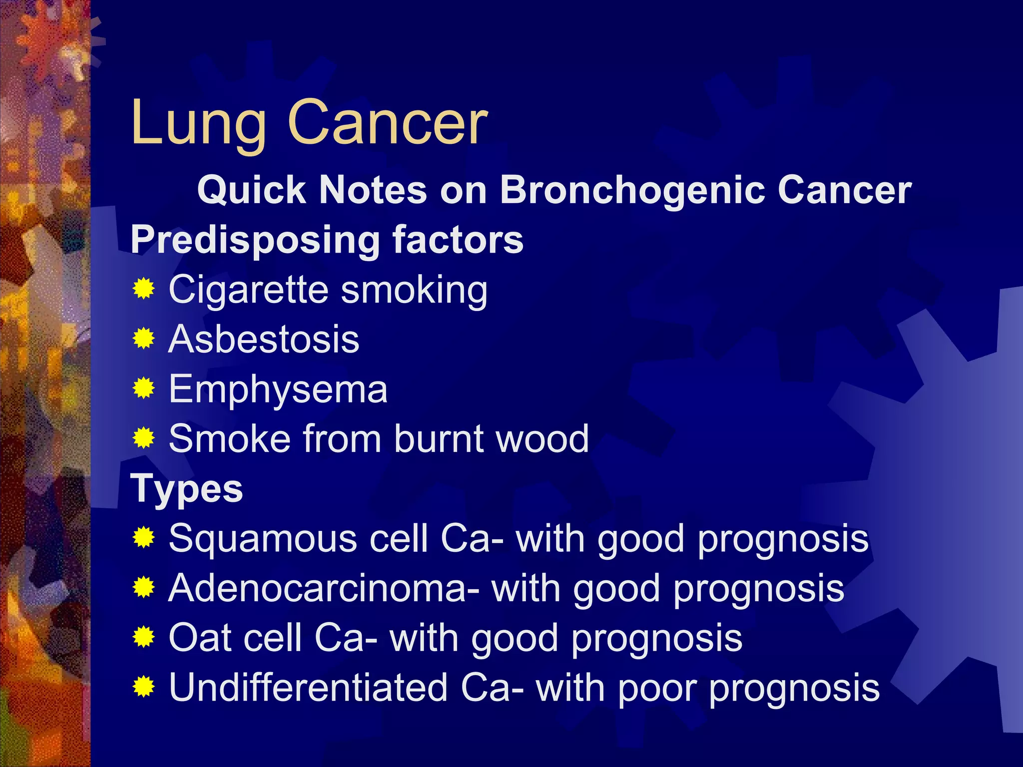
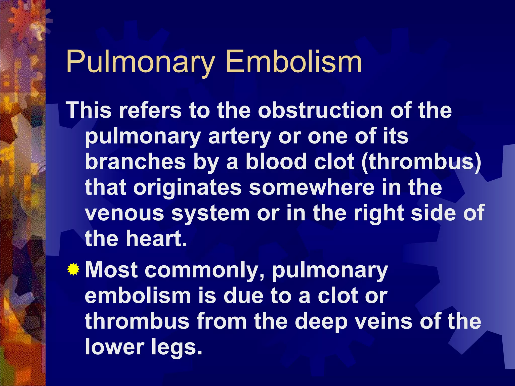
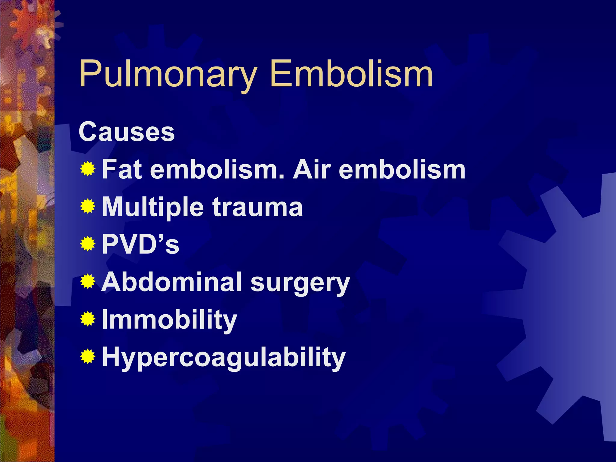
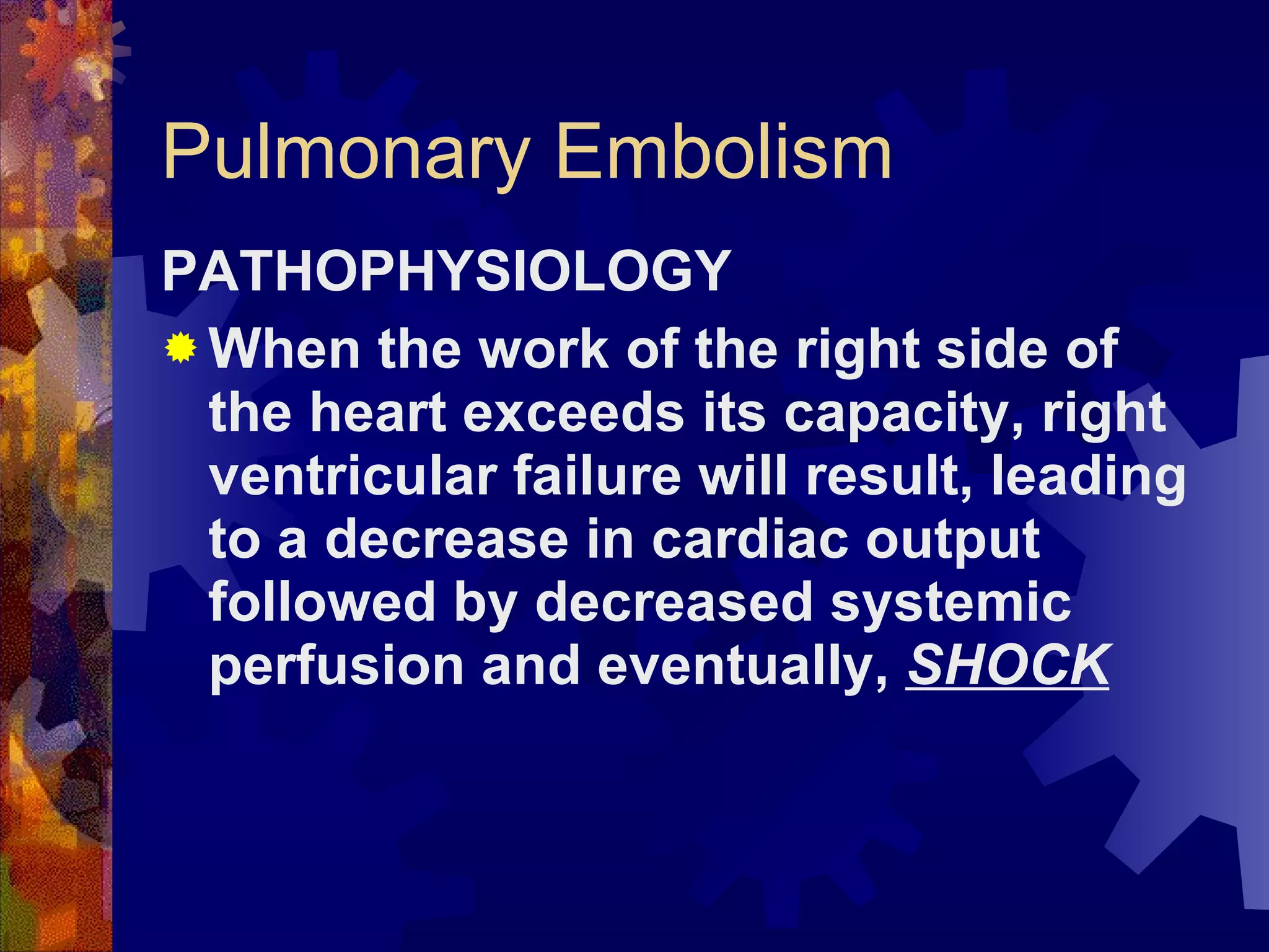
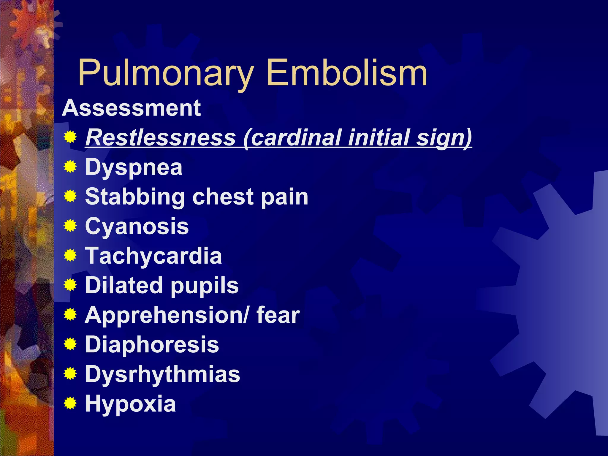
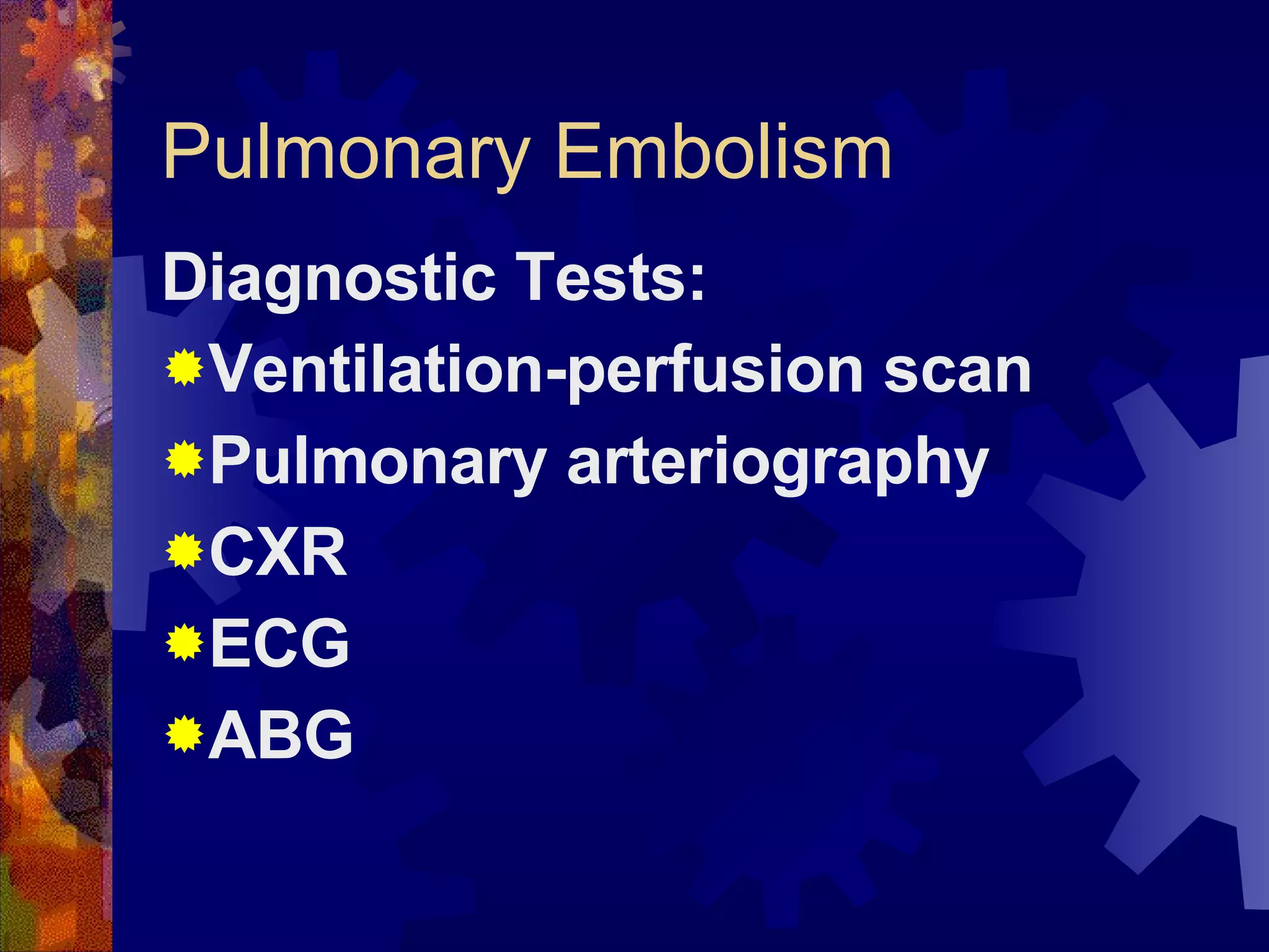
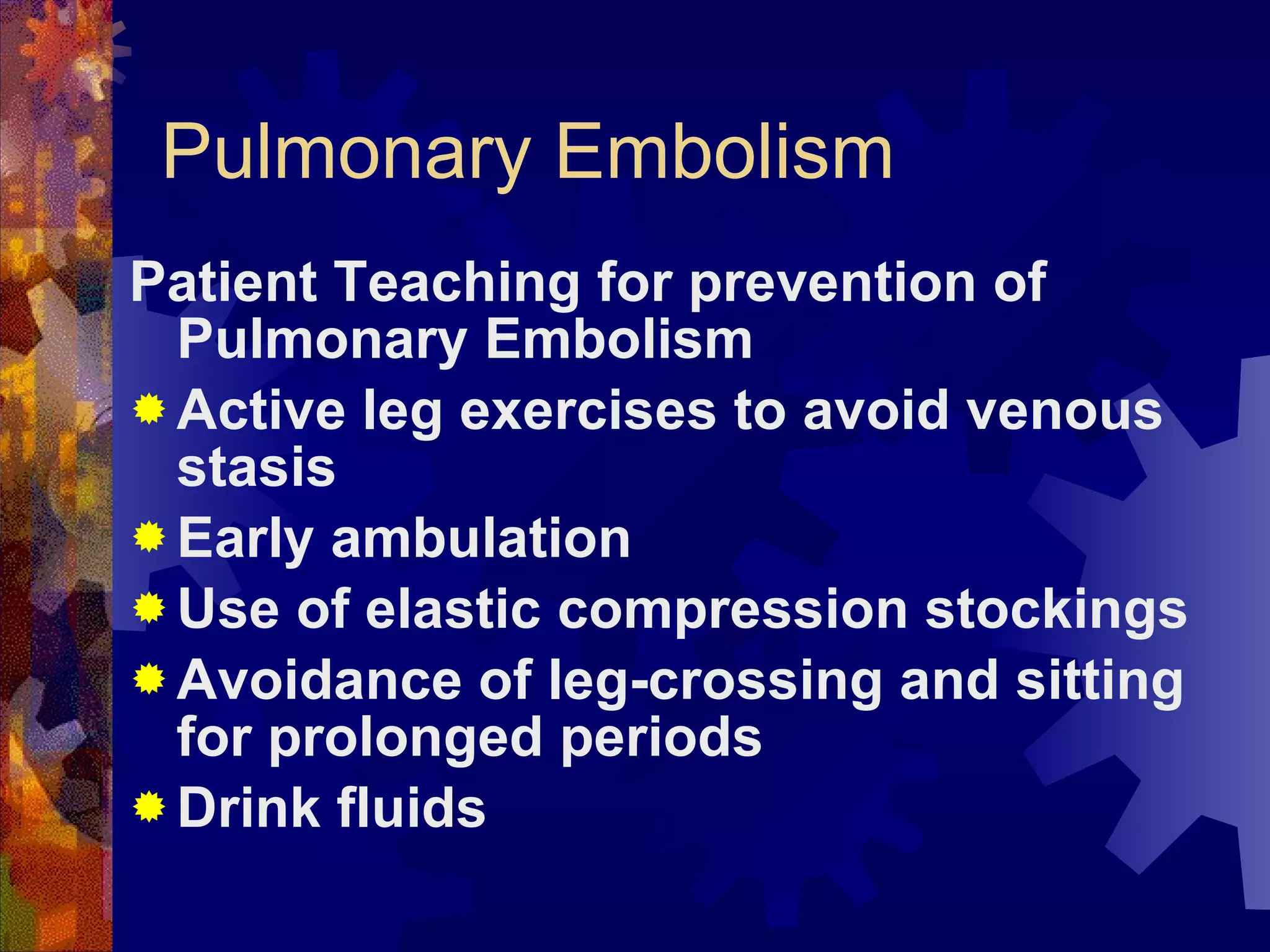
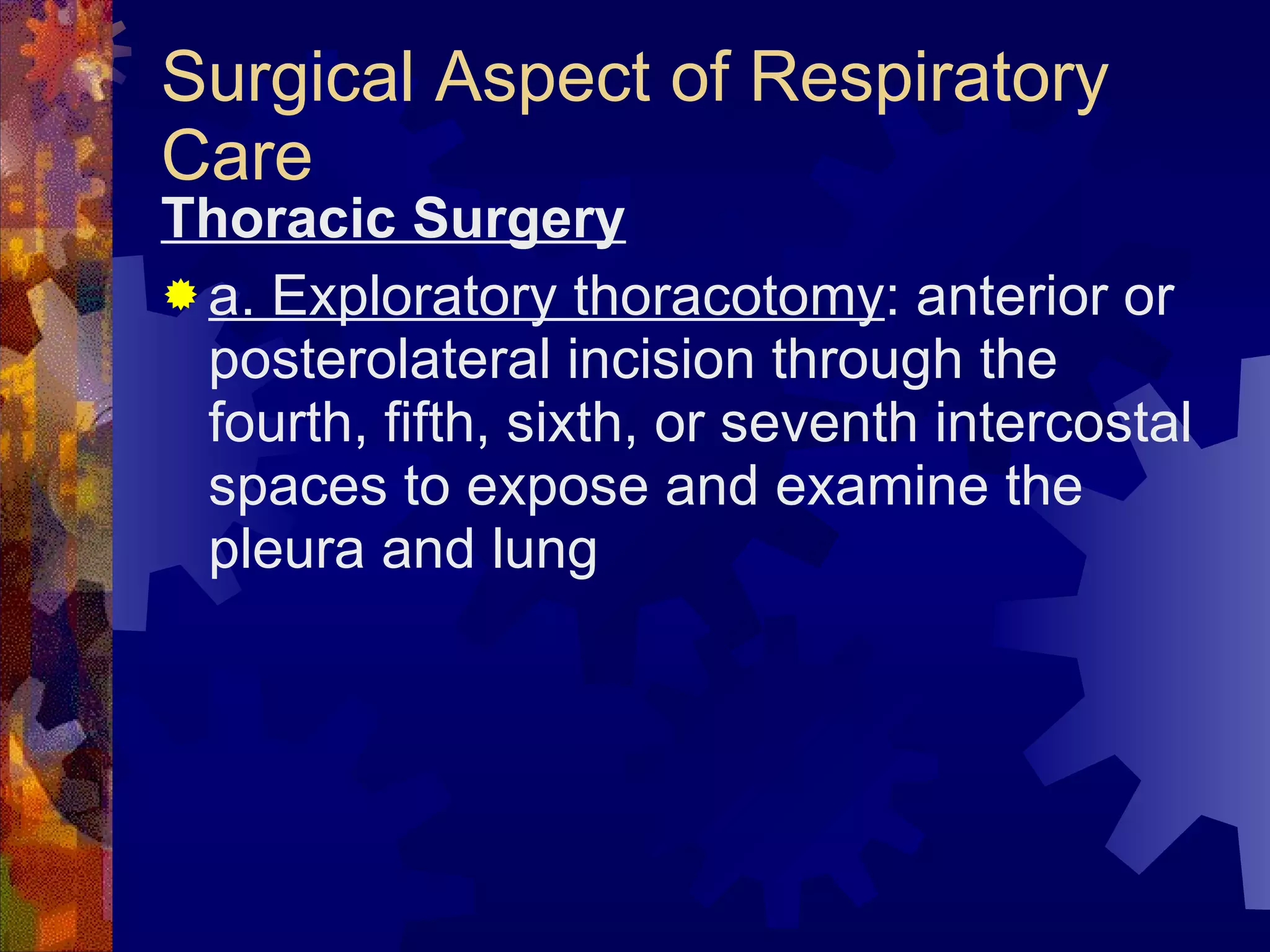
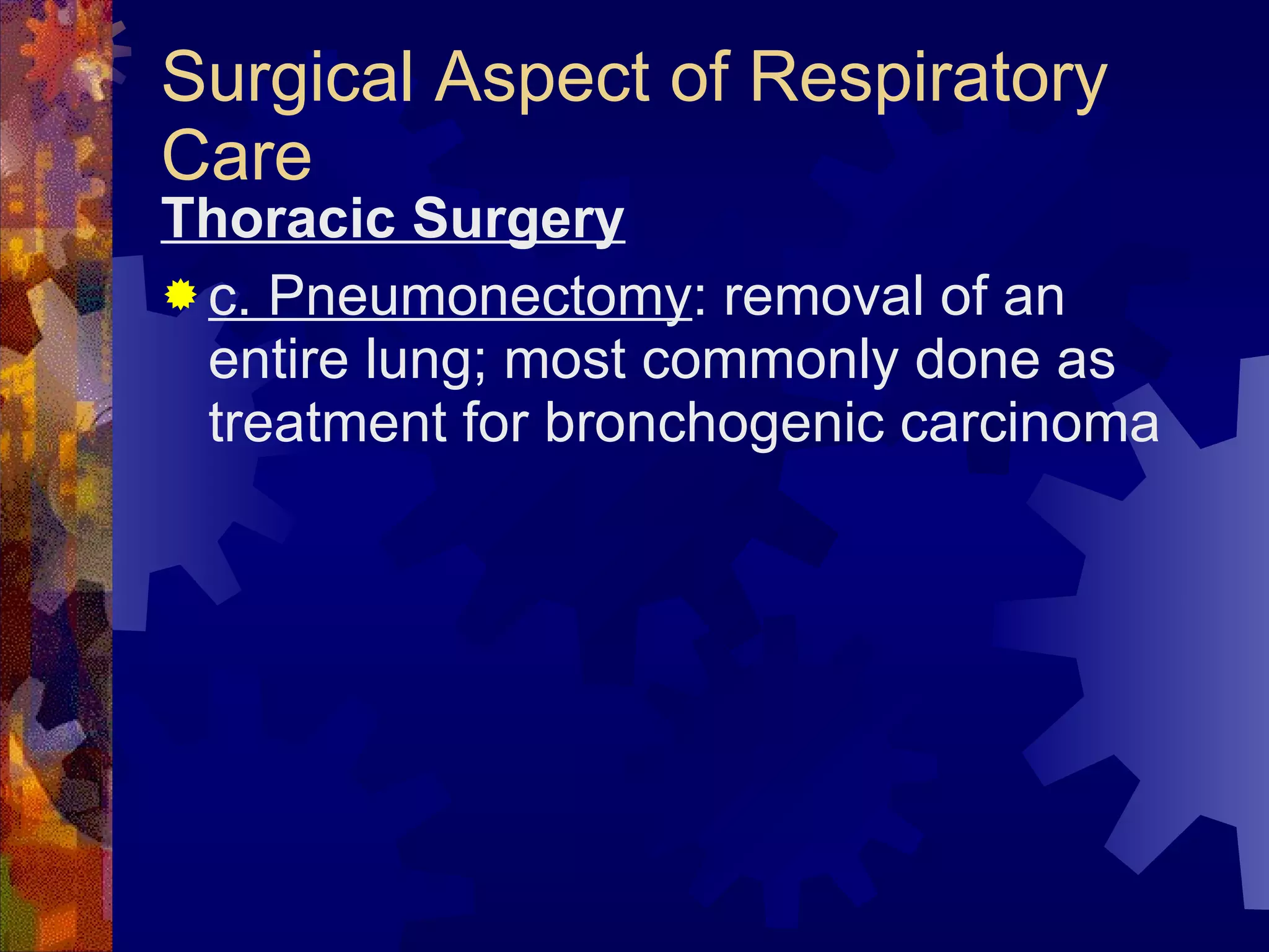
Medical and Surgical Nursing Review The Respiratory System Nurse Licensure Examination Review pinoynursing.webkotoh.com
 
Outline Of Review Concepts: Review of the relevant respiratory anatomy Review of the relevant respiratory physiology The respiratory assessment Common laboratory examinations
Outline Of Review Concepts: Review of the common respiratory problems and the nursing management Review of common respiratory diseases Upper respiratory conditions Lower respiratory conditions
Respiratory Anatomy & Physiology The respiratory system consists of two main parts - the upper and the lower tracts
Respiratory Anatomy & Physiology The UPPER respiratory system consists of: 1. nose 2. mouth 3. pharynx 4. larynx
Respiratory Anatomy & Physiology The LOWER respiratory system consists of: 1. Trachea 2. Bronchus 3. Bronchioles 4. Respiratory unit
Upper Respiratory Tract
The Nose This is the first part of the upper respiratory system that contains nasal bones and cartilages There are numerous hairs called vibrissae There are numerous superficial blood vessels in the nasal mucosa
The Nose The functions of the nose are: 1. To filter the air 2. To humidify the air 3. To aid in phonation 4. Olfaction
The Pharynx The pharynx is a musculo - membranous tube that is composed of three parts 1. Nasopharynx 2. Oropharynx 3. Laryngopharynx
The Pharynx The pharynx functions : 1. As passageway for both air and foods (in the oropharynx) 2. To protect the lower airway
The Larynx Also called the voice box Made of cartilage and membranes and connects the pharynx to the trachea
The Larynx Functions of the larynx: 1. Vocalization 2. Keeps the patency of the upper airway 3. Protects the lower airway
The Paranasal sinuses These are four paired bony cavities that are lined with nasal mucosa and ciliated pseudostratified columnar epithelium Named after their location - frontal, ethmoidal, sphenoidal and maxillary
The Paranasal sinuses The function of the sinuses: Resonating chambers in speech
The Lower Respiratory System The lower respiratory system consists of 1. Trachea 2. Main bronchus 3. Bronchial tree 4. Lungs- 3R/ 2L The trachea   to the terminal bronchioles is called the conducting airway The respiratory bronchioles   to the alveoli is called the respiratory acinus
The Trachea A cartilaginous tube measures 10-12 centimeters Composed of  about 20 C-shaped cartilages, incomplete posteriorly
The Trachea The function of the trachea is to conduct air towards the lungs The mucosa is lined up with mucus and cilia to trap particles and carry them towards the upper airway
The Bronchus The right and left primary bronchi begin at the carina The function is for air passage
The Primary Bronchus RIGHT BRONCHUS Wider Shorter More Vertical LEFT BRONCHUS Narrower Longer More horizontal
The Bronchioles The primary bronchus further divides into secondary, then tertiary then into bronchioles The terminal bronchiole is the last part of the conducting airway
The Respiratory Acinus The respiratory acinus is the chief respiratory unit It consists of  1. Respiratory bronchiole 2. Alveolar duct 3. alveolar sac
The Respiratory Acinus The respiratory acinus is the chief respiratory unit The function of the respiratory acinus is gas exchange through the respiratory membrane
The Respiratory Acinus The respiratory membrane is composed of two epithelial cells 1.The type 1 pneumocyte - most abundant, thin and flat. This is where gas exchange occurs 2. The type 2 pneumocyte - secretes the lung surfactant
The Respiratory Acinus A type III pneomocyte is just the macrophage that ingests foreign material and acts as an important defense mechanism
Accessory Structures The PLEURA Epithelial serous membrane lining the lung parenchyma Composed of two parts- the visceral and parietal pleurae The space in between is the pleural space containing a minute amount of fluid for lubrication
Accessory Structures The Thoracic cavity The chest wall composed of the sternum and the rib cage The cavity is separated by the diaphragm, the most important respiratory muscle
Accessory Structures The Mediastinum The space between the lungs, which includes the heart and pericardium, the aorta and the vena cavae.
GENERAL FUNCTIONS OF THE Respiratory System Gas exchange through ventilation, external respiration and cellular respiration Oxygen and carbon dioxide transport
The Assessment HISTORY Reason for seeking care Present illness Previous illness Family history Social history
The Assessment PHYSICAL EXAMINATION Skin- cyanosis, pallor Nail clubbing Cough and sputum production Inspect - palpate - percuss - auscultate the thorax
The Assessment LABORATORY EXAMINATION 1. ABG analysis 2. Sputum analysis 3. Direct visualization - bronchoscopy 4. Indirect visualization - CXR, CT and MRI 5. Pulmonary function test
ABG Analysis This test helps to evaluate gas exchange in the lungs by measuring the gas pressures and pH of an arterial sample
ABG Analysis Pre-test: choose site carefully, perform the Allen’s test, secure equipments- syringe, needle, container with ice Intra-test: Obtain a 5 mL specimen from the artery (brachial, femoral and radial) Post-test: Apply firm pressure for 5 minutes, label specimen correctly, place in the container with ice
ABG Analysis ABG normal values PaO2 80-100 mmHg PaCO2 35-45 mmHg pH   7.35- 7.45 HCO3 22- 26 mEq/L O2 Sat 95-99%
Sputum Analysis This test analyzes the sample of sputum to diagnose respiratory diseases, identify organism, and identify abnormal cells
Sputum Analysis Pre-test: Encourage to increase fluid intake Intra-test:  rinse mouth with WATER only,  instruct the patient to take 3 deep breaths and force a deep cough, steam nebulization, collect early morning sputum Post-test: provide oral hygiene, label specimen correctly
Pulse Oximetry Non-invasive method of continuously monitoring the oxygen saturation of hemoglobin A sensor or probe is attached to the earlobe, forehead, fingertip or the bridge of the nose
Bronchoscopy A direct inspection of the trachea and bronchi through a flexible fiber-optic or a rigid bronchoscope Done to  determine location of pathologic lesions, to remove foreign objects, to collect tissue specimen and remove secretions/aspirated materials
Bronchoscopy Pre-test: Consent, NPO x 6h, teaching Intra-test: position supine or sitting upright in a chair, administer sedative, gag reflex will be abolished, remove dentures Post-test: NPO until gag reflex returns, position SEMI-fowler’s with head turned to sides, hoarseness is temporary, CXR after the procedure, keep tracheostomy set and suction x 24 hours
Thoracentesis Pleural fluid aspiration for obtaining a specimen of pleural fluid for analysis, relief of lung compression and biopsy specimen collection
Thoracentesis Pre-test: Consent Intra-test: position the patient sitting with arms on a table or side-lying fowler’s, instruct not to cough, breathe deeply or move Post-test: position unaffected side to allow lung expansion of the affected side, CXR obtained,  maintain pressure dressing and monitor respiratory status
Pulmonary Function Tests Volume and capacity tests aid diagnosis in patient with suspected pulmonary dysfunction Evaluates ventilatory function Determines whether obstructive or restrictive disease Can be utilized as screening test
Pulmonary Function Test Lung Volumes Tidal volume Inspiratory reserve volume Expiratory reeve volume Residual volume
Pulmonary Function Test Lung capacities Inspiratory capacity Vital capacity Functional residual capacity Total lung capacity
Pulmonary Function Test Pre-test: Teaching, no smoking for 3 days, only light meal 4 hours before the test Intra-test: position sitting, bronchodilator, nose-clip and mouthpiece, fatigue and dyspnea during the test  Post-test: adequate rest periods, loosen tight clothing
Common Respiratory Problems and the common interventions
Dyspnea Breathing difficulty Associated with many conditions- CHF, MG, GBS, Muscular dystrophy, obstruction, etc…
Dyspnea General nursing interventions: 1. Fowler’s position to promote maximum lung expansion and promote comfort. An alternative position is the ORTHOPNEIC position 2. O2 usually via nasal cannula 3. Provide comfort and distractions
Cough and sputum production Cough is a protective reflex Sputum production has many stimuli Thick, yellow, green or rust-colored    bacterial pneumonia Profuse, Pink, frothy    pulmonary edema Scant, pink-tinged, mucoid    Lung tumor
Cough and sputum production General nursing Intervention 1. Provide adequate hydration 2. Administer aerosolized solutions 3. advise smoking cessation 4. oral hygiene
Cyanosis Bluish discoloration of the skin A LATE indicator of hypoxia Appears when the unoxygenated hemoglobin is more than 5 grams/dL Central cyanosis   observe color on the undersurface of tongue and lips Peripheral cyanosis   observe the nail beds, earlobes
Cyanosis Interventions: Check for airway patency Oxygen therapy Positioning Suctioning Chest physiotherapy Check for gas poisoning Measures to increased hemoglobin
Hemoptysis Expectoration of blood from the respiratory tract Common causes: Pulmo infection, Lung CA, Bronchiectasis, Pulmo emboli Bleeding from stomach    acidic pH, coffee ground material
Hemoptysis Interventions: Keep patent airway Determine the cause Suction and oxygen therapy Administer Fibrin stabilizers like aminocaproic acid and tranexamic acid
Epistaxis Bleeding from the nose caused by rupture of tiny, distended vessels in the mucus membrane  Most common site- anterior septum Causes 1. trauma 2. infection 3. Hypertension 4. blood dyscrasias , nasal tumor, cardio diseases
Epistaxis Nursing Interventions 1. Position patient: Upright, leaning forward, tilted   prevents swallowing and aspiration 2. Apply direct pressure. Pinch nose against the middle septum x 5-10 minutes 3. If unrelieved, administer topical vasoconstrictors, silver nitrate, gel foams 4. Assist in electrocautery and nasal packing for posterior bleeding
CONDITIONS OF THE UPPER AIRWAY Upper airway infections 1. Rhinitis- allergic, non-allergic and infectious 2. Sinusitis- acute and chronic 3. Pharyngitis- acute and chronic
CONDITIONS OF THE UPPER AIRWAY Upper airway infections 1. Rhinitis- Assessment findings Rhinorrhea Nasal congestion Nasal itchiness Sneezing Headache
CONDITIONS OF THE UPPER AIRWAY Upper airway infections 2. sinusitis- Assessment findings Facial pain Tenderness over the paranasal sinuses Purulent nasal discharges Ear pain, headache, dental pain Decreased sense of smell
CONDITIONS OF THE UPPER AIRWAY Upper airway infections 3. Pharyngitis- Assessment findings Fiery-red pharyngeal membrane White-purple flecked exudates Enlarged and tender cervical lymph nodes Fever malaise ,sore throat Difficulty swallowing Cough may be absent
CONDITIONS OF THE UPPER AIRWAY Upper airway infections- Laboratory tests 1. CBC 2. Culture
CONDITIONS OF THE UPPER AIRWAY Upper airway infections: Nursing Interventions 1. Maintain Patent Airway Increase fluid intake to loosen secretions Utilize room vaporizers or steam inhalation Administer medications to relieve nasal congestion
CONDITIONS OF THE UPPER AIRWAY Upper airway infections: Nursing Interventions 2. Promote comfort Administer prescribed analgesics Administer topical analgesics Warm gargles for the relief of sore throat Provide oral hygiene
CONDITIONS OF THE UPPER AIRWAY Upper airway infections: Nursing Interventions 3. Promote communication Instruct patient to refrain from speaking as much as possible Provide writing materials
CONDITIONS OF THE UPPER AIRWAY Upper airway infections: Nursing Interventions 4. Administer prescribed antibiotics Monitor for possible complications like meningitis, otitis media, abscess formation 5. Assist in surgical intervention
CONDITIONS OF THE UPPER AIRWAY Upper airway infection: Tonsillitis Infection and inflammation of the tonsils Most common organism- Group A- beta hemolytic streptococcus (GABS)
CONDITIONS OF THE UPPER AIRWAY Upper airway infection: Tonsillitis ASSESSMENT FINDINGS Sore throat and mouth breathing Fever Difficulty swallowing Enlarged, reddish tonsils Foul-smelling breath
CONDITIONS OF THE UPPER AIRWAY Upper airway infection: Tonsillitis Laboratory test 1. CBC 2. throat culture
CONDITIONS OF THE UPPER AIRWAY Upper airway infection: Tonsillitis MEDICAL management 1. Antibiotics- penicillin 2. Tonsillectomy for chronic cases and abscess formation
CONDITIONS OF THE UPPER AIRWAY Upper airway infection: Tonsillitis NURSING INTERVENTION for tonsillectomy 1. Pre-operative care Consent Routine  pre-op surgical care
CONDITIONS OF THE UPPER AIRWAY Upper airway infection: Tonsillitis NURSING INTERVENTION for tonsillectomy 2. POST-operative care Position: Most comfortable is PRONE, with head turned to side Maintain oral airway, until gag reflex returns
CONDITIONS OF THE UPPER AIRWAY Upper airway infection: Tonsillitis NURSING INTERVENTION for tonsillectomy 2. POST-operative care Apply ICE collar to the neck to reduce edema Advise patient to refrain from talking and coughing Ice chips are given when there is no bleeding and gag reflex returns
CONDITIONS OF THE UPPER AIRWAY Upper airway infection: Tonsillitis NURSING INTERVENTION for tonsillectomy 2. POST-operative care Notify physician if a. Patient swallows frequently b. vomiting of large amount of bright red or dark blood c. PR increased, restless and Temp is increased
Laryngeal Cancer A malignant tumor of the larynx More frequent in men 50-70 years old RISK FACTORS 1. Smoking 2. Alcohol 3. Exposure to chemicals 4. Straining of voice 5. chronic laryngitis 6. Deficiency of Riboflavin 7. family history
Laryngeal Cancer Growth can be anywhere in the larynx 1. Supraglottic- above the vocal cords 2. glottic- vocal cord area 3. infraglottic- below the vocal cords Most tumors are found in the glottic area
Laryngeal Cancer ASSESSMENT FINDINGS Hoarseness of more than TWO weeks duration Cough and sore throat  Burning and pain in the throat especially after consuming HOT liquids and citrus foods Neck lump Dysphagia, dyspnea, foul breath, CLAD
Laryngeal Cancer LABORATORY FINDINGS 1. Indirect laryngoscopy 2. direct laryngoscopy 3. Biopsy 4. CT and MRI Most commonly- squamos carcinoma
Laryngeal Cancer MEDICAL MANAGEMENT Radiation therapy Chemotherapy Surgery Partial laryngectomy Supraglottic laryngectomy Hemilaryngectomy Total laryngectomy
Laryngeal Cancer NURSING MANAGEMENT: PRE-operative 1. Provide the patient pre-operative teachings Clarify misconceptions Tell that the natural voice will be lost Teach communication alternatives Collaborate with other team members
Laryngeal Cancer NURSING MANAGEMENT 2. reduce patient ANXIETY Provide opportunities for patient and family members to ask questions Referrals to previous patients with laryngeal cancers and cancer groups
Laryngeal Cancer NURSING MANAGEMENT: POST-op 3. Maintain PATENT Airway Position patient: Semi or High Fowler’s Suction secretions Encourage to deep breath, turn and cough
Laryngeal Cancer NURSING MANAGEMENT: POST-op 4. Administer care of the laryngectomy tube Suction as needed Cleanse the stoma with saline Administer humidified oxygen Laryngectomy tube is usually removed within 3-6 weeks after surgery
Laryngeal Cancer NURSING MANAGEMENT: POST-op 5. Promote alternative communication methods Call bell or hand bell Magic Slate Hand signals Collaborate with speech therapist
Laryngeal Cancer NURSING MANAGEMENT: POST-op 6. Promote adequate Nutrition NPO after operation No foods or drinks per orem for 10 days IVF, TPN are alternative nutrition routes Start oral feedings with thick liquids, avoid sweet foods
Laryngeal Cancer NURSING MANAGEMENT: POST-op 7. Promote positive body image and self-esteem Encourage verbalization of feelings Allow independence in self-care
Laryngeal Cancer NURSING MANAGEMENT: POST-op 8. Monitor for COMPLICATIONS Respiratory Distress Suction Coughing and deep breathing Humidified oxygen Alert the surgeon
Laryngeal Cancer NURSING MANAGEMENT: POST-op 8. Monitor for Complications Hemorrhage Monitor for bleeding  Monitor vital signs Apply direct pressure over the bleeding artery Summon assistance and alert the surgeon
Laryngeal Cancer NURSING MANAGEMENT: POST-op 8. Monitor for COMPLICATIONS Wound infection and breakdown Monitor for increased temperature, purulent drainage and increased redness/tenderness Administer antibiotics Clean and change dressing OD
Laryngeal Cancer NURSING MANAGEMENT: HOME CARE Humidification system at home is needed AVOID swimming Cover the stoma with hands or plastic bib over the opening Advise beauty salons to avoid hair sprays, powders and loose hair near the opening Oral hygiene frequently
Acute Respiratory Failure  Sudden and life-threatening deterioration of the gas-exchange function of the lungs Occurs when the lungs no longer meet the body’s metabolic needs
Acute Respiratory Failure  Defined clinically as: 1. PaO2 of less than 50 mmHg 2. PaCO2 of greater than 5o mmHg 3. Arterial pH of less than 7.35
Acute Respiratory Failure  CAUSES CNS depression- head trauma, sedatives CVS diseases- MI, CHF, pulmonary emboli Airway irritants- smoke, fumes Endocrine and metabolic disorders- myxedema, metabolic alkalosis Thoracic abnormalities-  chest trauma, pneumothorax
Acute Respiratory Failure  PATHOPHYSIOLOGY Decreased Respiratory Drive Brain injury, sedatives, metabolic disorders    impair the normal response of the brain to normal respiratory stimulation
Acute Respiratory Failure  PATHOPHYSIOLOGY Dysfunction of the chest wall Dystrophy, MS disorders, peripheral nerve disorders   disrupt the impulse transmission from the nerve to the diaphragm   abnormal ventilation
Acute Respiratory Failure  PATHOPHYSIOLOGY Dysfunction of the Lung Parenchyma Pleural effusion, hemothorax, pneumothorax, obstruction   interfere ventilation   prevent lung expansion
Acute Respiratory Failure  ASSESSMENT FINDINGS Restlessness dyspnea Cyanosis Altered respiration Altered mentation Tachycardia Cardiac arrhythmias Respiratory arrest
Acute Respiratory Failure  DIAGNOSTIC FINDINGS Pulmonary function test- pH below 7.35 CXR- pulmonary infiltrates ECG- arrhythmias
Acute Respiratory Failure  MEDICAL TREATMENT Intubation Mechanical ventilation Antibiotics Steroids Bronchodilators
Acute Respiratory Failure  NURSING INTERVENTIONS 1. Maintain patent airway  2. Administer O2 to maintain Pa02 at more than 50 mmHg 3.  Suction airways as required 4. Monitor serum electrolyte levels 5. Administer care of patient on mechanical ventilation
COPD These are group of disorders associated with recurrent or persistent obstruction of air passage and airflow, usually irreversible.
COPD The most common cause of COPD is cigarette smoking. Asthma, Chronic bronchitis, Emphysema and Bronchiectasis are the common disorders.
COPD The general pathophysiology: In COPD there is airflow limitation that is both progressive and associated with abnormal inflammatory response of the lungs to stimuli, usually smoke, particles and dust
ASTHMA The acute episode of airway obstruction is characterized by airway hyperactivity to various stimuli that results in recurrent wheezing brought about by edema and bronchospasm
Asthma Pathophysiology Immunologic/allergic reaction results in histamine release, which produces three main airway responses a. Edema of mucous membranes b. Spasm of the smooth muscle of bronchi and bronchioles c. Accumulation of tenacious secretions
Asthma Assessment Findings Assessment findings 1. Family history of allergies 2. Client history of eczema
Asthma Assessment Findings Assessment findings 3. Pulmonary signs and symptoms- Respiratory distress: slow onset of shortness of breath,  expiratory wheeze , prolonged expiratory phase, air trapping (barrel chest if chronic),  use of accessory muscles, irritability (from hypoxia), diaphoresis, cough, anxiety, weak pulse, diaphoresis  and change in sensorium if severe attack
Asthma Assessment Findings Assessment findings 4.  Use of accessory muscles of respiration, inspiratory retractions, prolonged I:E ratio 5. Cardiovascular symptoms: tachycardia, ECG changes, hypertension, decreased cardiac contractility, pulsus paradoxus 6. CNS manifestations: anxiety, restlessness, fear and disorientation
Emphysema There is progressive and irreversible alveolocapillary destruction with abnormal alveolar enlargement causing alveolar wall destruction. The result is INCREASED lung compliance, DECREASED oxygen diffusion and INCREASED airway resistance!
Emphysema These changes cause a state of carbon dioxide retention, hypoxia, and respiratory acidosis.
Emphysema Cigarette smoking Heredity, Bronchial asthma Aging process   Disequilibrium between ELASTASE & ANTIELASTASE (alpha-1-antitrypsin) Destruction of distal airways and alveoli Overdistention of ALVEOLI Hyper-inflated and pale lungs Air traping, decreased gas exchange and Retention of CO2   Hypoxia Respiratory acidosis
Emphysema Assessment 1. Anorexia, fatigue, weight loss 2. Feeling of breathlessness, cough, sputum production, flaring of the nostrils, use of accessory muscles of respiration, increased rate and depth of breathing, dyspnea
Emphysema Assessment 3. Decreased respiratory excursion, resonance to hyper-resonance, decreased breath sounds with prolonged expiration, normal or decreased fremitus 4. Diagnostic tests: pCO2 elevated or normal; PO2 normal or slightly decreased
Chronic bronchitis Chronic inflammation of the bronchial air passageway characterized by the presence of cough and  sputum production for at least 3 months in each 2 consecutive years. Excessive production of mucus in the bronchi with accompanying persistent cough.
Chronic Bronchitis pathophysiology Characteristic changes include hypertrophy/ hyperplasia of the mucus-secreting glands in the bronchi, decreased ciliary activity, chronic inflammation, and narrowing of the small airways.
Chronic Bronchitis Assessment I. Productive (copious) cough, dyspnea on exertion, use of accessory muscles of respiration, scattered rales and rhonchi 2. Feeling of epigastric fullness, cyanosis, distended neck veins, ankle edema 3. Diagnostic tests: increased pCO2 decreased PO2
Bronchiectasis Permanent abnormal dilation of the bronchi with destruction of muscular and elastic structure of the bronchial wall
Bronchiectasis  Caused by bacterial infection; recurrent lower respiratory tract infections; congenital defects (altered bronchial structures); lung tumors
Bronchiectasis 1. Chronic cough with production of mucopurulent sputum,  hemoptysis,  exertional dyspnea, wheezing 2. Anorexia, fatigue, weight loss 3. Diagnostic tests a. Bronchoscopy reveals sources and sites of secretions b. Possible elevation of WBC
COPD Management Independent and Collaborative Management 1. Rest- To reduce oxygen demands of tissues 2. Increase fluid intake -To liquefy mucus secretions 3. Good oral care- To remove sputum and prevent infection
COPD Management Independent and Collaborative Management 4. Diet:  High caloric diet  provides source of energy High protein  diet helps maintain integrity of alveolar walls Moderate fats Low carbohydrate  diet limits carbon dioxide production (natural end product). The client has difficulty exhaling carbon dioxide.
COPD Management Independent and Collaborative Management 5. O2 therapy  1 to 3 lpm ( 2 lpm is safest ) Do not give high concentration of oxygen. The drive for breathing may be depressed.
COPD Management Independent and Collaborative Management 6 . Avoid cigarette smoking, alcohol, and environmental pollutants-These inhibit mucociliary function. 7. CPT  –percussion, vibration, postural drainage
COPD Management Independent and Collaborative Management 8. Bronchial hygiene measures Steam inhalation Aerosol inhalation Medimist inhalation
COPD Management Pharmacotherapy 1. Expectorants (guaiafenessin)/ mucolytic (mucomyst/mucosolvan) 2. Antitussives Dextrometorphan Codeine Observe for drowsiness Avoid activities that involve mental alertness, e.g driving, operating electrical machines Cause decrease peristalsis thereby constipation
COPD Management Pharmacotherapy 3. Bronchodilators Aminophylline (Theophylline) Ventolin (Salbutamol) Bricanyl (Terbutaline) Alupent (Metaproterenol) Observe for tachycardia
COPD Management Pharmacotherapy 4. Antihistamine Benadryl (Diphenhydramine) Observe for drowsiness 5. Steroids Anti-inflammatory effect 6. Antimicrobials
Flail Chest Complication of chest trauma occurring when 3 or more adjacent ribs are fractured at two or more sites, resulting in free-floating rib segments.
Flail Chest Chest wall is no longer able to provide the bony structure necessary to maintain adequate ventilation; consequently the flail portion and underlying tissue move paradoxically (in opposition) to the rest of the chest cage and lungs.
Flail Chest The flail portion is sucked in on inspiration and bulges out on expiration. Result is hypoxia, hypercarbia, and increased retained secretions. Caused by trauma (sternal rib fracture with possible costochondral separations).
Flail Chest PATHOPHYSIOLOGY During inspiration, as the chest expands, the detached part of the rib segment  (flail segment) moves in a “paradoxical” manner  The chest is pulled INWARD during inspiration, reducing the amount of air that can be drawn into the lungs The chest bulges OUTWARD during expiration because the intrathoracic pressure exceeds atmospheric pressure. The patient has impaired exhalation
Flail Chest This paradoxical action will lead to: Increased dead space Reduced alveolar ventilation Decreased lung compliance Hypoxemia and respiratory acidosis Hypotension, inadequate tissue perfusion can also follow
Flail Chest Assessment findings 1. Severe dyspnea; rapid, shallow, grunty breathing; paradoxical chest motion.  The chest will move INWARDS on inhalation and OUTWARDS on exhalation. 2. Cyanosis, possible neck vein distension, tachycardia, hypotension 3. Diagnostic tests a. PO2 decreased b. pCO2 elevated c. pH decreased
Flail Chest Nursing interventions 1. Maintain an open airway: suction secretions, blood from nose, throat, mouth, and via endotracheal tube; note changes in amount, color, and characteristics. 2. Monitor mechanical ventilation  3. Encourage turning, coughing, and deep breathing. 4. Monitor for signs of shock: HYPOTENSION, TACHYCARDIA
Flail Chest Medical management: SUPPORTIVE 1. Internal stabilization with a volume-cycled ventilator 2. Drug therapy (narcotics, sedatives)
Pneumothorax Partial or complete collapse of the lung due to an accumulation of air or fluid in the pleural space
Pneumothorax Types a . Spontaneous pneumothorax : the most common type of closed pneumothorax; air accumulates within the pleural space without an obvious cause. Rupture of a small bleb on the visceral pleura most frequently produces this type of pneumothorax.
Pneumothorax Types b.  Open pneumothorax : air enters the pleural space through an opening in the chest wall; usually caused by stabbing or gunshot wound.
Pneumothorax Types c.  Tension pneumothorax : air enters the pleural space with each inspiration but cannot escape; causes increased intrathoracic pressure and  shifting of the mediastinal contents to the unaffected side (mediastinal shift ).
Pneumothorax Assessment findings 1. Sudden sharp pain in the chest, dyspnea, diminished or  absent breath sounds on affected side ,  tracheal shift to the opposite side  (tension pneumothorax accompanied by mediastinal shift) 2. Weak, rapid pulse; anxiety; diaphoresis
Pneumothorax Assessment findings 3. Diagnostic tests a. Chest x-ray reveals area and degree of pneumothorax b. pCO2 elevated c. pH decreased
Pneumothorax Nursing interventions 1. Provide nursing care for the client with an endotracheal tube: suction secretions, vomitus, blood from nose, mouth, throat, or via endotracheal tube; monitor mechanical ventilation.
Pneumothorax Nursing interventions 2. Restore/promote adequate respiratory function. a. Assist with thoracentesis and provide appropriate nursing care.  b. Assist with insertion of a chest tube to water- seal drainage and provide appropriate nursing care. c. Continuously evaluate respiratory patterns and report any changes.
Pneumothorax Nursing interventions 3. Provide relief/control of pain. a. Administer narcotics/analgesics/sedatives as ordered and monitor effects. b. Position client in high-Fowler’s position.
Atelectasis Collapse of part or all of a lung due to bronchial obstruction May be caused by  intrabronchial obstruction tumors, bronchospasm foreign bodies extrabronchial compression (tumors, enlarged lymph nodes); or endobronchial disease (bronchogenic carcinoma, inflammatory structures)
Atelectasis Assessment findings 1. Signs and symptoms may be absent depending upon degree of collapse and rapidity with which bronchial obstruction occurs 2.  Dyspnea, decreased breath sounds on affected side, decreased respiratory excursion, dullness to flatness upon percussion over affected area
Atelectasis Assessment findings 3. Cyanosis, tachycardia, tachypnea, elevated temperature, weakness, pain over affected area
Atelectasis Assessment findings 4. Diagnostic tests a. Bronchoscopy: may or may not reveal an obstruction b. Chest x-ray shows diminished size of affected lung and lack of radiance over atelectatic area c. pO2 decreased
Pleural Effusion Defined broadly as a collection of fluid in the pleural space A symptom, not a disease; may be produced by numerous conditions
Pleural Effusion General Classification Transudative effusion: accumulation of protein-poor, cell-poor fluid Exudative effusion: accumulation of protein rich fluid
Pleural Effusion Assessment findings 1. Dyspnea, dullness over affected area upon percussion, absent or decreased breath sounds over affected area, pleural pain, dry cough, pleural friction rub 2. Pallor, fatigue, fever, and night sweats (with empyema)
Pleural Effusion Assessment findings 3. Diagnostic tests a. Chest x-ray positive if greater than 250 cc pleural fluid b. Pleural biopsy may reveal bronchogenic carcinoma c. Thoracentesis may contain blood if cause is cancer, pulmonary infarction, or tuberculosis; positive for specific organism in  empyema.
Pleural Effusion Nursing interventions: In general: 1. Assist with repeated thoracentesis. 2. Administer narcotics/sedatives as ordered to decrease pain. 3. Assist with instillation of medication into pleural space (reposition client every 15 minutes to distribute the drug within the pleurae). 4. Place client in high-Fowler’s position to promote ventilation.
Pleural Effusion Medical management 1. Identification and treatment of the Underlying cause 2. Thoracentesis 3. Drug therapy a. Antibiotics: either systemic or inserted directly into pleural space b. Fibrinolytic enzymes: trypsin, streptokinase-. streptodornase to decrease thickness of pus and dissolve fibrin clots 4. Closed chest drainage 5. Surgery: open drainage
Pneumonia An inflammation of the alveolar spaces of the lung, resulting in consolidation of lung tissue as the alveoli fill with exudates The various types of pneumonias are classified according to the offending organism. Pneumonia can also be classified as COMMUNITY Acquired Pneumonia (CAP) and Hospital acquired pneumonia (HAP)
Pneumonia PATHOPHYSIOLOGIC FINDINGS ARE:  HYPERTROPHY OF MUCOUS MEMBRANE Increased sputum production Wheezing Dyspnea Cough Rales Ronchi
Pneumonia PATHOPHYSIOLOGIC FINDINGS ARE:  INCREASED CAPILLARY PERMEABILITY Increased Fluid Exudation Consolidation-tissue that solidifies as a result of collapsed alveoli Hypoxemia
Pneumonia PATHOPHYSIOLOGIC FINDINGS ARE:  INFLAMMATION OF THE PLEURA Chest pain Pleural effusion Dullness Decreased Breath sounds Increased tactile fremitus
Pneumonia PATHOPHYSIOLOGIC FINDINGS ARE:  HYPOVENTILATION Decreased  Chest expansion Respiratory acidosis Depressed PROTECTIVE MECHANISM Increased  WBC (leukocytosis) Increased  RR and Fever
Pneumonia Assessment findings Cough with greenish to rust-colored sputum production rapid, shallow respirations with an expiratory grunt nasal flaring; intercostal rib retraction; use of accessory muscles of respiration rales or crackles (early) progressing to coarse (later). Tactile fremitus is INCREASED!
Pneumonia Assessment findings Fever, chills, chest pain, weakness, generalized malaise Tachycardia, cyanosis, profuse perspiration, abdominal distension Rapid shallow breathing
Pneumonia Diagnostic tests a. Chest x-ray shows consolidation over affected areas b. WBC increased c. pO2 decreased d. Sputum specimen- culture reveal particular causative organism
Pneumonia 1. Facilitate adequate ventilation. a. Administer oxygen as needed and assess its effectiveness. b.  Place client in Fowler’s position . c. Turn and reposition frequently clients who are immobilized/obtunded. d. Administer analgesics as ordered to relieve pain associated with breathing e. Auscultate breath sounds every 2—4 hours. f. Monitor ABGs.
Pneumonia GENERAL Nursing interventions 2. Facilitate removal of secretions general hydration deep breathing and coughing Suctioning Expectorants aerosol treatments via nebulizer, humidification of inhaled air chest physical therapy
Pneumonia GENERAL Nursing interventions 3. Observe color, characteristics of sputum and report any changes; encourage client to perform good oral hygiene after expectoration.
Pneumonia GENERAL Nursing interventions 4. Provide adequate rest and relief/control of pain. a. Provide bed rest with limited physical activity. b. Limit visits and minimize conversations. c. Plan for uninterrupted rest periods. d. Institute nursing care in blocks to ensure periods of rest. e. Maintain pleasant and restful environment
Pneumonia GENERAL Nursing interventions 5. Administer antibiotics as ordered. Monitor effects and possible toxicity. 6. Prevent transmission (respiratory isolation may be required for clients with staphylococcal pneumonia). 7. Control fever and chills: monitor temperature and administer  antipyretics as ordered, maintain increased fluid intake, provide frequent clothing and linen changes.
Pneumonia GENERAL Nursing interventions 8. Provide client teaching and discharge planning concerning prevention of recurrence. a. Medication regimen/antibiotic therapy b. Need for adequate rest,  c. Need to continue deep breathing and coughing
Pneumonia GENERAL Nursing interventions 8. Provide client teaching and discharge planning concerning prevention of recurrence. d. Availability of vaccines e. Techniques that prevent transmission (use of tissues when coughing, adequate disposal of secretions) f. Avoidance of persons with known respiratory infections g. Need to report signs and symptoms of respiratory infection
Lung Cancer Primary pulmonary tumors arise from the bronchial epithelium and are therefore referred to as bronchogenic carcinomas. FACTORS:  Possibly caused by inhaled carcinogens (primarily cigarette smoke but also asbestos, nickel, iron oxides, air silicone pollution; preexisting pulmonary disorders PTB, COPD)
Lung Cancer Assessment findings Persistent cough (may be productive or blood tinged) chest pain dyspnea unilateral wheezing, friction rub, possible unilateral paralysis of the diaphragm Fatigue, anorexia, nausea, vomiting, pallor
Lung Cancer Diagnostic tests. a. Chest x-ray may show presence of tumor or evidence of metastasis to surrounding structures b. Sputum for cytology reveals malignant cells c. Bronchoscopy: biopsy reveals malignancy d. Thoracentesis: pleural fluid contains malignant cells e. Biopsy of  lymph nodes may reveal metastasis
Lung Cancer 1. Provide support and guidance to client as needed. 2. Provide relief/control of pain. 3. Administer medications as ordered and monitor effects/side effects. 4. Control nausea: administer medications as ordered, provide good oral hygiene, provide small and more frequent feedings.
Lung Cancer 5. Provide nursing care for a client with a thoracotomy. 6. Provide client teaching and discharge planning concerning a. Disease process, diagnostic and therapeutic interventions b. Side effects of radiation and chemotherapy c. Realistic information about prognosis
Lung Cancer Medical management 1. Radiation therapy 2. Chemotherapy: usually includes cyclophosphamide, methotrexate, vincristine, doxorubicin, and procarbazine; concurrently in some combination 3. Surgery: when entire tumor can be removed
Lung Cancer Quick Notes on Bronchogenic Cancer Predisposing factors Cigarette smoking Asbestosis Emphysema Smoke from burnt wood Types Squamous cell Ca- with good prognosis Adenocarcinoma- with good prognosis Oat cell Ca- with good prognosis Undifferentiated Ca- with poor prognosis
Lung Cancer Quick Notes on Bronchogenic Cancer Nursing Interventions Patent airway O2 / Aerosol therapy Deep breathing exercises Relief of pain Protection from infection Adequate nutrition  Chest tube management
Lung Cancer Quick Notes on Bronchogenic Cancer Surgery Pneumonectomy= Removal of a lung (either left or right) Lobectomy =Removal of a lobe. Segmentectomy= Removal of a segment. Wedge resection =Removal of the entire tumor regardless of the segment. Decortication= Stripping off of fibrinous membrane enclosing the lung Thoracoplasty= Removal of rib/s. Usually done after pneumonectomy, to reduce the size of the empty thorax thereby prevent mediastinal shift.
Pulmonary Embolism This refers to the obstruction of the pulmonary artery or one of its branches by a blood clot (thrombus) that originates somewhere in the venous system or in the right side of the heart. Most commonly, pulmonary embolism is due to a clot or thrombus from the deep veins of the lower legs.
Pulmonary Embolism Causes Fat embolism. Air embolism Multiple trauma PVD’s  Abdominal surgery Immobility Hypercoagulability
Pulmonary Embolism PATHOPHYSIOLOGY The thrombus that travels from any part of the venous system  obstructs either completely or partially . Then the lungs will have inadequate blood supply, with resultant increase in dead space in the lungs Gas exchange will be impaired or absent in the involved area
Pulmonary Embolism PATHOPHYSIOLOGY The regional pulmonary vasculature will constrict causing increased resistance, increased pulmonary arterial pressure and then increase workload of the right side of the heart.
Pulmonary Embolism PATHOPHYSIOLOGY When the work of the right side of the heart exceeds its capacity, right ventricular failure will result, leading to a decrease in cardiac output followed by decreased systemic perfusion and eventually,  SHOCK
Pulmonary Embolism Assessment Restlessness (cardinal initial sign) Dyspnea Stabbing chest pain Cyanosis Tachycardia Dilated pupils Apprehension/ fear Diaphoresis Dysrhythmias Hypoxia
Pulmonary Embolism Diagnostic Tests: Ventilation-perfusion scan Pulmonary arteriography CXR ECG ABG
Pulmonary Embolism Nursing Interventions Oxygen therapy STAT Early ambulation postop Monitor obese patient Do not massage legs Relieve pain- analgesics HOB elevated Heparin (2 weeks) then Coumadin (3-6 months)
Pulmonary Embolism Patient Teaching for prevention of Pulmonary Embolism Active leg exercises to avoid venous stasis Early ambulation Use of elastic compression stockings Avoidance of leg-crossing and sitting for prolonged periods Drink fluids
Surgical Aspect of Respiratory Care Thoracic Surgery a. Exploratory thoracotomy : anterior or posterolateral incision through the fourth, fifth, sixth, or seventh intercostal spaces to expose and examine the pleura and lung
Surgical Aspect of Respiratory Care Thoracic Surgery b. Lobectomy : removal of one lobe of a lung; treatment for bronchiectasis, bronchogenic carcinoma, emphysematous blebs, lung abscesses
Surgical Aspect of Respiratory Care Thoracic Surgery c. Pneumonectomy : removal of an entire lung; most commonly done as treatment for bronchogenic carcinoma
Surgical Aspect of Respiratory Care Thoracic Surgery d. Segmental resection : removal of one or more segments of lung; most often done as treatment for bronchiectasis
Surgical Aspect of Respiratory Care Thoracic Surgery e. Wedge resection : removal of lesions that occupy only part of a segment of lung tissue; for excision of small nodules or to obtain a biopsy
Surgical Aspect of Respiratory Care Nursing interventions:  PREOPERATIVE 1. Provide routine pre-op care. 2. Perform a complete physical assessment of the lungs to obtain baseline data. 3. Explain expected post-op measures: care of incision site, oxygen, suctioning, chest tubes (except if pneumonectomy performed)
Surgical Aspect of Respiratory Care Nursing interventions: PREOPERATIVE 4. Teach client adequate splinting of incision with hands or pillow for turning, coughing, and deep breathing. 5. Demonstrate ROM exercises for affected side. 6. Provide chest physical therapy to help remove secretions.
Surgical Aspect of Respiratory Care Nursing interventions:  POSTOPERATIVE 1. Provide routine post-op care. 2. Promote adequate ventilation. a. Perform complete physical assessment of lungs and compare with pre-op findings. b. Auscultate lung fields every 1—2 hours. c.  Encourage turning, coughing, and deep breathing every 1—2 hours after pain relief obtained.
Surgical Aspect of Respiratory Care Nursing interventions: POSTOPERATIVE 2. Promote adequate ventilation. d. Perform tracheobronchial suctioning if needed. e. Assess for proper maintenance of chest drainage system (except after pneumonectomy). f. Monitor ABGs and report significant changes. g. Place client in semi-Fowler’s position
Surgical Aspect of Respiratory Care Nursing interventions: POSTOPERATIVE If  pneumonectomy  is performed, follow surgeon’s orders about positioning,  often on back or  OPERATIVE SIDE If  Lobectomy , patient is usually positioned on the  UNOPERATIVE SIDE
Surgical Aspect of Respiratory Care Nursing interventions: POSTOPERATIVE 3. Provide pain relief. a. Administer narcotics/analgesics prior to turning, coughing, and deep breathing. b. Assist with splinting while turning, coughing, deep breathing.
Surgical Aspect of Respiratory Care Nursing interventions: POSTOPERATIVE 4. Provide client teaching and discharge planning concerning a. Need to continue with coughing/deep breathing for 6—8 weeks post-op and to continue ROM exercises b. Importance of adequate rest with gradual increases in activity levels
Surgical Aspect of Respiratory Care Nursing interventions:  POSTOPERATIVE 4. Provide client teaching and discharge planning concerning c. High-protein diet with inclusion of adequate fluids  d. Chest physical therapy e. Good oral hygiene f.  Need to avoid persons with known upper respiratory infection g. Adverse signs and symptoms  h. Avoidance of crowds and poorly ventilated areas.

Respiratory System

  • 1.
    Medical and SurgicalNursing Review The Respiratory System Nurse Licensure Examination Review pinoynursing.webkotoh.com
  • 2.
  • 3.
    Outline Of ReviewConcepts: Review of the relevant respiratory anatomy Review of the relevant respiratory physiology The respiratory assessment Common laboratory examinations
  • 4.
    Outline Of ReviewConcepts: Review of the common respiratory problems and the nursing management Review of common respiratory diseases Upper respiratory conditions Lower respiratory conditions
  • 5.
    Respiratory Anatomy &Physiology The respiratory system consists of two main parts - the upper and the lower tracts
  • 6.
    Respiratory Anatomy &Physiology The UPPER respiratory system consists of: 1. nose 2. mouth 3. pharynx 4. larynx
  • 7.
    Respiratory Anatomy &Physiology The LOWER respiratory system consists of: 1. Trachea 2. Bronchus 3. Bronchioles 4. Respiratory unit
  • 8.
  • 9.
    The Nose Thisis the first part of the upper respiratory system that contains nasal bones and cartilages There are numerous hairs called vibrissae There are numerous superficial blood vessels in the nasal mucosa
  • 10.
    The Nose Thefunctions of the nose are: 1. To filter the air 2. To humidify the air 3. To aid in phonation 4. Olfaction
  • 11.
    The Pharynx Thepharynx is a musculo - membranous tube that is composed of three parts 1. Nasopharynx 2. Oropharynx 3. Laryngopharynx
  • 12.
    The Pharynx Thepharynx functions : 1. As passageway for both air and foods (in the oropharynx) 2. To protect the lower airway
  • 13.
    The Larynx Alsocalled the voice box Made of cartilage and membranes and connects the pharynx to the trachea
  • 14.
    The Larynx Functionsof the larynx: 1. Vocalization 2. Keeps the patency of the upper airway 3. Protects the lower airway
  • 15.
    The Paranasal sinusesThese are four paired bony cavities that are lined with nasal mucosa and ciliated pseudostratified columnar epithelium Named after their location - frontal, ethmoidal, sphenoidal and maxillary
  • 16.
    The Paranasal sinusesThe function of the sinuses: Resonating chambers in speech
  • 17.
    The Lower RespiratorySystem The lower respiratory system consists of 1. Trachea 2. Main bronchus 3. Bronchial tree 4. Lungs- 3R/ 2L The trachea  to the terminal bronchioles is called the conducting airway The respiratory bronchioles  to the alveoli is called the respiratory acinus
  • 18.
    The Trachea Acartilaginous tube measures 10-12 centimeters Composed of about 20 C-shaped cartilages, incomplete posteriorly
  • 19.
    The Trachea Thefunction of the trachea is to conduct air towards the lungs The mucosa is lined up with mucus and cilia to trap particles and carry them towards the upper airway
  • 20.
    The Bronchus Theright and left primary bronchi begin at the carina The function is for air passage
  • 21.
    The Primary BronchusRIGHT BRONCHUS Wider Shorter More Vertical LEFT BRONCHUS Narrower Longer More horizontal
  • 22.
    The Bronchioles Theprimary bronchus further divides into secondary, then tertiary then into bronchioles The terminal bronchiole is the last part of the conducting airway
  • 23.
    The Respiratory AcinusThe respiratory acinus is the chief respiratory unit It consists of 1. Respiratory bronchiole 2. Alveolar duct 3. alveolar sac
  • 24.
    The Respiratory AcinusThe respiratory acinus is the chief respiratory unit The function of the respiratory acinus is gas exchange through the respiratory membrane
  • 25.
    The Respiratory AcinusThe respiratory membrane is composed of two epithelial cells 1.The type 1 pneumocyte - most abundant, thin and flat. This is where gas exchange occurs 2. The type 2 pneumocyte - secretes the lung surfactant
  • 26.
    The Respiratory AcinusA type III pneomocyte is just the macrophage that ingests foreign material and acts as an important defense mechanism
  • 27.
    Accessory Structures ThePLEURA Epithelial serous membrane lining the lung parenchyma Composed of two parts- the visceral and parietal pleurae The space in between is the pleural space containing a minute amount of fluid for lubrication
  • 28.
    Accessory Structures TheThoracic cavity The chest wall composed of the sternum and the rib cage The cavity is separated by the diaphragm, the most important respiratory muscle
  • 29.
    Accessory Structures TheMediastinum The space between the lungs, which includes the heart and pericardium, the aorta and the vena cavae.
  • 30.
    GENERAL FUNCTIONS OFTHE Respiratory System Gas exchange through ventilation, external respiration and cellular respiration Oxygen and carbon dioxide transport
  • 31.
    The Assessment HISTORYReason for seeking care Present illness Previous illness Family history Social history
  • 32.
    The Assessment PHYSICALEXAMINATION Skin- cyanosis, pallor Nail clubbing Cough and sputum production Inspect - palpate - percuss - auscultate the thorax
  • 33.
    The Assessment LABORATORYEXAMINATION 1. ABG analysis 2. Sputum analysis 3. Direct visualization - bronchoscopy 4. Indirect visualization - CXR, CT and MRI 5. Pulmonary function test
  • 34.
    ABG Analysis Thistest helps to evaluate gas exchange in the lungs by measuring the gas pressures and pH of an arterial sample
  • 35.
    ABG Analysis Pre-test:choose site carefully, perform the Allen’s test, secure equipments- syringe, needle, container with ice Intra-test: Obtain a 5 mL specimen from the artery (brachial, femoral and radial) Post-test: Apply firm pressure for 5 minutes, label specimen correctly, place in the container with ice
  • 36.
    ABG Analysis ABGnormal values PaO2 80-100 mmHg PaCO2 35-45 mmHg pH 7.35- 7.45 HCO3 22- 26 mEq/L O2 Sat 95-99%
  • 37.
    Sputum Analysis Thistest analyzes the sample of sputum to diagnose respiratory diseases, identify organism, and identify abnormal cells
  • 38.
    Sputum Analysis Pre-test:Encourage to increase fluid intake Intra-test: rinse mouth with WATER only, instruct the patient to take 3 deep breaths and force a deep cough, steam nebulization, collect early morning sputum Post-test: provide oral hygiene, label specimen correctly
  • 39.
    Pulse Oximetry Non-invasivemethod of continuously monitoring the oxygen saturation of hemoglobin A sensor or probe is attached to the earlobe, forehead, fingertip or the bridge of the nose
  • 40.
    Bronchoscopy A directinspection of the trachea and bronchi through a flexible fiber-optic or a rigid bronchoscope Done to determine location of pathologic lesions, to remove foreign objects, to collect tissue specimen and remove secretions/aspirated materials
  • 41.
    Bronchoscopy Pre-test: Consent,NPO x 6h, teaching Intra-test: position supine or sitting upright in a chair, administer sedative, gag reflex will be abolished, remove dentures Post-test: NPO until gag reflex returns, position SEMI-fowler’s with head turned to sides, hoarseness is temporary, CXR after the procedure, keep tracheostomy set and suction x 24 hours
  • 42.
    Thoracentesis Pleural fluidaspiration for obtaining a specimen of pleural fluid for analysis, relief of lung compression and biopsy specimen collection
  • 43.
    Thoracentesis Pre-test: ConsentIntra-test: position the patient sitting with arms on a table or side-lying fowler’s, instruct not to cough, breathe deeply or move Post-test: position unaffected side to allow lung expansion of the affected side, CXR obtained, maintain pressure dressing and monitor respiratory status
  • 44.
    Pulmonary Function TestsVolume and capacity tests aid diagnosis in patient with suspected pulmonary dysfunction Evaluates ventilatory function Determines whether obstructive or restrictive disease Can be utilized as screening test
  • 45.
    Pulmonary Function TestLung Volumes Tidal volume Inspiratory reserve volume Expiratory reeve volume Residual volume
  • 46.
    Pulmonary Function TestLung capacities Inspiratory capacity Vital capacity Functional residual capacity Total lung capacity
  • 47.
    Pulmonary Function TestPre-test: Teaching, no smoking for 3 days, only light meal 4 hours before the test Intra-test: position sitting, bronchodilator, nose-clip and mouthpiece, fatigue and dyspnea during the test Post-test: adequate rest periods, loosen tight clothing
  • 48.
    Common Respiratory Problemsand the common interventions
  • 49.
    Dyspnea Breathing difficultyAssociated with many conditions- CHF, MG, GBS, Muscular dystrophy, obstruction, etc…
  • 50.
    Dyspnea General nursinginterventions: 1. Fowler’s position to promote maximum lung expansion and promote comfort. An alternative position is the ORTHOPNEIC position 2. O2 usually via nasal cannula 3. Provide comfort and distractions
  • 51.
    Cough and sputumproduction Cough is a protective reflex Sputum production has many stimuli Thick, yellow, green or rust-colored  bacterial pneumonia Profuse, Pink, frothy  pulmonary edema Scant, pink-tinged, mucoid  Lung tumor
  • 52.
    Cough and sputumproduction General nursing Intervention 1. Provide adequate hydration 2. Administer aerosolized solutions 3. advise smoking cessation 4. oral hygiene
  • 53.
    Cyanosis Bluish discolorationof the skin A LATE indicator of hypoxia Appears when the unoxygenated hemoglobin is more than 5 grams/dL Central cyanosis  observe color on the undersurface of tongue and lips Peripheral cyanosis  observe the nail beds, earlobes
  • 54.
    Cyanosis Interventions: Checkfor airway patency Oxygen therapy Positioning Suctioning Chest physiotherapy Check for gas poisoning Measures to increased hemoglobin
  • 55.
    Hemoptysis Expectoration ofblood from the respiratory tract Common causes: Pulmo infection, Lung CA, Bronchiectasis, Pulmo emboli Bleeding from stomach  acidic pH, coffee ground material
  • 56.
    Hemoptysis Interventions: Keeppatent airway Determine the cause Suction and oxygen therapy Administer Fibrin stabilizers like aminocaproic acid and tranexamic acid
  • 57.
    Epistaxis Bleeding fromthe nose caused by rupture of tiny, distended vessels in the mucus membrane Most common site- anterior septum Causes 1. trauma 2. infection 3. Hypertension 4. blood dyscrasias , nasal tumor, cardio diseases
  • 58.
    Epistaxis Nursing Interventions1. Position patient: Upright, leaning forward, tilted  prevents swallowing and aspiration 2. Apply direct pressure. Pinch nose against the middle septum x 5-10 minutes 3. If unrelieved, administer topical vasoconstrictors, silver nitrate, gel foams 4. Assist in electrocautery and nasal packing for posterior bleeding
  • 59.
    CONDITIONS OF THEUPPER AIRWAY Upper airway infections 1. Rhinitis- allergic, non-allergic and infectious 2. Sinusitis- acute and chronic 3. Pharyngitis- acute and chronic
  • 60.
    CONDITIONS OF THEUPPER AIRWAY Upper airway infections 1. Rhinitis- Assessment findings Rhinorrhea Nasal congestion Nasal itchiness Sneezing Headache
  • 61.
    CONDITIONS OF THEUPPER AIRWAY Upper airway infections 2. sinusitis- Assessment findings Facial pain Tenderness over the paranasal sinuses Purulent nasal discharges Ear pain, headache, dental pain Decreased sense of smell
  • 62.
    CONDITIONS OF THEUPPER AIRWAY Upper airway infections 3. Pharyngitis- Assessment findings Fiery-red pharyngeal membrane White-purple flecked exudates Enlarged and tender cervical lymph nodes Fever malaise ,sore throat Difficulty swallowing Cough may be absent
  • 63.
    CONDITIONS OF THEUPPER AIRWAY Upper airway infections- Laboratory tests 1. CBC 2. Culture
  • 64.
    CONDITIONS OF THEUPPER AIRWAY Upper airway infections: Nursing Interventions 1. Maintain Patent Airway Increase fluid intake to loosen secretions Utilize room vaporizers or steam inhalation Administer medications to relieve nasal congestion
  • 65.
    CONDITIONS OF THEUPPER AIRWAY Upper airway infections: Nursing Interventions 2. Promote comfort Administer prescribed analgesics Administer topical analgesics Warm gargles for the relief of sore throat Provide oral hygiene
  • 66.
    CONDITIONS OF THEUPPER AIRWAY Upper airway infections: Nursing Interventions 3. Promote communication Instruct patient to refrain from speaking as much as possible Provide writing materials
  • 67.
    CONDITIONS OF THEUPPER AIRWAY Upper airway infections: Nursing Interventions 4. Administer prescribed antibiotics Monitor for possible complications like meningitis, otitis media, abscess formation 5. Assist in surgical intervention
  • 68.
    CONDITIONS OF THEUPPER AIRWAY Upper airway infection: Tonsillitis Infection and inflammation of the tonsils Most common organism- Group A- beta hemolytic streptococcus (GABS)
  • 69.
    CONDITIONS OF THEUPPER AIRWAY Upper airway infection: Tonsillitis ASSESSMENT FINDINGS Sore throat and mouth breathing Fever Difficulty swallowing Enlarged, reddish tonsils Foul-smelling breath
  • 70.
    CONDITIONS OF THEUPPER AIRWAY Upper airway infection: Tonsillitis Laboratory test 1. CBC 2. throat culture
  • 71.
    CONDITIONS OF THEUPPER AIRWAY Upper airway infection: Tonsillitis MEDICAL management 1. Antibiotics- penicillin 2. Tonsillectomy for chronic cases and abscess formation
  • 72.
    CONDITIONS OF THEUPPER AIRWAY Upper airway infection: Tonsillitis NURSING INTERVENTION for tonsillectomy 1. Pre-operative care Consent Routine pre-op surgical care
  • 73.
    CONDITIONS OF THEUPPER AIRWAY Upper airway infection: Tonsillitis NURSING INTERVENTION for tonsillectomy 2. POST-operative care Position: Most comfortable is PRONE, with head turned to side Maintain oral airway, until gag reflex returns
  • 74.
    CONDITIONS OF THEUPPER AIRWAY Upper airway infection: Tonsillitis NURSING INTERVENTION for tonsillectomy 2. POST-operative care Apply ICE collar to the neck to reduce edema Advise patient to refrain from talking and coughing Ice chips are given when there is no bleeding and gag reflex returns
  • 75.
    CONDITIONS OF THEUPPER AIRWAY Upper airway infection: Tonsillitis NURSING INTERVENTION for tonsillectomy 2. POST-operative care Notify physician if a. Patient swallows frequently b. vomiting of large amount of bright red or dark blood c. PR increased, restless and Temp is increased
  • 76.
    Laryngeal Cancer Amalignant tumor of the larynx More frequent in men 50-70 years old RISK FACTORS 1. Smoking 2. Alcohol 3. Exposure to chemicals 4. Straining of voice 5. chronic laryngitis 6. Deficiency of Riboflavin 7. family history
  • 77.
    Laryngeal Cancer Growthcan be anywhere in the larynx 1. Supraglottic- above the vocal cords 2. glottic- vocal cord area 3. infraglottic- below the vocal cords Most tumors are found in the glottic area
  • 78.
    Laryngeal Cancer ASSESSMENTFINDINGS Hoarseness of more than TWO weeks duration Cough and sore throat Burning and pain in the throat especially after consuming HOT liquids and citrus foods Neck lump Dysphagia, dyspnea, foul breath, CLAD
  • 79.
    Laryngeal Cancer LABORATORYFINDINGS 1. Indirect laryngoscopy 2. direct laryngoscopy 3. Biopsy 4. CT and MRI Most commonly- squamos carcinoma
  • 80.
    Laryngeal Cancer MEDICALMANAGEMENT Radiation therapy Chemotherapy Surgery Partial laryngectomy Supraglottic laryngectomy Hemilaryngectomy Total laryngectomy
  • 81.
    Laryngeal Cancer NURSINGMANAGEMENT: PRE-operative 1. Provide the patient pre-operative teachings Clarify misconceptions Tell that the natural voice will be lost Teach communication alternatives Collaborate with other team members
  • 82.
    Laryngeal Cancer NURSINGMANAGEMENT 2. reduce patient ANXIETY Provide opportunities for patient and family members to ask questions Referrals to previous patients with laryngeal cancers and cancer groups
  • 83.
    Laryngeal Cancer NURSINGMANAGEMENT: POST-op 3. Maintain PATENT Airway Position patient: Semi or High Fowler’s Suction secretions Encourage to deep breath, turn and cough
  • 84.
    Laryngeal Cancer NURSINGMANAGEMENT: POST-op 4. Administer care of the laryngectomy tube Suction as needed Cleanse the stoma with saline Administer humidified oxygen Laryngectomy tube is usually removed within 3-6 weeks after surgery
  • 85.
    Laryngeal Cancer NURSINGMANAGEMENT: POST-op 5. Promote alternative communication methods Call bell or hand bell Magic Slate Hand signals Collaborate with speech therapist
  • 86.
    Laryngeal Cancer NURSINGMANAGEMENT: POST-op 6. Promote adequate Nutrition NPO after operation No foods or drinks per orem for 10 days IVF, TPN are alternative nutrition routes Start oral feedings with thick liquids, avoid sweet foods
  • 87.
    Laryngeal Cancer NURSINGMANAGEMENT: POST-op 7. Promote positive body image and self-esteem Encourage verbalization of feelings Allow independence in self-care
  • 88.
    Laryngeal Cancer NURSINGMANAGEMENT: POST-op 8. Monitor for COMPLICATIONS Respiratory Distress Suction Coughing and deep breathing Humidified oxygen Alert the surgeon
  • 89.
    Laryngeal Cancer NURSINGMANAGEMENT: POST-op 8. Monitor for Complications Hemorrhage Monitor for bleeding Monitor vital signs Apply direct pressure over the bleeding artery Summon assistance and alert the surgeon
  • 90.
    Laryngeal Cancer NURSINGMANAGEMENT: POST-op 8. Monitor for COMPLICATIONS Wound infection and breakdown Monitor for increased temperature, purulent drainage and increased redness/tenderness Administer antibiotics Clean and change dressing OD
  • 91.
    Laryngeal Cancer NURSINGMANAGEMENT: HOME CARE Humidification system at home is needed AVOID swimming Cover the stoma with hands or plastic bib over the opening Advise beauty salons to avoid hair sprays, powders and loose hair near the opening Oral hygiene frequently
  • 92.
    Acute Respiratory Failure Sudden and life-threatening deterioration of the gas-exchange function of the lungs Occurs when the lungs no longer meet the body’s metabolic needs
  • 93.
    Acute Respiratory Failure Defined clinically as: 1. PaO2 of less than 50 mmHg 2. PaCO2 of greater than 5o mmHg 3. Arterial pH of less than 7.35
  • 94.
    Acute Respiratory Failure CAUSES CNS depression- head trauma, sedatives CVS diseases- MI, CHF, pulmonary emboli Airway irritants- smoke, fumes Endocrine and metabolic disorders- myxedema, metabolic alkalosis Thoracic abnormalities- chest trauma, pneumothorax
  • 95.
    Acute Respiratory Failure PATHOPHYSIOLOGY Decreased Respiratory Drive Brain injury, sedatives, metabolic disorders  impair the normal response of the brain to normal respiratory stimulation
  • 96.
    Acute Respiratory Failure PATHOPHYSIOLOGY Dysfunction of the chest wall Dystrophy, MS disorders, peripheral nerve disorders  disrupt the impulse transmission from the nerve to the diaphragm  abnormal ventilation
  • 97.
    Acute Respiratory Failure PATHOPHYSIOLOGY Dysfunction of the Lung Parenchyma Pleural effusion, hemothorax, pneumothorax, obstruction  interfere ventilation  prevent lung expansion
  • 98.
    Acute Respiratory Failure ASSESSMENT FINDINGS Restlessness dyspnea Cyanosis Altered respiration Altered mentation Tachycardia Cardiac arrhythmias Respiratory arrest
  • 99.
    Acute Respiratory Failure DIAGNOSTIC FINDINGS Pulmonary function test- pH below 7.35 CXR- pulmonary infiltrates ECG- arrhythmias
  • 100.
    Acute Respiratory Failure MEDICAL TREATMENT Intubation Mechanical ventilation Antibiotics Steroids Bronchodilators
  • 101.
    Acute Respiratory Failure NURSING INTERVENTIONS 1. Maintain patent airway 2. Administer O2 to maintain Pa02 at more than 50 mmHg 3. Suction airways as required 4. Monitor serum electrolyte levels 5. Administer care of patient on mechanical ventilation
  • 102.
    COPD These aregroup of disorders associated with recurrent or persistent obstruction of air passage and airflow, usually irreversible.
  • 103.
    COPD The mostcommon cause of COPD is cigarette smoking. Asthma, Chronic bronchitis, Emphysema and Bronchiectasis are the common disorders.
  • 104.
    COPD The generalpathophysiology: In COPD there is airflow limitation that is both progressive and associated with abnormal inflammatory response of the lungs to stimuli, usually smoke, particles and dust
  • 105.
    ASTHMA The acuteepisode of airway obstruction is characterized by airway hyperactivity to various stimuli that results in recurrent wheezing brought about by edema and bronchospasm
  • 106.
    Asthma Pathophysiology Immunologic/allergicreaction results in histamine release, which produces three main airway responses a. Edema of mucous membranes b. Spasm of the smooth muscle of bronchi and bronchioles c. Accumulation of tenacious secretions
  • 107.
    Asthma Assessment FindingsAssessment findings 1. Family history of allergies 2. Client history of eczema
  • 108.
    Asthma Assessment FindingsAssessment findings 3. Pulmonary signs and symptoms- Respiratory distress: slow onset of shortness of breath, expiratory wheeze , prolonged expiratory phase, air trapping (barrel chest if chronic), use of accessory muscles, irritability (from hypoxia), diaphoresis, cough, anxiety, weak pulse, diaphoresis and change in sensorium if severe attack
  • 109.
    Asthma Assessment FindingsAssessment findings 4. Use of accessory muscles of respiration, inspiratory retractions, prolonged I:E ratio 5. Cardiovascular symptoms: tachycardia, ECG changes, hypertension, decreased cardiac contractility, pulsus paradoxus 6. CNS manifestations: anxiety, restlessness, fear and disorientation
  • 110.
    Emphysema There isprogressive and irreversible alveolocapillary destruction with abnormal alveolar enlargement causing alveolar wall destruction. The result is INCREASED lung compliance, DECREASED oxygen diffusion and INCREASED airway resistance!
  • 111.
    Emphysema These changescause a state of carbon dioxide retention, hypoxia, and respiratory acidosis.
  • 112.
    Emphysema Cigarette smokingHeredity, Bronchial asthma Aging process Disequilibrium between ELASTASE & ANTIELASTASE (alpha-1-antitrypsin) Destruction of distal airways and alveoli Overdistention of ALVEOLI Hyper-inflated and pale lungs Air traping, decreased gas exchange and Retention of CO2 Hypoxia Respiratory acidosis
  • 113.
    Emphysema Assessment 1.Anorexia, fatigue, weight loss 2. Feeling of breathlessness, cough, sputum production, flaring of the nostrils, use of accessory muscles of respiration, increased rate and depth of breathing, dyspnea
  • 114.
    Emphysema Assessment 3.Decreased respiratory excursion, resonance to hyper-resonance, decreased breath sounds with prolonged expiration, normal or decreased fremitus 4. Diagnostic tests: pCO2 elevated or normal; PO2 normal or slightly decreased
  • 115.
    Chronic bronchitis Chronicinflammation of the bronchial air passageway characterized by the presence of cough and sputum production for at least 3 months in each 2 consecutive years. Excessive production of mucus in the bronchi with accompanying persistent cough.
  • 116.
    Chronic Bronchitis pathophysiologyCharacteristic changes include hypertrophy/ hyperplasia of the mucus-secreting glands in the bronchi, decreased ciliary activity, chronic inflammation, and narrowing of the small airways.
  • 117.
    Chronic Bronchitis AssessmentI. Productive (copious) cough, dyspnea on exertion, use of accessory muscles of respiration, scattered rales and rhonchi 2. Feeling of epigastric fullness, cyanosis, distended neck veins, ankle edema 3. Diagnostic tests: increased pCO2 decreased PO2
  • 118.
    Bronchiectasis Permanent abnormaldilation of the bronchi with destruction of muscular and elastic structure of the bronchial wall
  • 119.
    Bronchiectasis Causedby bacterial infection; recurrent lower respiratory tract infections; congenital defects (altered bronchial structures); lung tumors
  • 120.
    Bronchiectasis 1. Chroniccough with production of mucopurulent sputum, hemoptysis, exertional dyspnea, wheezing 2. Anorexia, fatigue, weight loss 3. Diagnostic tests a. Bronchoscopy reveals sources and sites of secretions b. Possible elevation of WBC
  • 121.
    COPD Management Independentand Collaborative Management 1. Rest- To reduce oxygen demands of tissues 2. Increase fluid intake -To liquefy mucus secretions 3. Good oral care- To remove sputum and prevent infection
  • 122.
    COPD Management Independentand Collaborative Management 4. Diet: High caloric diet provides source of energy High protein diet helps maintain integrity of alveolar walls Moderate fats Low carbohydrate diet limits carbon dioxide production (natural end product). The client has difficulty exhaling carbon dioxide.
  • 123.
    COPD Management Independentand Collaborative Management 5. O2 therapy 1 to 3 lpm ( 2 lpm is safest ) Do not give high concentration of oxygen. The drive for breathing may be depressed.
  • 124.
    COPD Management Independentand Collaborative Management 6 . Avoid cigarette smoking, alcohol, and environmental pollutants-These inhibit mucociliary function. 7. CPT –percussion, vibration, postural drainage
  • 125.
    COPD Management Independentand Collaborative Management 8. Bronchial hygiene measures Steam inhalation Aerosol inhalation Medimist inhalation
  • 126.
    COPD Management Pharmacotherapy1. Expectorants (guaiafenessin)/ mucolytic (mucomyst/mucosolvan) 2. Antitussives Dextrometorphan Codeine Observe for drowsiness Avoid activities that involve mental alertness, e.g driving, operating electrical machines Cause decrease peristalsis thereby constipation
  • 127.
    COPD Management Pharmacotherapy3. Bronchodilators Aminophylline (Theophylline) Ventolin (Salbutamol) Bricanyl (Terbutaline) Alupent (Metaproterenol) Observe for tachycardia
  • 128.
    COPD Management Pharmacotherapy4. Antihistamine Benadryl (Diphenhydramine) Observe for drowsiness 5. Steroids Anti-inflammatory effect 6. Antimicrobials
  • 129.
    Flail Chest Complicationof chest trauma occurring when 3 or more adjacent ribs are fractured at two or more sites, resulting in free-floating rib segments.
  • 130.
    Flail Chest Chestwall is no longer able to provide the bony structure necessary to maintain adequate ventilation; consequently the flail portion and underlying tissue move paradoxically (in opposition) to the rest of the chest cage and lungs.
  • 131.
    Flail Chest Theflail portion is sucked in on inspiration and bulges out on expiration. Result is hypoxia, hypercarbia, and increased retained secretions. Caused by trauma (sternal rib fracture with possible costochondral separations).
  • 132.
    Flail Chest PATHOPHYSIOLOGYDuring inspiration, as the chest expands, the detached part of the rib segment (flail segment) moves in a “paradoxical” manner The chest is pulled INWARD during inspiration, reducing the amount of air that can be drawn into the lungs The chest bulges OUTWARD during expiration because the intrathoracic pressure exceeds atmospheric pressure. The patient has impaired exhalation
  • 133.
    Flail Chest Thisparadoxical action will lead to: Increased dead space Reduced alveolar ventilation Decreased lung compliance Hypoxemia and respiratory acidosis Hypotension, inadequate tissue perfusion can also follow
  • 134.
    Flail Chest Assessmentfindings 1. Severe dyspnea; rapid, shallow, grunty breathing; paradoxical chest motion. The chest will move INWARDS on inhalation and OUTWARDS on exhalation. 2. Cyanosis, possible neck vein distension, tachycardia, hypotension 3. Diagnostic tests a. PO2 decreased b. pCO2 elevated c. pH decreased
  • 135.
    Flail Chest Nursinginterventions 1. Maintain an open airway: suction secretions, blood from nose, throat, mouth, and via endotracheal tube; note changes in amount, color, and characteristics. 2. Monitor mechanical ventilation 3. Encourage turning, coughing, and deep breathing. 4. Monitor for signs of shock: HYPOTENSION, TACHYCARDIA
  • 136.
    Flail Chest Medicalmanagement: SUPPORTIVE 1. Internal stabilization with a volume-cycled ventilator 2. Drug therapy (narcotics, sedatives)
  • 137.
    Pneumothorax Partial orcomplete collapse of the lung due to an accumulation of air or fluid in the pleural space
  • 138.
    Pneumothorax Types a. Spontaneous pneumothorax : the most common type of closed pneumothorax; air accumulates within the pleural space without an obvious cause. Rupture of a small bleb on the visceral pleura most frequently produces this type of pneumothorax.
  • 139.
    Pneumothorax Types b. Open pneumothorax : air enters the pleural space through an opening in the chest wall; usually caused by stabbing or gunshot wound.
  • 140.
    Pneumothorax Types c. Tension pneumothorax : air enters the pleural space with each inspiration but cannot escape; causes increased intrathoracic pressure and shifting of the mediastinal contents to the unaffected side (mediastinal shift ).
  • 141.
    Pneumothorax Assessment findings1. Sudden sharp pain in the chest, dyspnea, diminished or absent breath sounds on affected side , tracheal shift to the opposite side (tension pneumothorax accompanied by mediastinal shift) 2. Weak, rapid pulse; anxiety; diaphoresis
  • 142.
    Pneumothorax Assessment findings3. Diagnostic tests a. Chest x-ray reveals area and degree of pneumothorax b. pCO2 elevated c. pH decreased
  • 143.
    Pneumothorax Nursing interventions1. Provide nursing care for the client with an endotracheal tube: suction secretions, vomitus, blood from nose, mouth, throat, or via endotracheal tube; monitor mechanical ventilation.
  • 144.
    Pneumothorax Nursing interventions2. Restore/promote adequate respiratory function. a. Assist with thoracentesis and provide appropriate nursing care. b. Assist with insertion of a chest tube to water- seal drainage and provide appropriate nursing care. c. Continuously evaluate respiratory patterns and report any changes.
  • 145.
    Pneumothorax Nursing interventions3. Provide relief/control of pain. a. Administer narcotics/analgesics/sedatives as ordered and monitor effects. b. Position client in high-Fowler’s position.
  • 146.
    Atelectasis Collapse ofpart or all of a lung due to bronchial obstruction May be caused by intrabronchial obstruction tumors, bronchospasm foreign bodies extrabronchial compression (tumors, enlarged lymph nodes); or endobronchial disease (bronchogenic carcinoma, inflammatory structures)
  • 147.
    Atelectasis Assessment findings1. Signs and symptoms may be absent depending upon degree of collapse and rapidity with which bronchial obstruction occurs 2. Dyspnea, decreased breath sounds on affected side, decreased respiratory excursion, dullness to flatness upon percussion over affected area
  • 148.
    Atelectasis Assessment findings3. Cyanosis, tachycardia, tachypnea, elevated temperature, weakness, pain over affected area
  • 149.
    Atelectasis Assessment findings4. Diagnostic tests a. Bronchoscopy: may or may not reveal an obstruction b. Chest x-ray shows diminished size of affected lung and lack of radiance over atelectatic area c. pO2 decreased
  • 150.
    Pleural Effusion Definedbroadly as a collection of fluid in the pleural space A symptom, not a disease; may be produced by numerous conditions
  • 151.
    Pleural Effusion GeneralClassification Transudative effusion: accumulation of protein-poor, cell-poor fluid Exudative effusion: accumulation of protein rich fluid
  • 152.
    Pleural Effusion Assessmentfindings 1. Dyspnea, dullness over affected area upon percussion, absent or decreased breath sounds over affected area, pleural pain, dry cough, pleural friction rub 2. Pallor, fatigue, fever, and night sweats (with empyema)
  • 153.
    Pleural Effusion Assessmentfindings 3. Diagnostic tests a. Chest x-ray positive if greater than 250 cc pleural fluid b. Pleural biopsy may reveal bronchogenic carcinoma c. Thoracentesis may contain blood if cause is cancer, pulmonary infarction, or tuberculosis; positive for specific organism in empyema.
  • 154.
    Pleural Effusion Nursinginterventions: In general: 1. Assist with repeated thoracentesis. 2. Administer narcotics/sedatives as ordered to decrease pain. 3. Assist with instillation of medication into pleural space (reposition client every 15 minutes to distribute the drug within the pleurae). 4. Place client in high-Fowler’s position to promote ventilation.
  • 155.
    Pleural Effusion Medicalmanagement 1. Identification and treatment of the Underlying cause 2. Thoracentesis 3. Drug therapy a. Antibiotics: either systemic or inserted directly into pleural space b. Fibrinolytic enzymes: trypsin, streptokinase-. streptodornase to decrease thickness of pus and dissolve fibrin clots 4. Closed chest drainage 5. Surgery: open drainage
  • 156.
    Pneumonia An inflammationof the alveolar spaces of the lung, resulting in consolidation of lung tissue as the alveoli fill with exudates The various types of pneumonias are classified according to the offending organism. Pneumonia can also be classified as COMMUNITY Acquired Pneumonia (CAP) and Hospital acquired pneumonia (HAP)
  • 157.
    Pneumonia PATHOPHYSIOLOGIC FINDINGSARE: HYPERTROPHY OF MUCOUS MEMBRANE Increased sputum production Wheezing Dyspnea Cough Rales Ronchi
  • 158.
    Pneumonia PATHOPHYSIOLOGIC FINDINGSARE: INCREASED CAPILLARY PERMEABILITY Increased Fluid Exudation Consolidation-tissue that solidifies as a result of collapsed alveoli Hypoxemia
  • 159.
    Pneumonia PATHOPHYSIOLOGIC FINDINGSARE: INFLAMMATION OF THE PLEURA Chest pain Pleural effusion Dullness Decreased Breath sounds Increased tactile fremitus
  • 160.
    Pneumonia PATHOPHYSIOLOGIC FINDINGSARE: HYPOVENTILATION Decreased Chest expansion Respiratory acidosis Depressed PROTECTIVE MECHANISM Increased WBC (leukocytosis) Increased RR and Fever
  • 161.
    Pneumonia Assessment findingsCough with greenish to rust-colored sputum production rapid, shallow respirations with an expiratory grunt nasal flaring; intercostal rib retraction; use of accessory muscles of respiration rales or crackles (early) progressing to coarse (later). Tactile fremitus is INCREASED!
  • 162.
    Pneumonia Assessment findingsFever, chills, chest pain, weakness, generalized malaise Tachycardia, cyanosis, profuse perspiration, abdominal distension Rapid shallow breathing
  • 163.
    Pneumonia Diagnostic testsa. Chest x-ray shows consolidation over affected areas b. WBC increased c. pO2 decreased d. Sputum specimen- culture reveal particular causative organism
  • 164.
    Pneumonia 1. Facilitateadequate ventilation. a. Administer oxygen as needed and assess its effectiveness. b. Place client in Fowler’s position . c. Turn and reposition frequently clients who are immobilized/obtunded. d. Administer analgesics as ordered to relieve pain associated with breathing e. Auscultate breath sounds every 2—4 hours. f. Monitor ABGs.
  • 165.
    Pneumonia GENERAL Nursinginterventions 2. Facilitate removal of secretions general hydration deep breathing and coughing Suctioning Expectorants aerosol treatments via nebulizer, humidification of inhaled air chest physical therapy
  • 166.
    Pneumonia GENERAL Nursinginterventions 3. Observe color, characteristics of sputum and report any changes; encourage client to perform good oral hygiene after expectoration.
  • 167.
    Pneumonia GENERAL Nursinginterventions 4. Provide adequate rest and relief/control of pain. a. Provide bed rest with limited physical activity. b. Limit visits and minimize conversations. c. Plan for uninterrupted rest periods. d. Institute nursing care in blocks to ensure periods of rest. e. Maintain pleasant and restful environment
  • 168.
    Pneumonia GENERAL Nursinginterventions 5. Administer antibiotics as ordered. Monitor effects and possible toxicity. 6. Prevent transmission (respiratory isolation may be required for clients with staphylococcal pneumonia). 7. Control fever and chills: monitor temperature and administer antipyretics as ordered, maintain increased fluid intake, provide frequent clothing and linen changes.
  • 169.
    Pneumonia GENERAL Nursinginterventions 8. Provide client teaching and discharge planning concerning prevention of recurrence. a. Medication regimen/antibiotic therapy b. Need for adequate rest, c. Need to continue deep breathing and coughing
  • 170.
    Pneumonia GENERAL Nursinginterventions 8. Provide client teaching and discharge planning concerning prevention of recurrence. d. Availability of vaccines e. Techniques that prevent transmission (use of tissues when coughing, adequate disposal of secretions) f. Avoidance of persons with known respiratory infections g. Need to report signs and symptoms of respiratory infection
  • 171.
    Lung Cancer Primarypulmonary tumors arise from the bronchial epithelium and are therefore referred to as bronchogenic carcinomas. FACTORS: Possibly caused by inhaled carcinogens (primarily cigarette smoke but also asbestos, nickel, iron oxides, air silicone pollution; preexisting pulmonary disorders PTB, COPD)
  • 172.
    Lung Cancer Assessmentfindings Persistent cough (may be productive or blood tinged) chest pain dyspnea unilateral wheezing, friction rub, possible unilateral paralysis of the diaphragm Fatigue, anorexia, nausea, vomiting, pallor
  • 173.
    Lung Cancer Diagnostictests. a. Chest x-ray may show presence of tumor or evidence of metastasis to surrounding structures b. Sputum for cytology reveals malignant cells c. Bronchoscopy: biopsy reveals malignancy d. Thoracentesis: pleural fluid contains malignant cells e. Biopsy of lymph nodes may reveal metastasis
  • 174.
    Lung Cancer 1.Provide support and guidance to client as needed. 2. Provide relief/control of pain. 3. Administer medications as ordered and monitor effects/side effects. 4. Control nausea: administer medications as ordered, provide good oral hygiene, provide small and more frequent feedings.
  • 175.
    Lung Cancer 5.Provide nursing care for a client with a thoracotomy. 6. Provide client teaching and discharge planning concerning a. Disease process, diagnostic and therapeutic interventions b. Side effects of radiation and chemotherapy c. Realistic information about prognosis
  • 176.
    Lung Cancer Medicalmanagement 1. Radiation therapy 2. Chemotherapy: usually includes cyclophosphamide, methotrexate, vincristine, doxorubicin, and procarbazine; concurrently in some combination 3. Surgery: when entire tumor can be removed
  • 177.
    Lung Cancer QuickNotes on Bronchogenic Cancer Predisposing factors Cigarette smoking Asbestosis Emphysema Smoke from burnt wood Types Squamous cell Ca- with good prognosis Adenocarcinoma- with good prognosis Oat cell Ca- with good prognosis Undifferentiated Ca- with poor prognosis
  • 178.
    Lung Cancer QuickNotes on Bronchogenic Cancer Nursing Interventions Patent airway O2 / Aerosol therapy Deep breathing exercises Relief of pain Protection from infection Adequate nutrition Chest tube management
  • 179.
    Lung Cancer QuickNotes on Bronchogenic Cancer Surgery Pneumonectomy= Removal of a lung (either left or right) Lobectomy =Removal of a lobe. Segmentectomy= Removal of a segment. Wedge resection =Removal of the entire tumor regardless of the segment. Decortication= Stripping off of fibrinous membrane enclosing the lung Thoracoplasty= Removal of rib/s. Usually done after pneumonectomy, to reduce the size of the empty thorax thereby prevent mediastinal shift.
  • 180.
    Pulmonary Embolism Thisrefers to the obstruction of the pulmonary artery or one of its branches by a blood clot (thrombus) that originates somewhere in the venous system or in the right side of the heart. Most commonly, pulmonary embolism is due to a clot or thrombus from the deep veins of the lower legs.
  • 181.
    Pulmonary Embolism CausesFat embolism. Air embolism Multiple trauma PVD’s Abdominal surgery Immobility Hypercoagulability
  • 182.
    Pulmonary Embolism PATHOPHYSIOLOGYThe thrombus that travels from any part of the venous system obstructs either completely or partially . Then the lungs will have inadequate blood supply, with resultant increase in dead space in the lungs Gas exchange will be impaired or absent in the involved area
  • 183.
    Pulmonary Embolism PATHOPHYSIOLOGYThe regional pulmonary vasculature will constrict causing increased resistance, increased pulmonary arterial pressure and then increase workload of the right side of the heart.
  • 184.
    Pulmonary Embolism PATHOPHYSIOLOGYWhen the work of the right side of the heart exceeds its capacity, right ventricular failure will result, leading to a decrease in cardiac output followed by decreased systemic perfusion and eventually, SHOCK
  • 185.
    Pulmonary Embolism AssessmentRestlessness (cardinal initial sign) Dyspnea Stabbing chest pain Cyanosis Tachycardia Dilated pupils Apprehension/ fear Diaphoresis Dysrhythmias Hypoxia
  • 186.
    Pulmonary Embolism DiagnosticTests: Ventilation-perfusion scan Pulmonary arteriography CXR ECG ABG
  • 187.
    Pulmonary Embolism NursingInterventions Oxygen therapy STAT Early ambulation postop Monitor obese patient Do not massage legs Relieve pain- analgesics HOB elevated Heparin (2 weeks) then Coumadin (3-6 months)
  • 188.
    Pulmonary Embolism PatientTeaching for prevention of Pulmonary Embolism Active leg exercises to avoid venous stasis Early ambulation Use of elastic compression stockings Avoidance of leg-crossing and sitting for prolonged periods Drink fluids
  • 189.
    Surgical Aspect ofRespiratory Care Thoracic Surgery a. Exploratory thoracotomy : anterior or posterolateral incision through the fourth, fifth, sixth, or seventh intercostal spaces to expose and examine the pleura and lung
  • 190.
    Surgical Aspect ofRespiratory Care Thoracic Surgery b. Lobectomy : removal of one lobe of a lung; treatment for bronchiectasis, bronchogenic carcinoma, emphysematous blebs, lung abscesses
  • 191.
    Surgical Aspect ofRespiratory Care Thoracic Surgery c. Pneumonectomy : removal of an entire lung; most commonly done as treatment for bronchogenic carcinoma
  • 192.
    Surgical Aspect ofRespiratory Care Thoracic Surgery d. Segmental resection : removal of one or more segments of lung; most often done as treatment for bronchiectasis
  • 193.
    Surgical Aspect ofRespiratory Care Thoracic Surgery e. Wedge resection : removal of lesions that occupy only part of a segment of lung tissue; for excision of small nodules or to obtain a biopsy
  • 194.
    Surgical Aspect ofRespiratory Care Nursing interventions: PREOPERATIVE 1. Provide routine pre-op care. 2. Perform a complete physical assessment of the lungs to obtain baseline data. 3. Explain expected post-op measures: care of incision site, oxygen, suctioning, chest tubes (except if pneumonectomy performed)
  • 195.
    Surgical Aspect ofRespiratory Care Nursing interventions: PREOPERATIVE 4. Teach client adequate splinting of incision with hands or pillow for turning, coughing, and deep breathing. 5. Demonstrate ROM exercises for affected side. 6. Provide chest physical therapy to help remove secretions.
  • 196.
    Surgical Aspect ofRespiratory Care Nursing interventions: POSTOPERATIVE 1. Provide routine post-op care. 2. Promote adequate ventilation. a. Perform complete physical assessment of lungs and compare with pre-op findings. b. Auscultate lung fields every 1—2 hours. c. Encourage turning, coughing, and deep breathing every 1—2 hours after pain relief obtained.
  • 197.
    Surgical Aspect ofRespiratory Care Nursing interventions: POSTOPERATIVE 2. Promote adequate ventilation. d. Perform tracheobronchial suctioning if needed. e. Assess for proper maintenance of chest drainage system (except after pneumonectomy). f. Monitor ABGs and report significant changes. g. Place client in semi-Fowler’s position
  • 198.
    Surgical Aspect ofRespiratory Care Nursing interventions: POSTOPERATIVE If pneumonectomy is performed, follow surgeon’s orders about positioning, often on back or OPERATIVE SIDE If Lobectomy , patient is usually positioned on the UNOPERATIVE SIDE
  • 199.
    Surgical Aspect ofRespiratory Care Nursing interventions: POSTOPERATIVE 3. Provide pain relief. a. Administer narcotics/analgesics prior to turning, coughing, and deep breathing. b. Assist with splinting while turning, coughing, deep breathing.
  • 200.
    Surgical Aspect ofRespiratory Care Nursing interventions: POSTOPERATIVE 4. Provide client teaching and discharge planning concerning a. Need to continue with coughing/deep breathing for 6—8 weeks post-op and to continue ROM exercises b. Importance of adequate rest with gradual increases in activity levels
  • 201.
    Surgical Aspect ofRespiratory Care Nursing interventions: POSTOPERATIVE 4. Provide client teaching and discharge planning concerning c. High-protein diet with inclusion of adequate fluids d. Chest physical therapy e. Good oral hygiene f. Need to avoid persons with known upper respiratory infection g. Adverse signs and symptoms h. Avoidance of crowds and poorly ventilated areas.