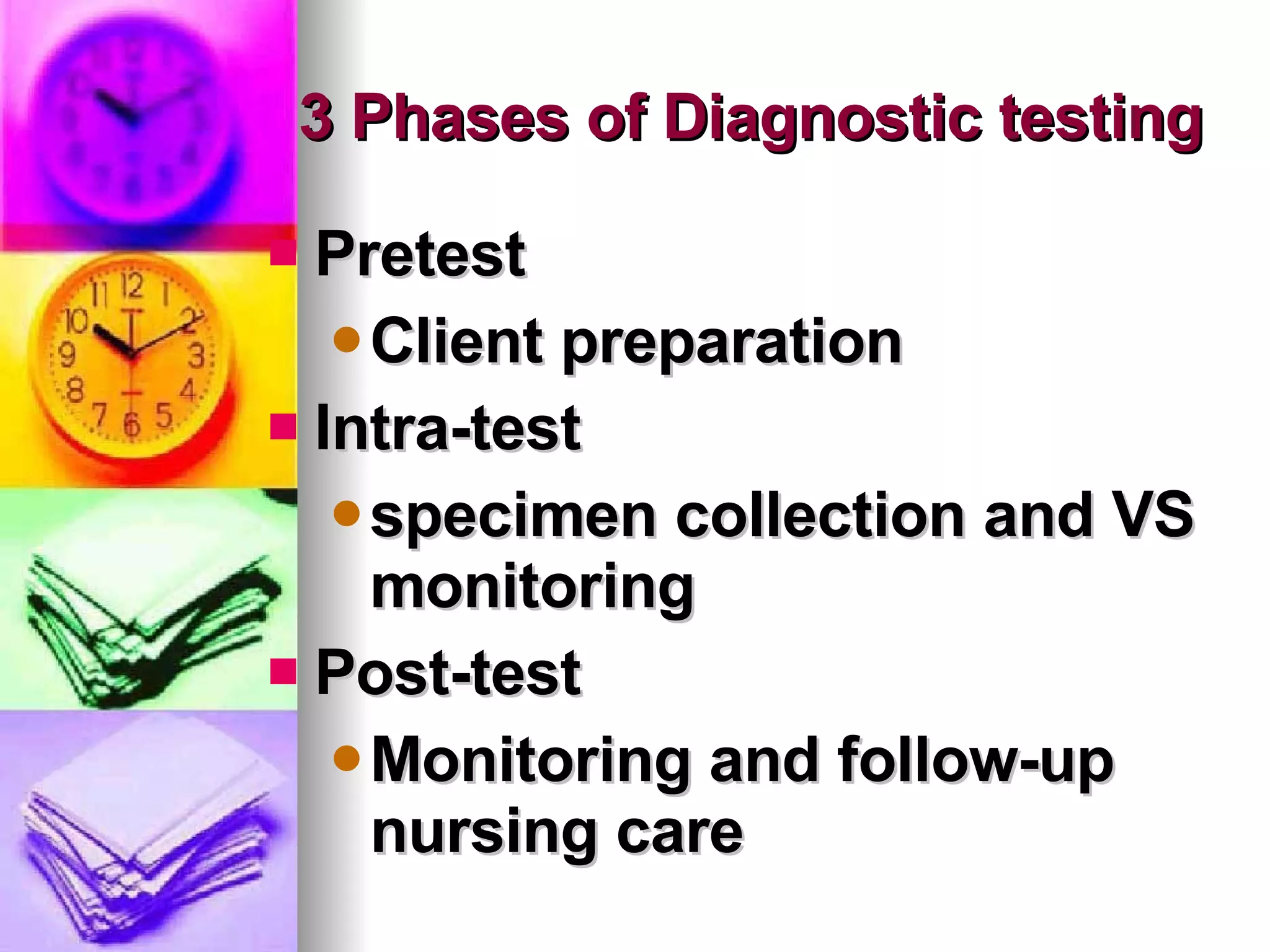
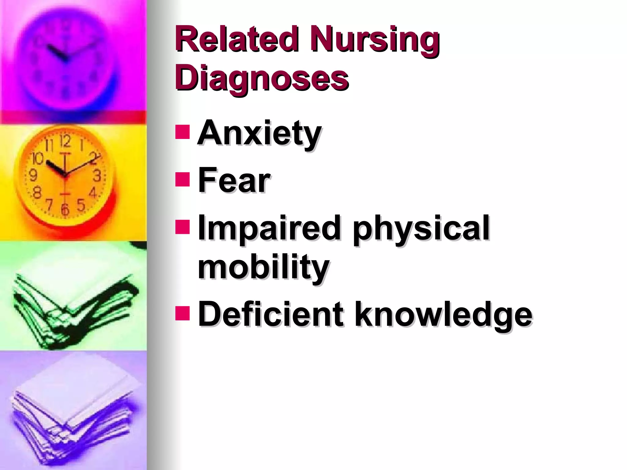
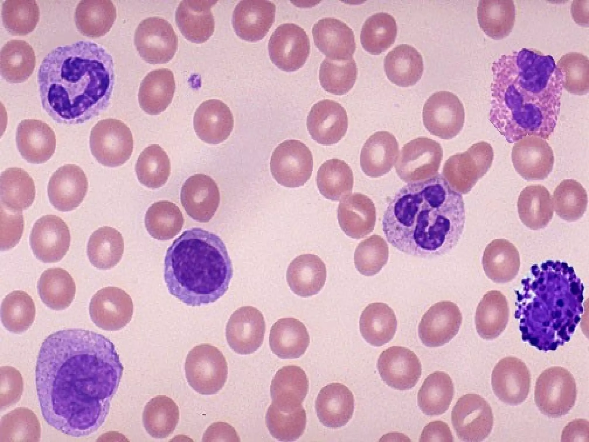
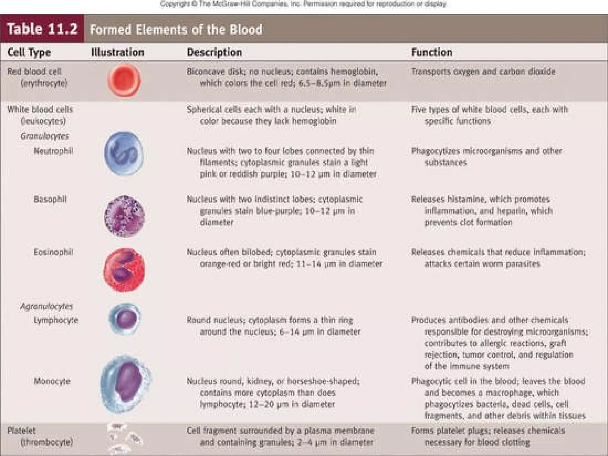
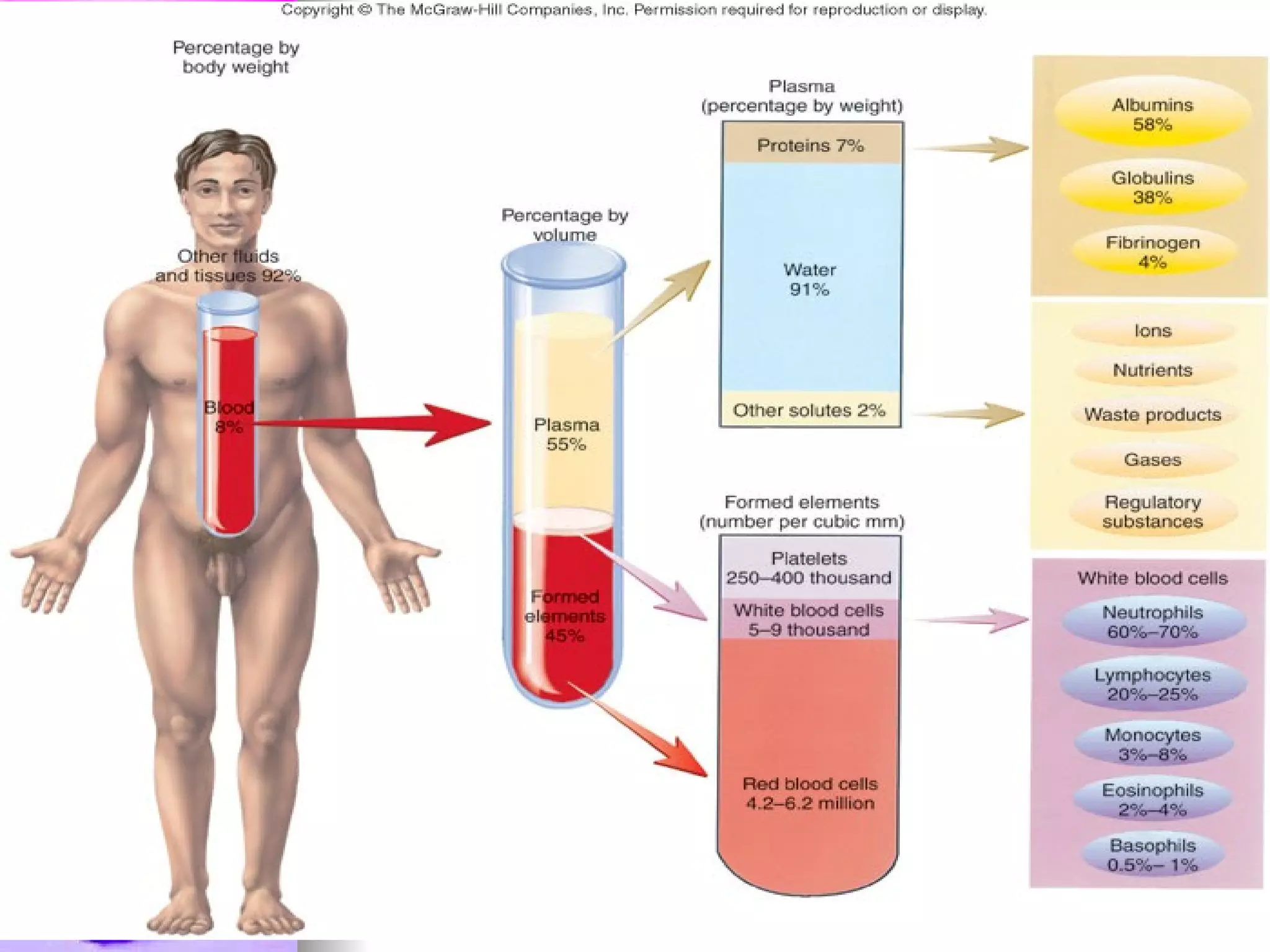
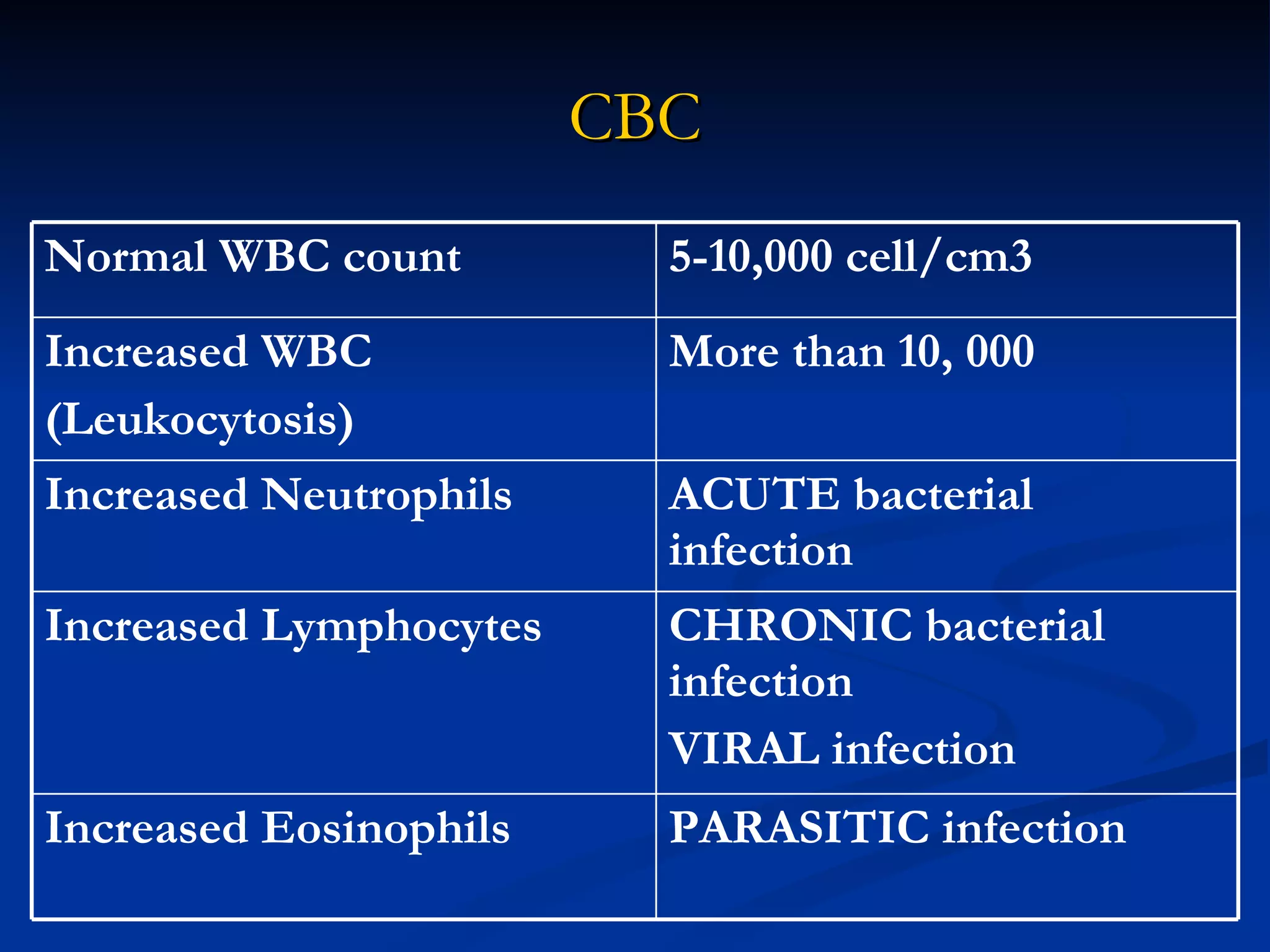
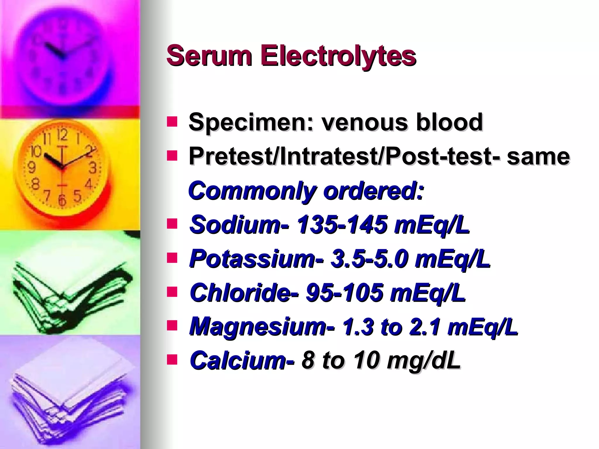
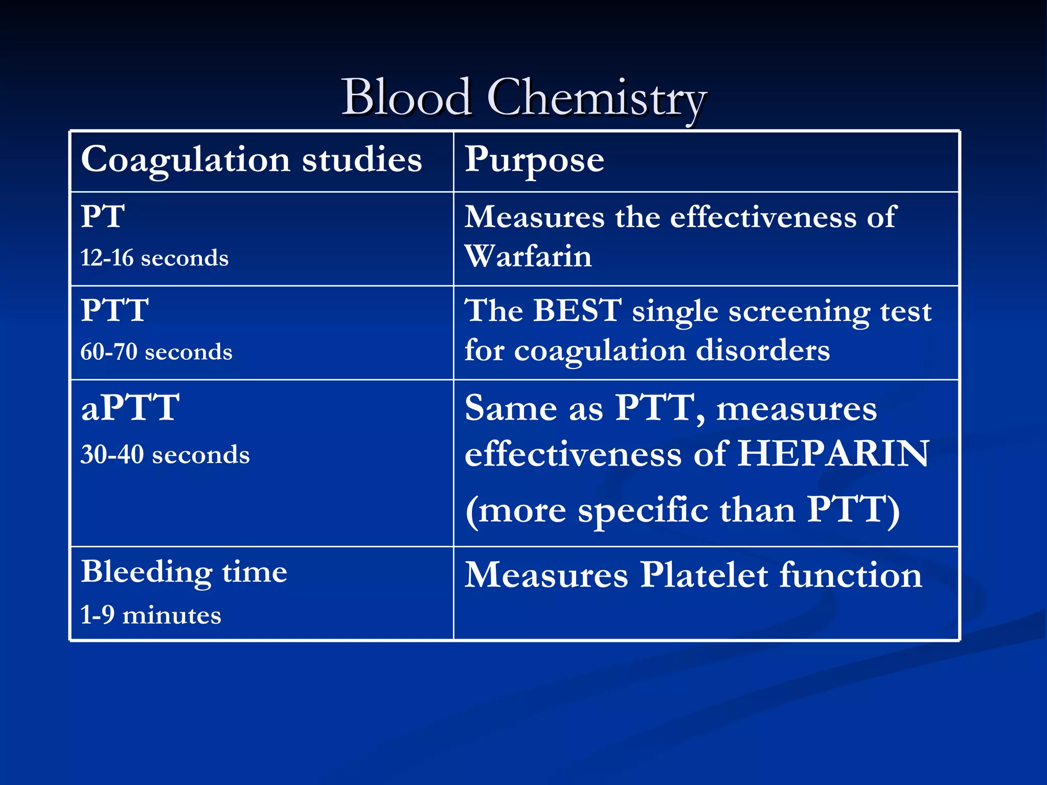
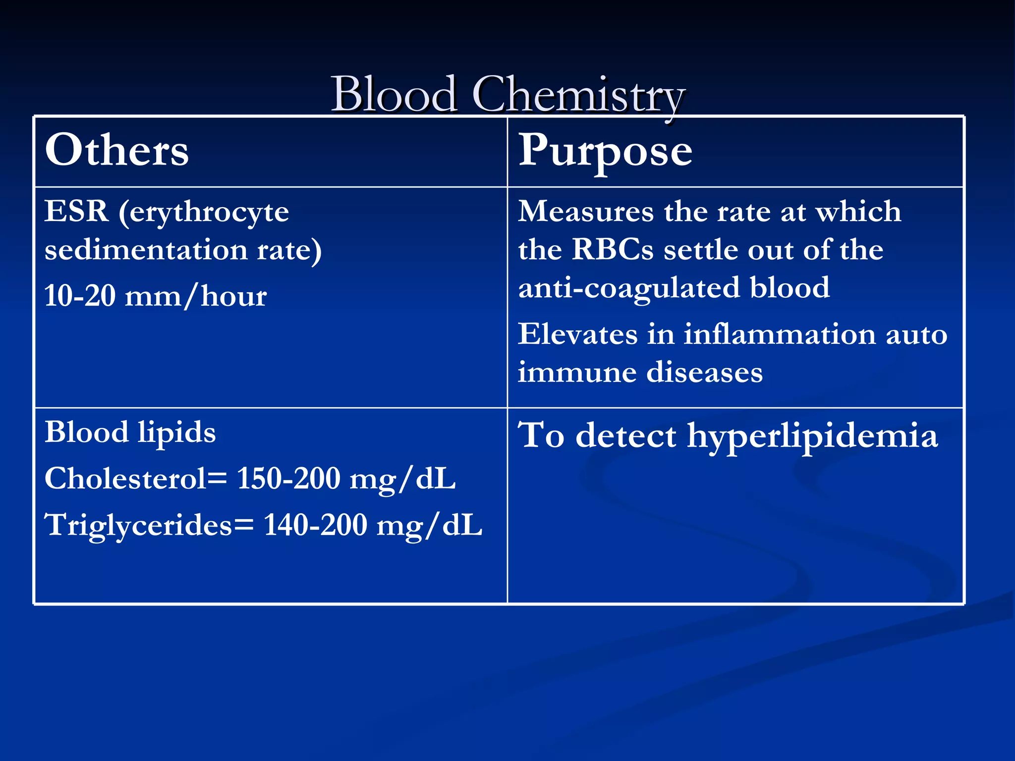
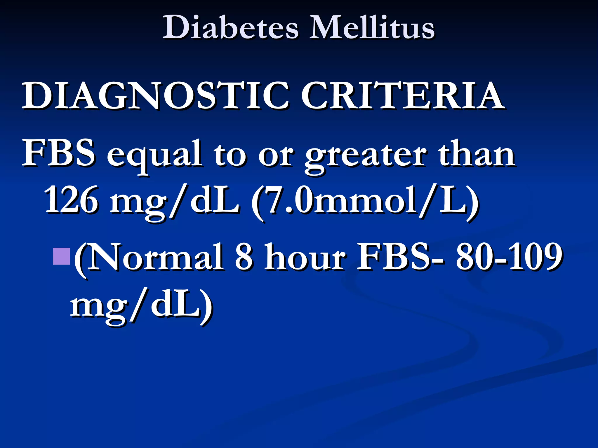
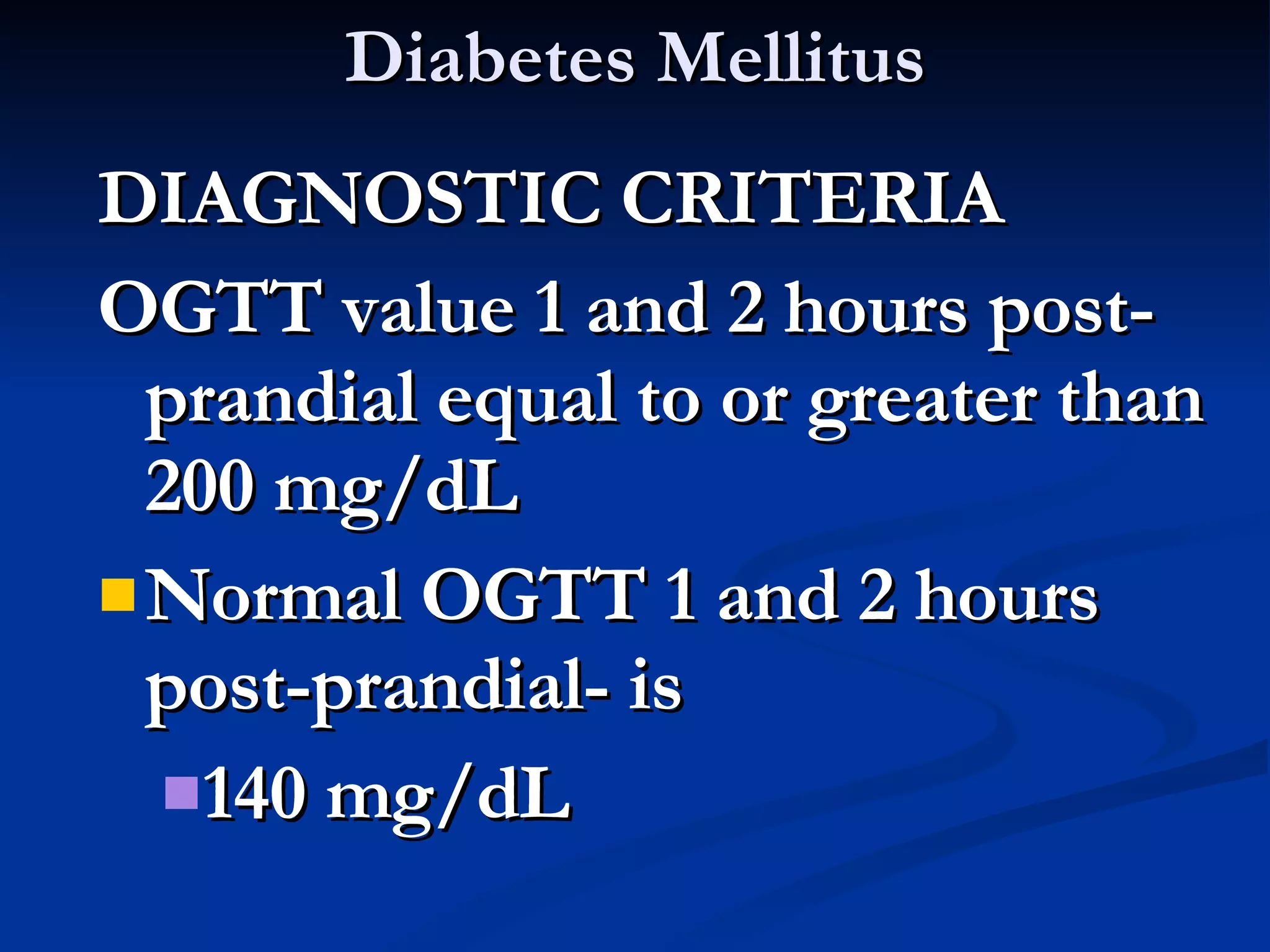
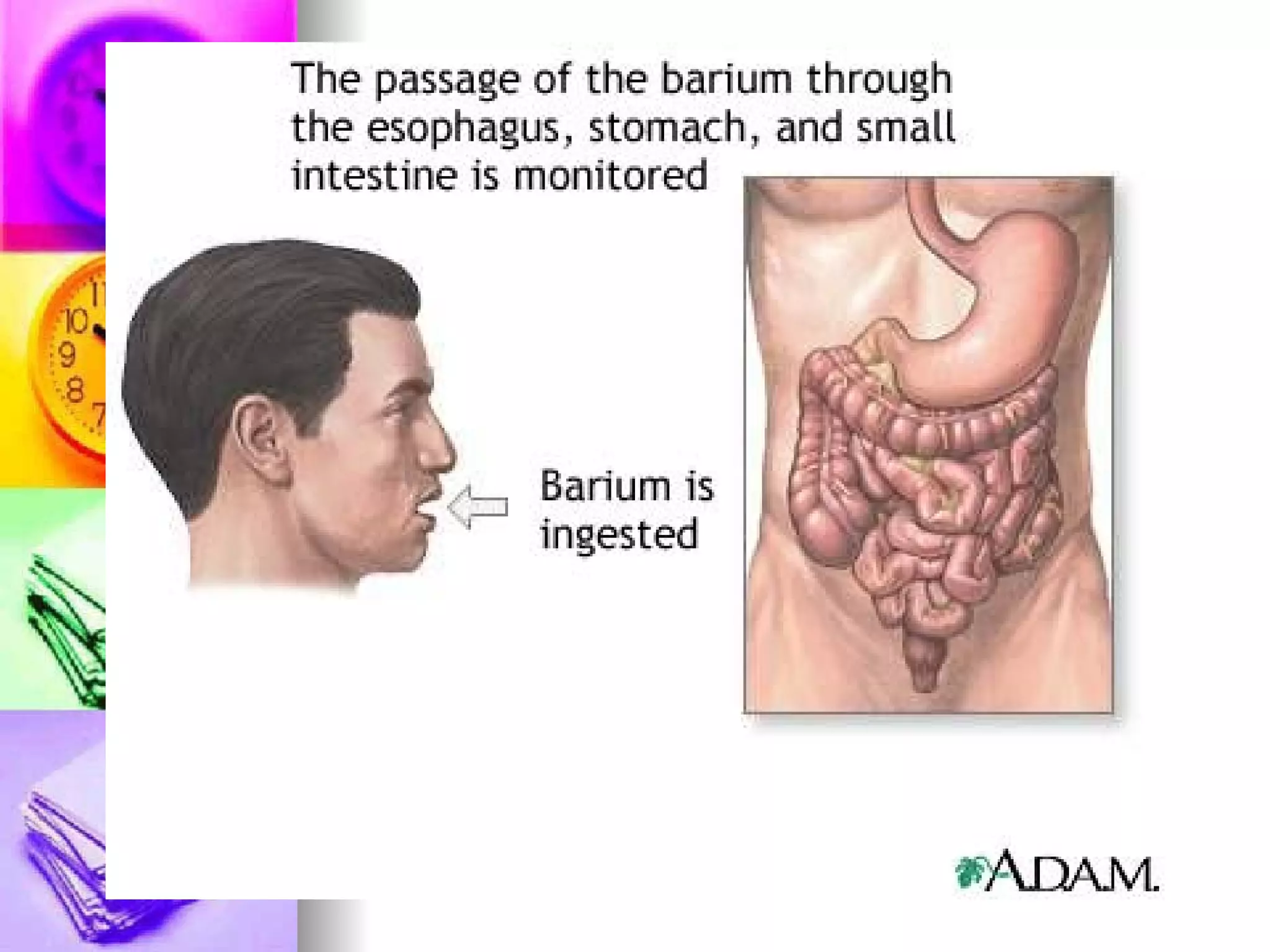
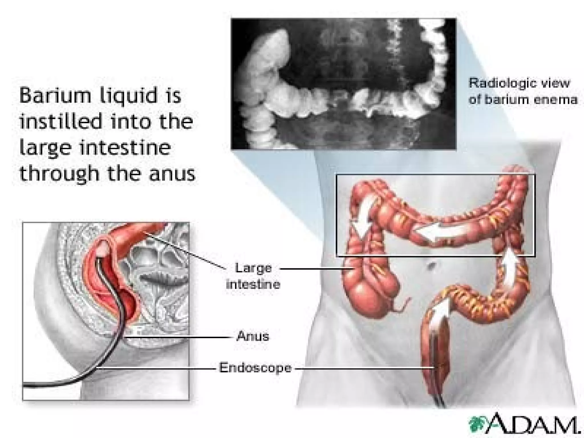
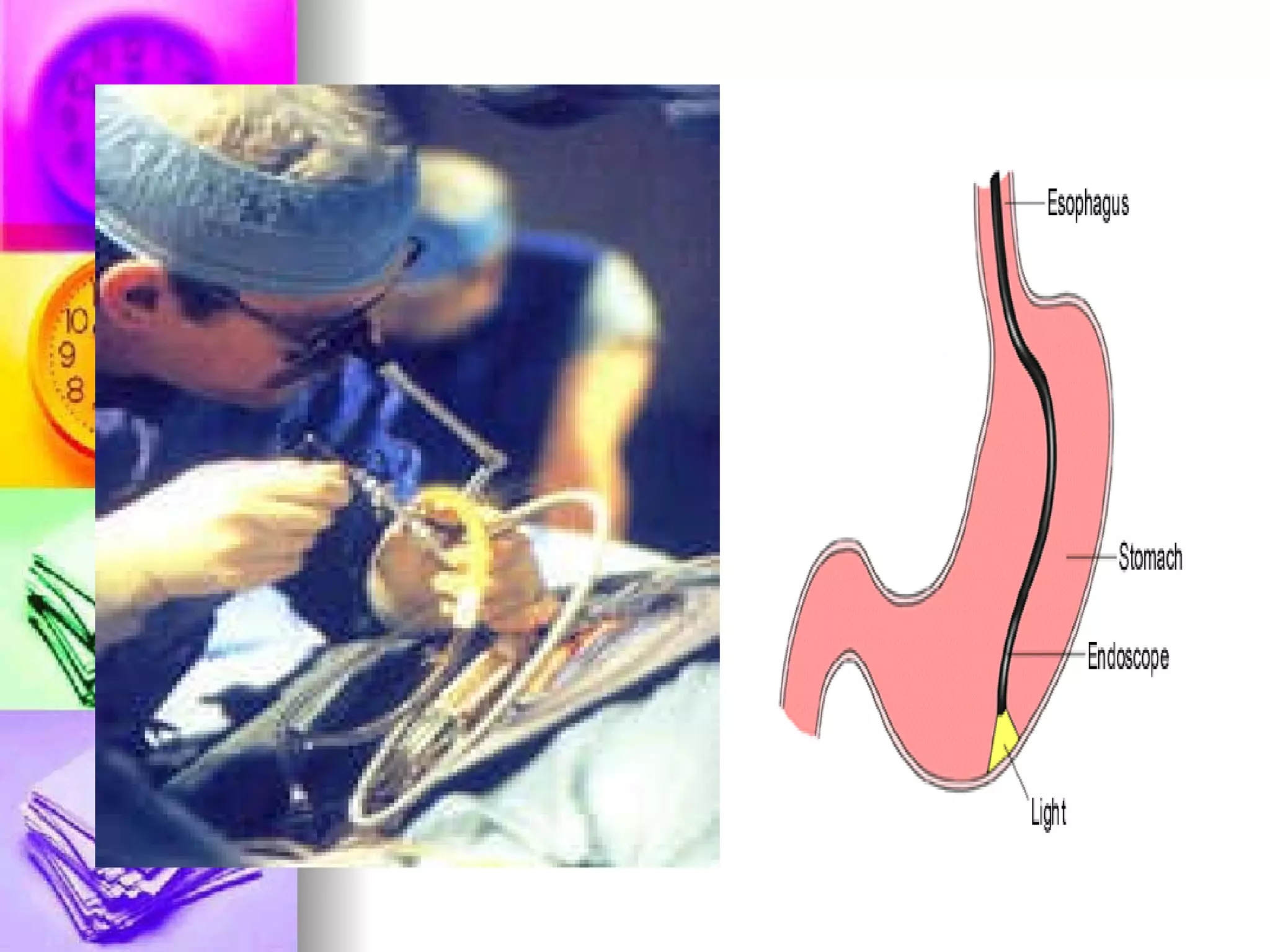
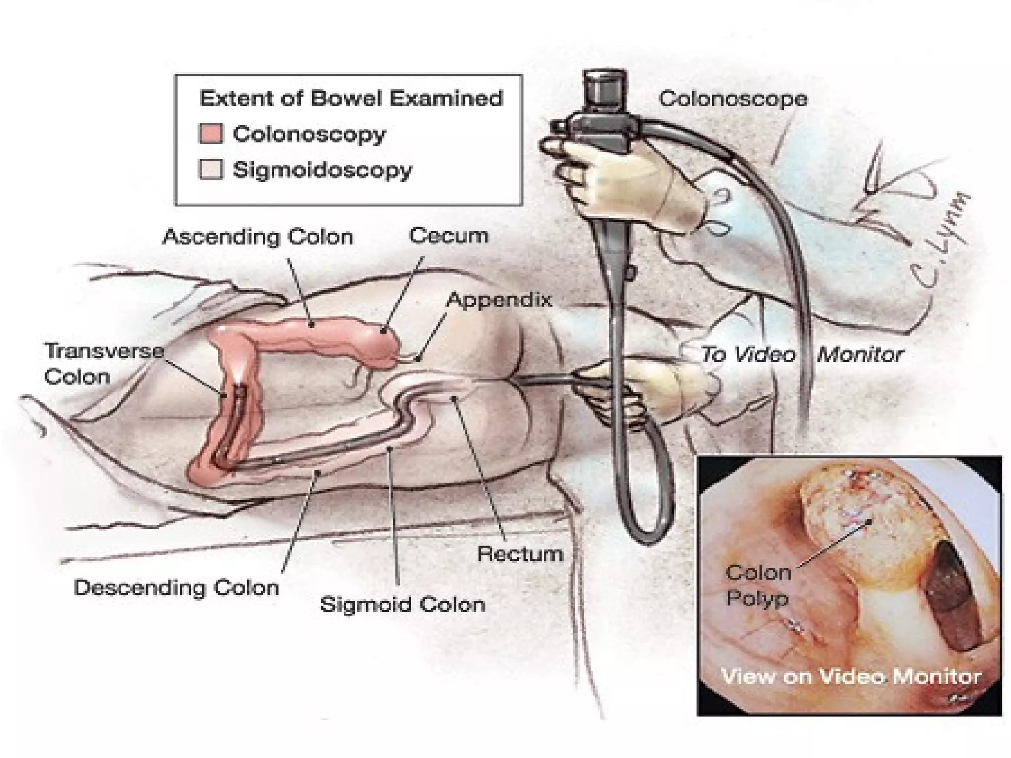
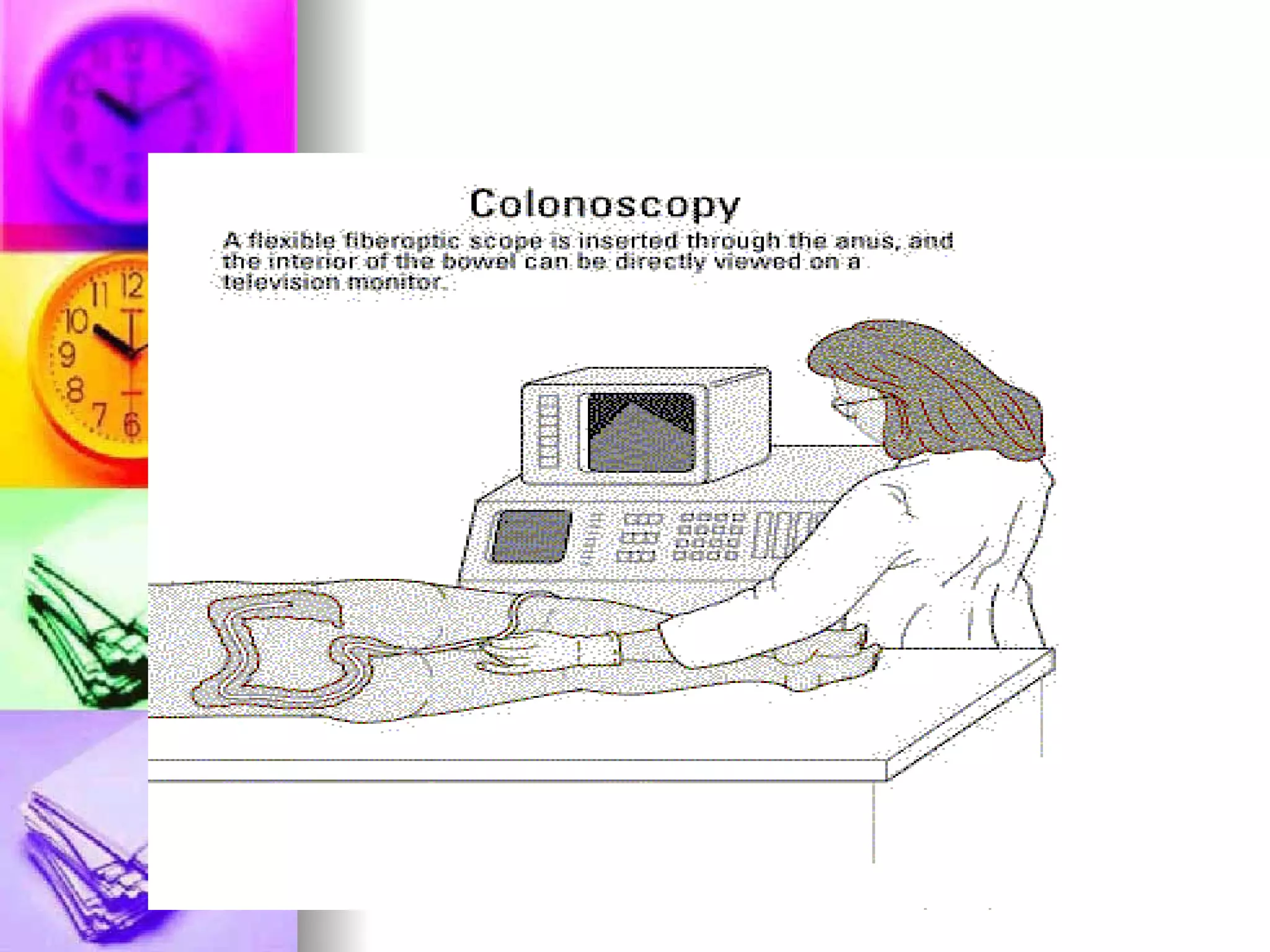
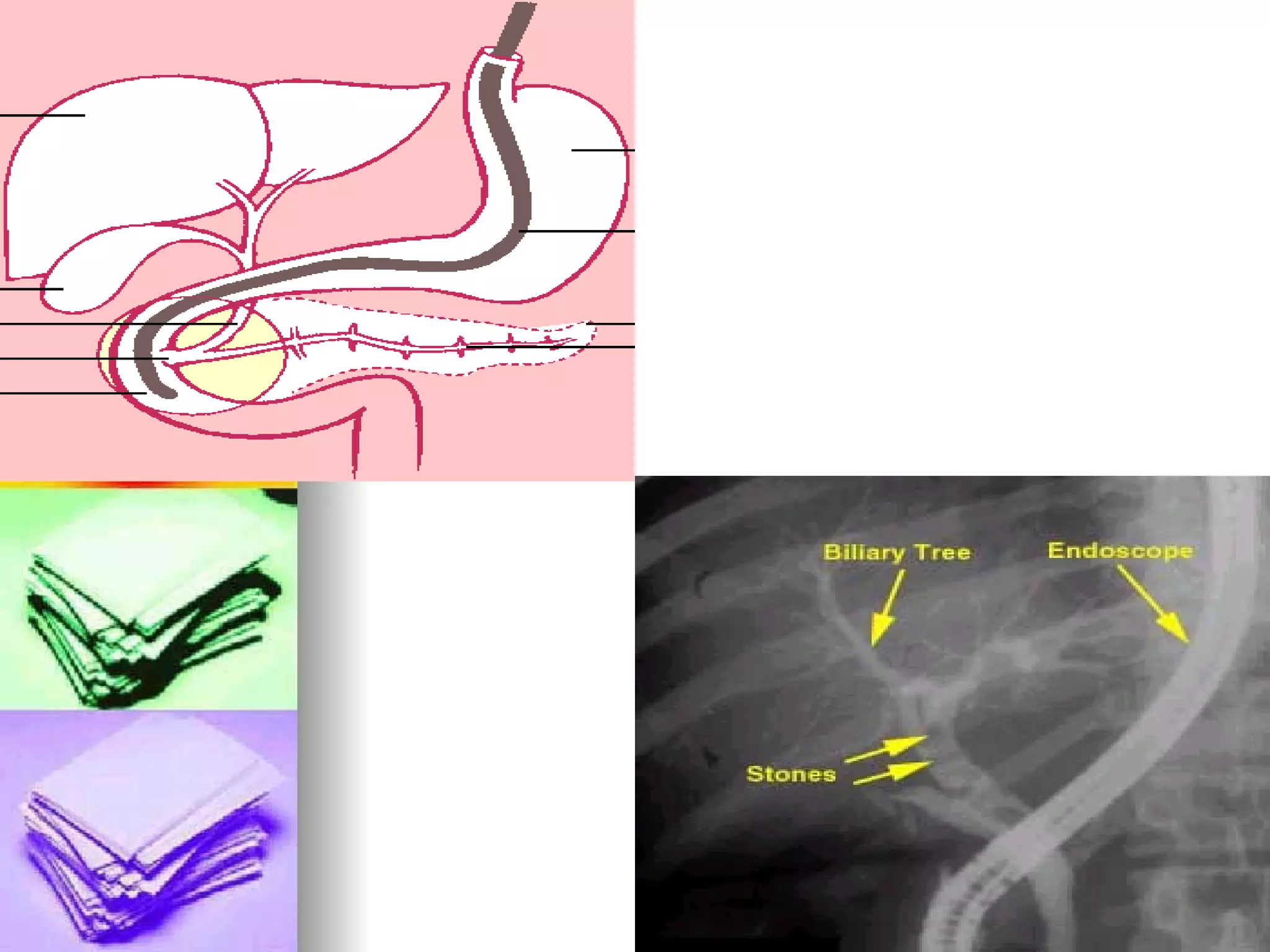
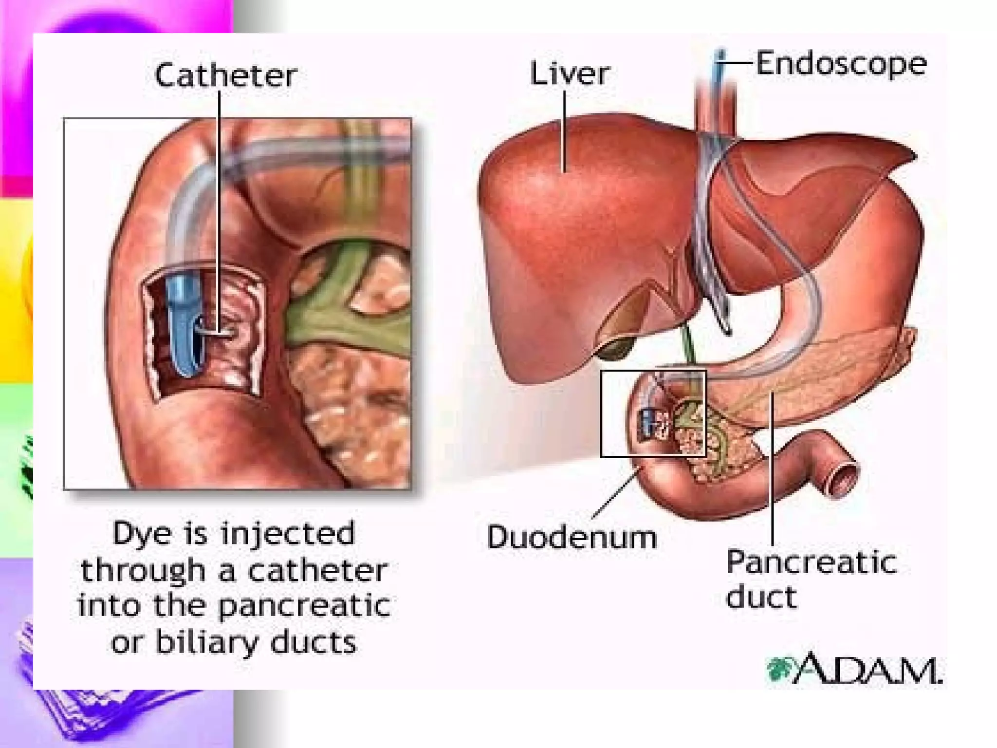
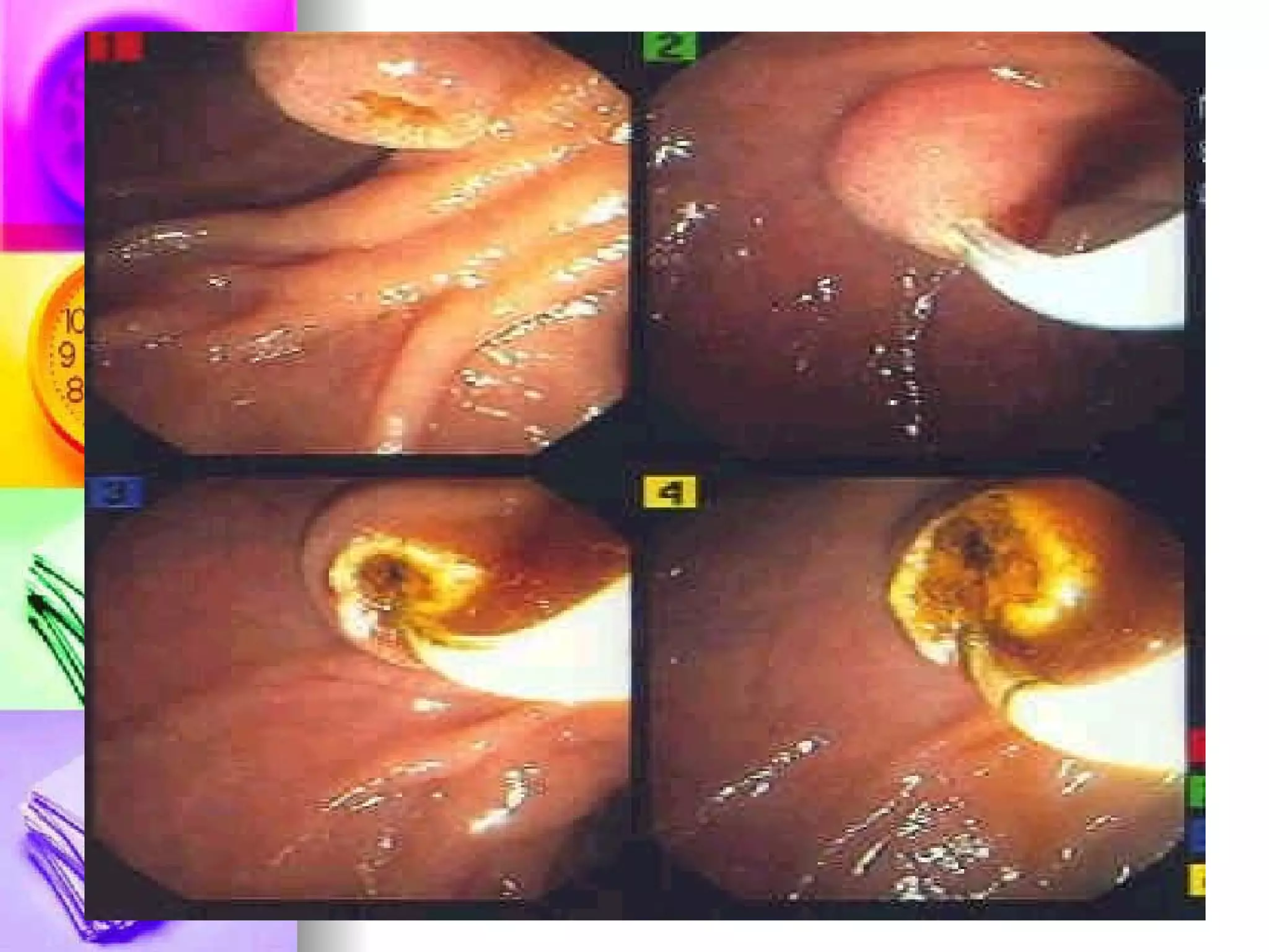
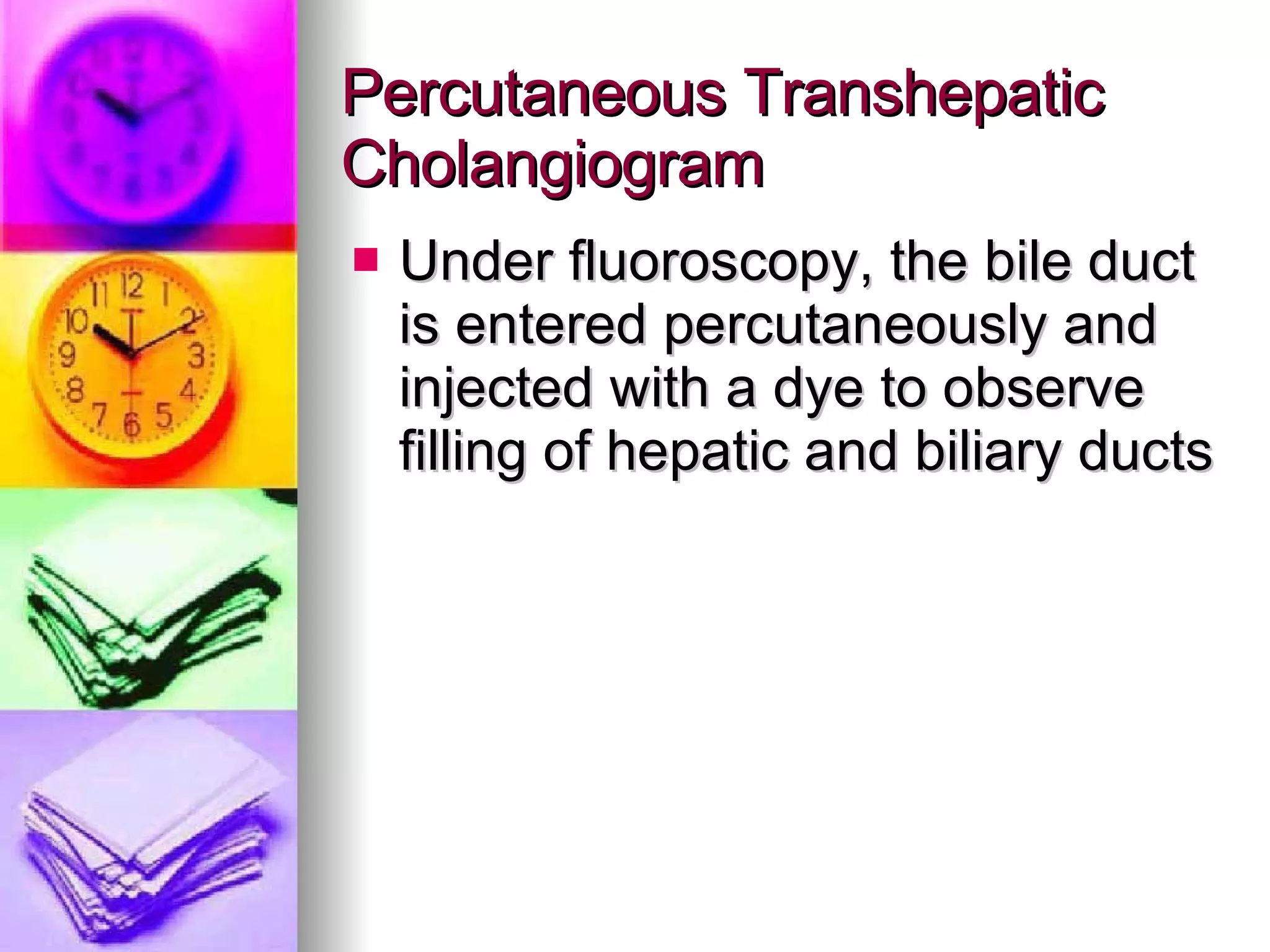
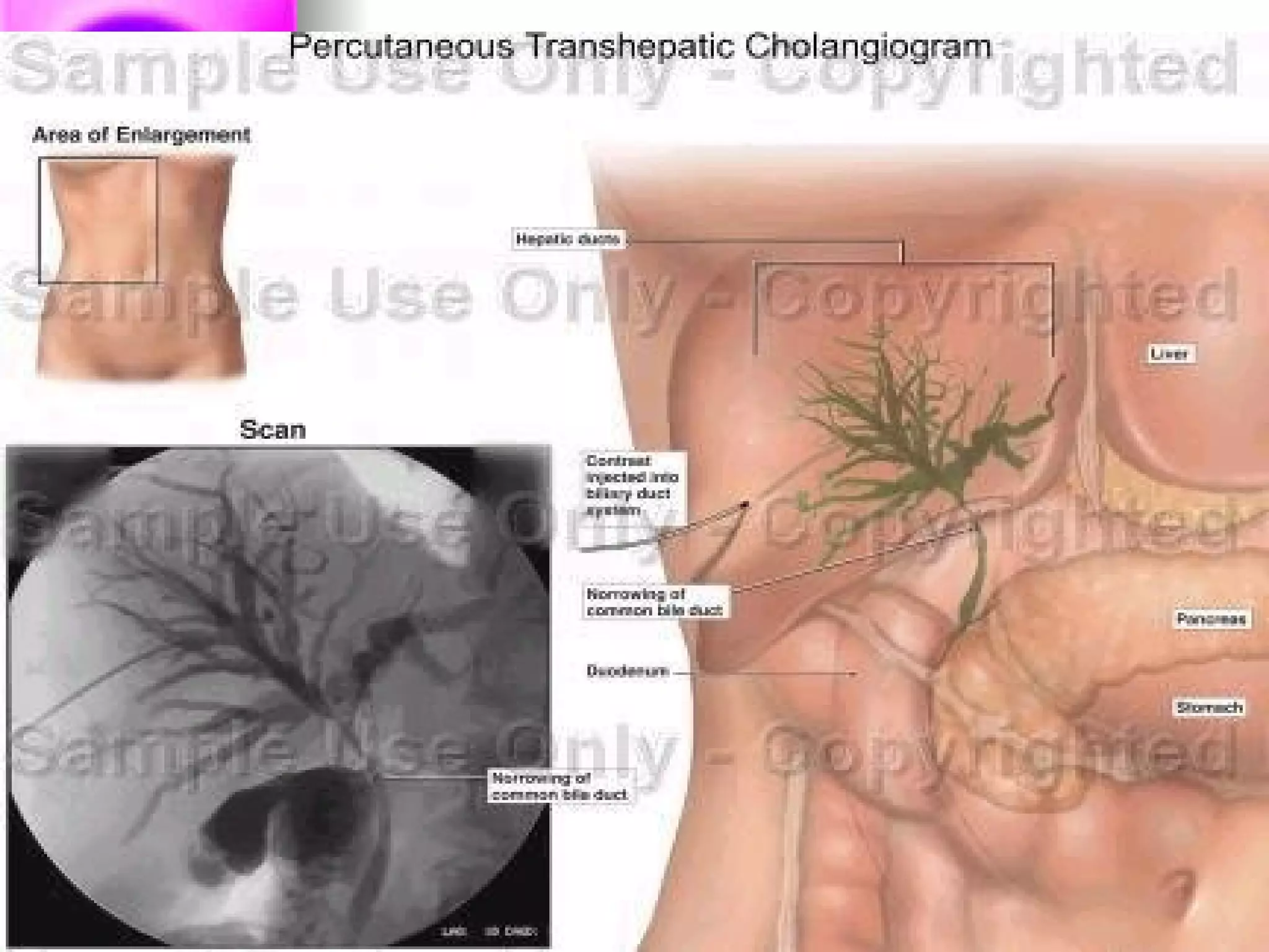
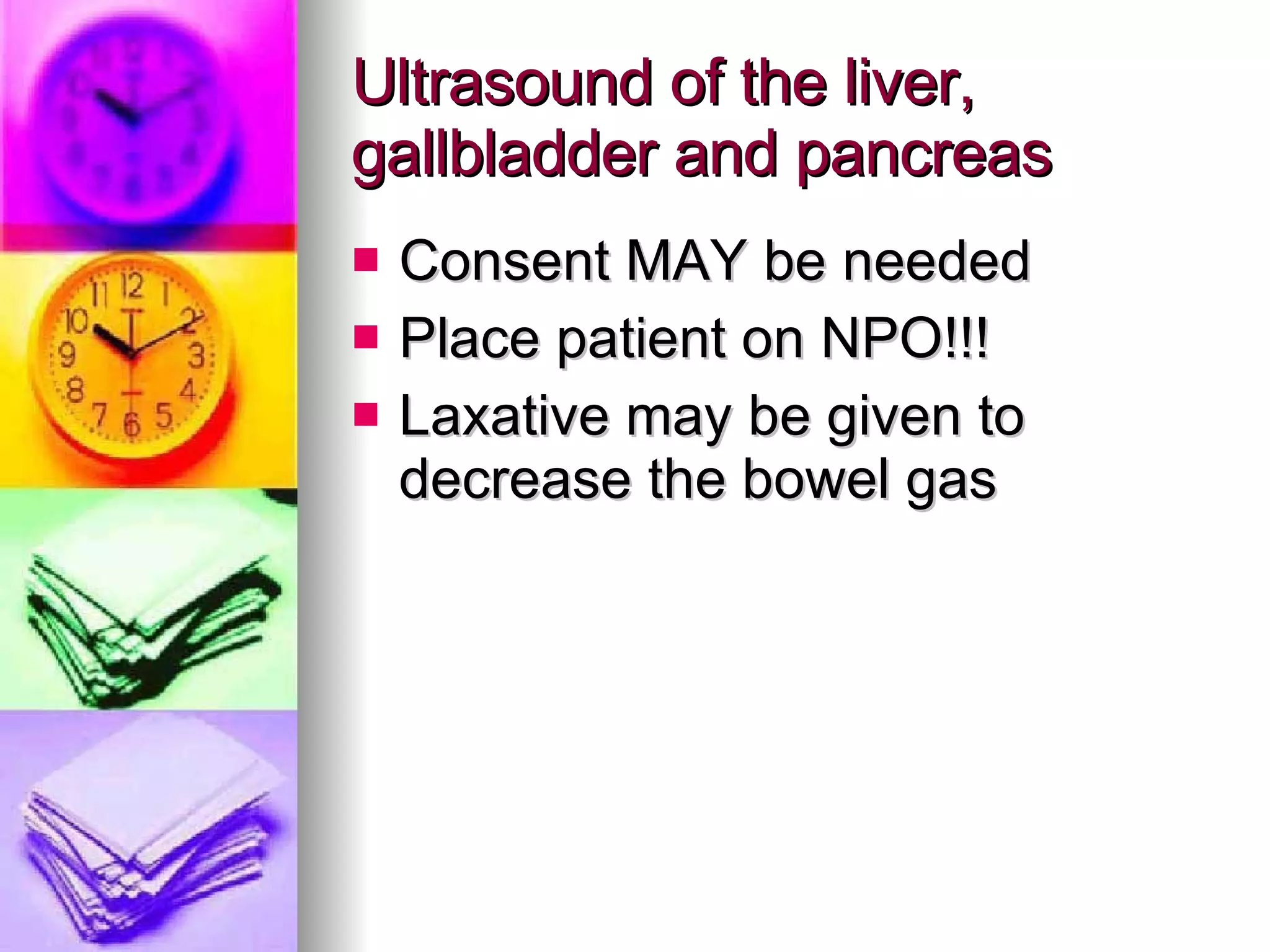
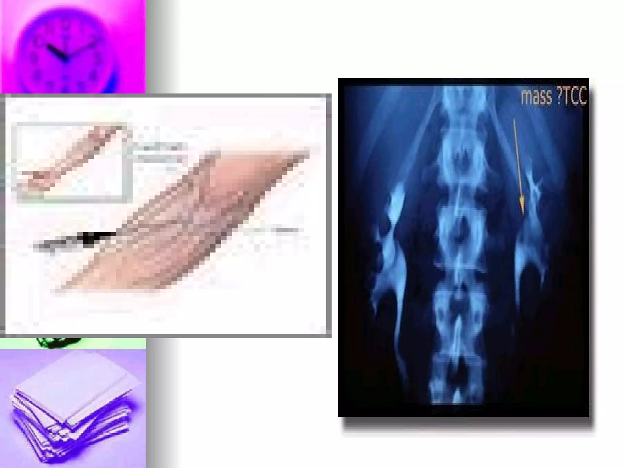
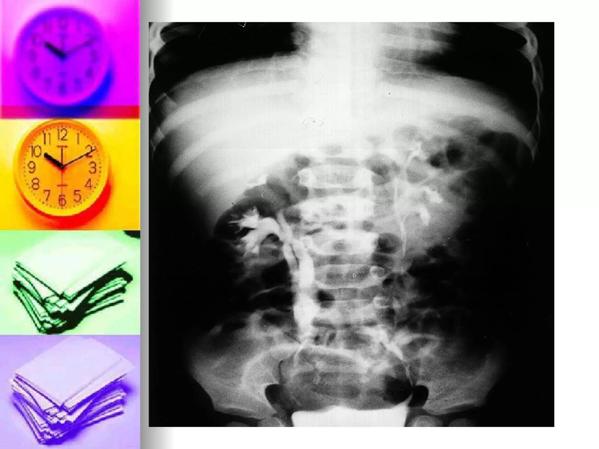
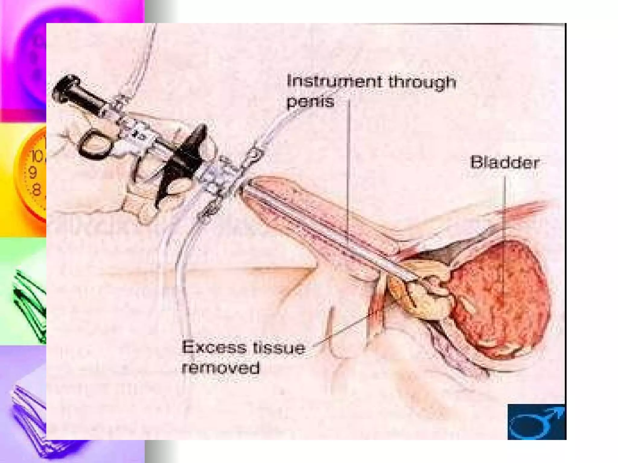
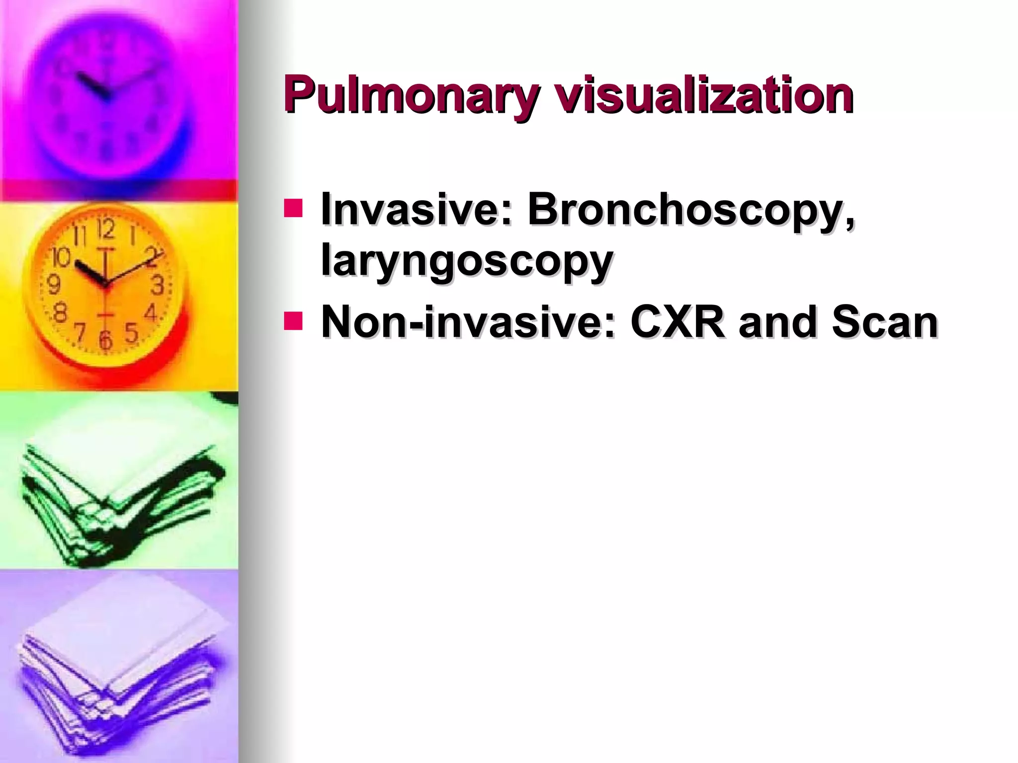
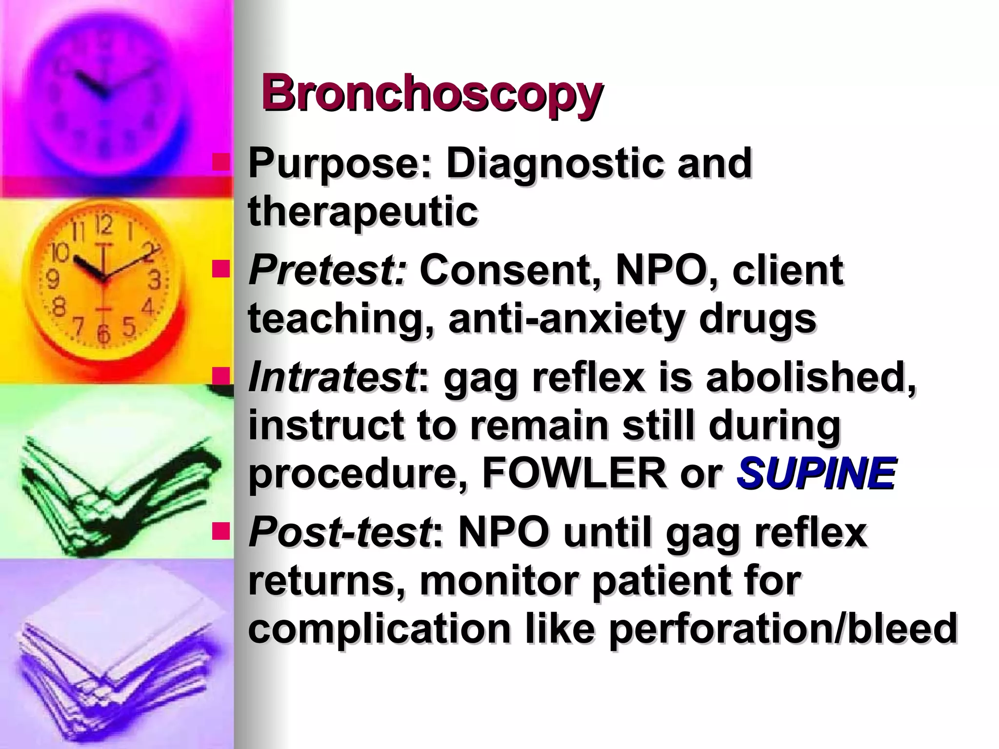
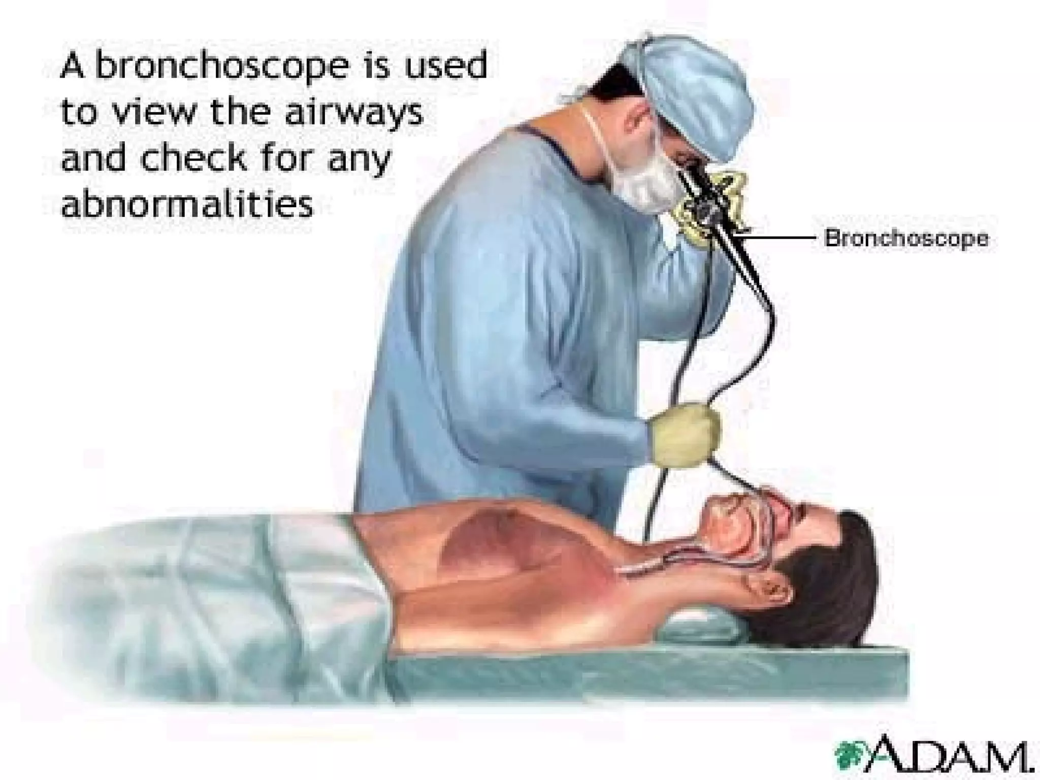
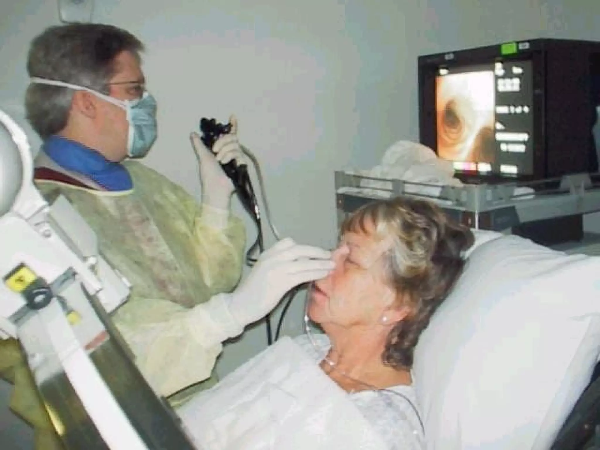
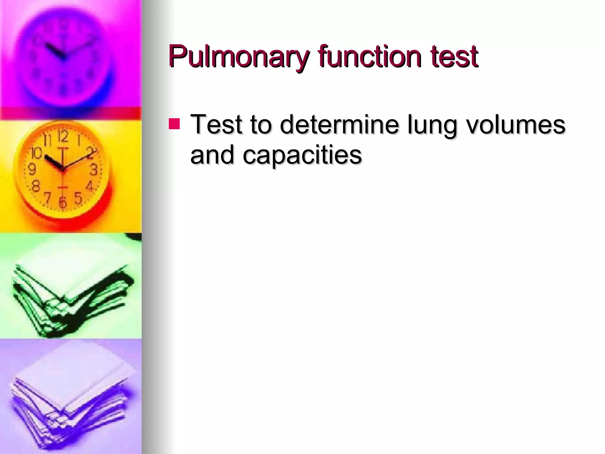
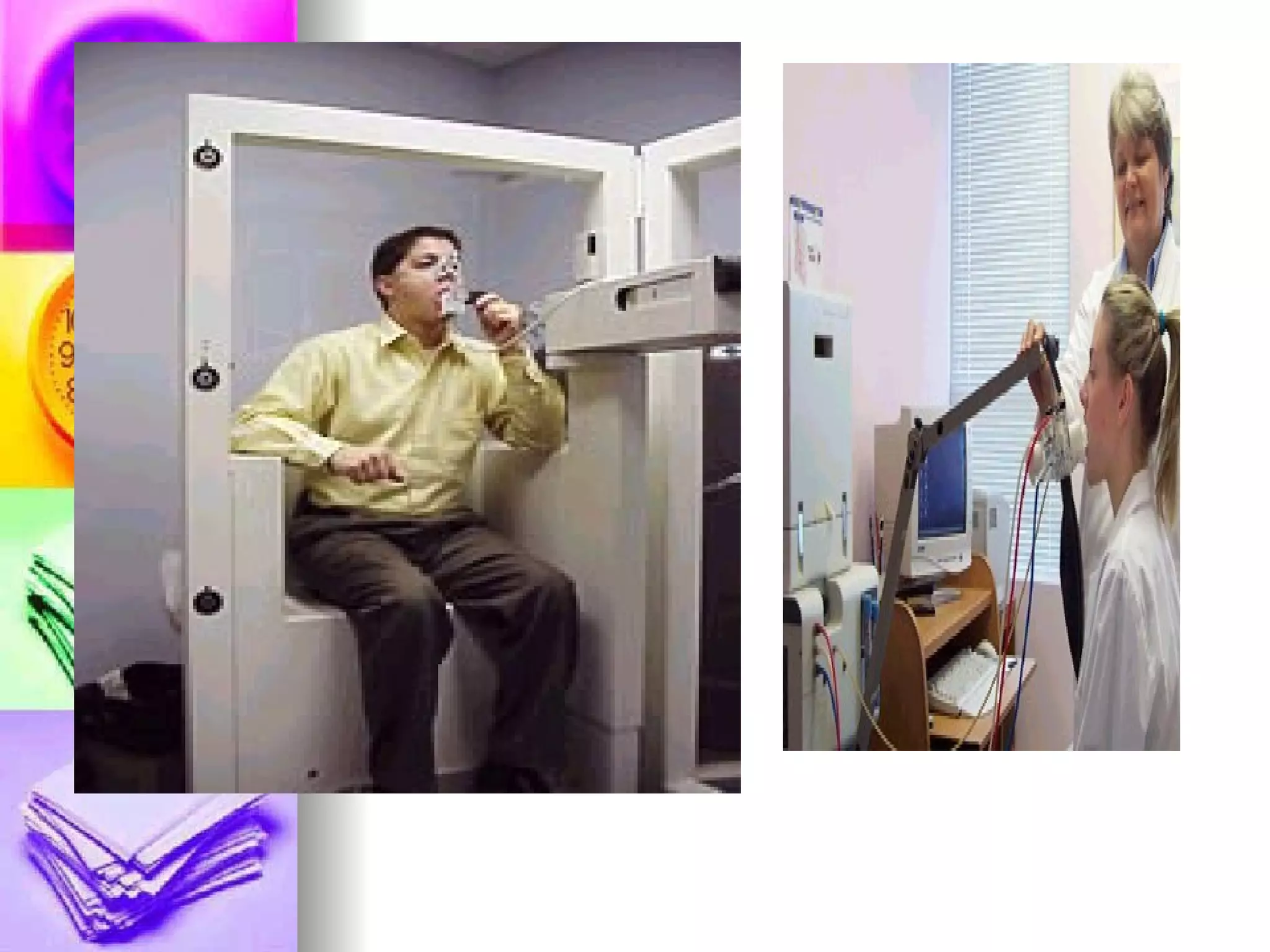
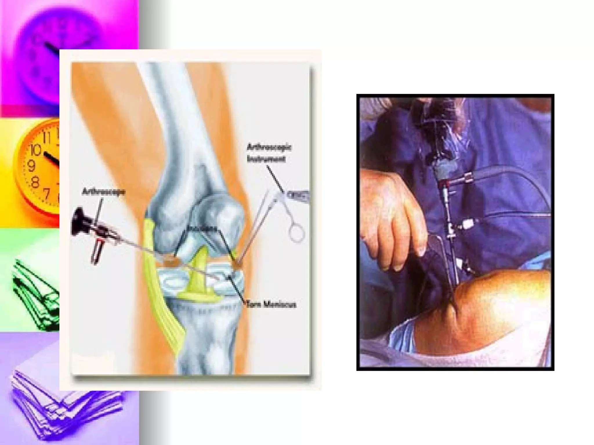
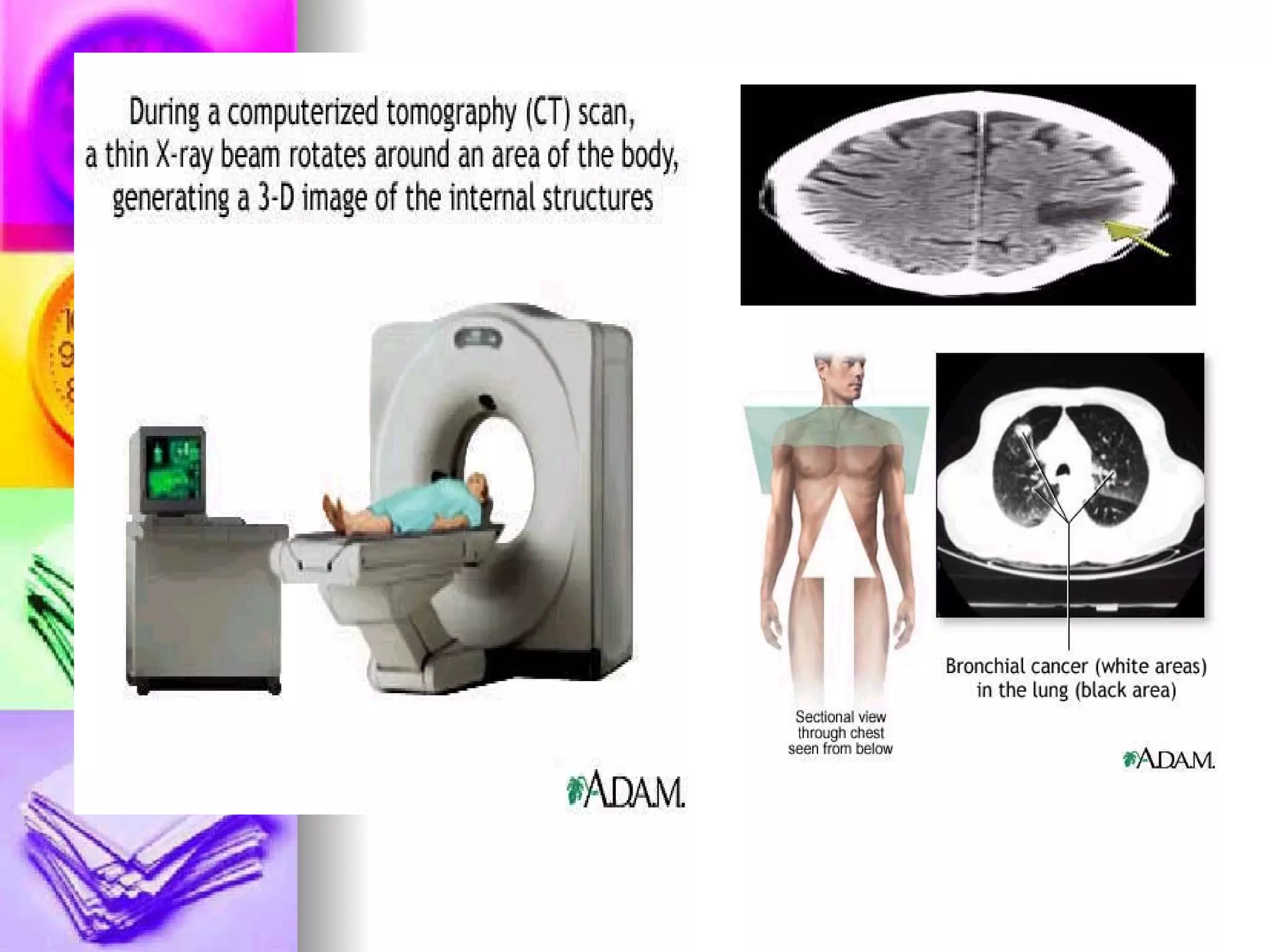
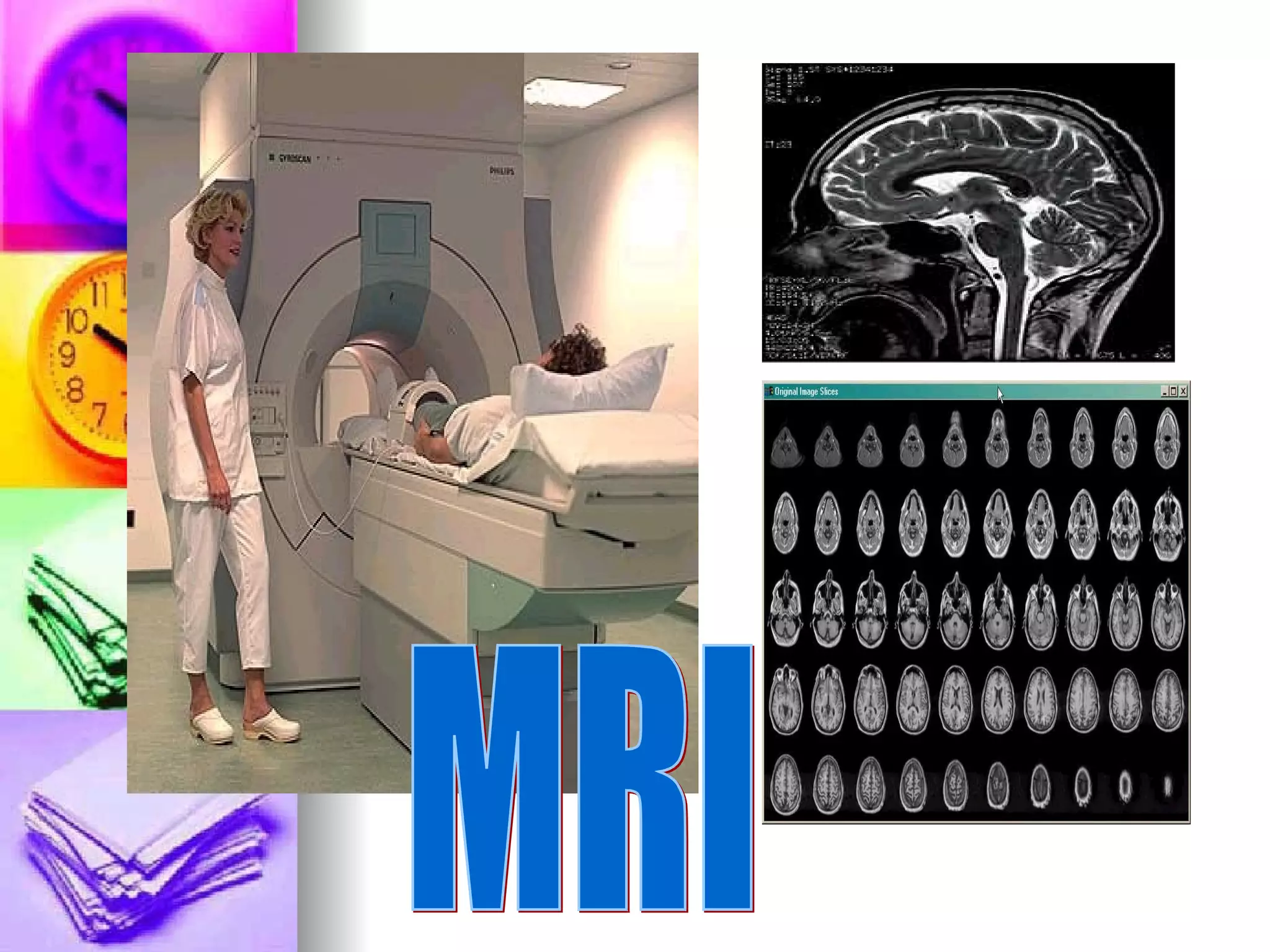
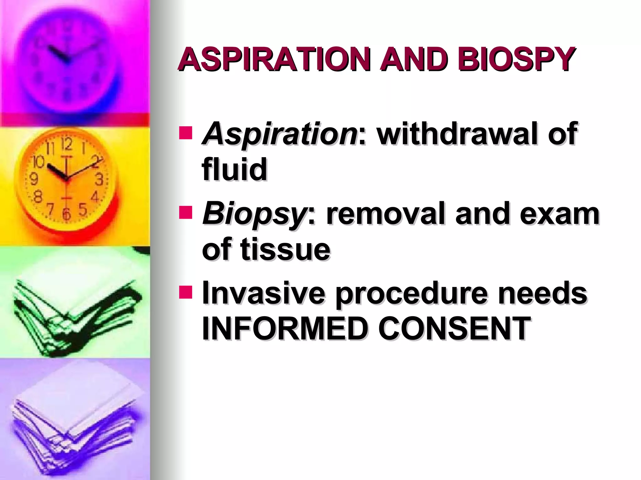
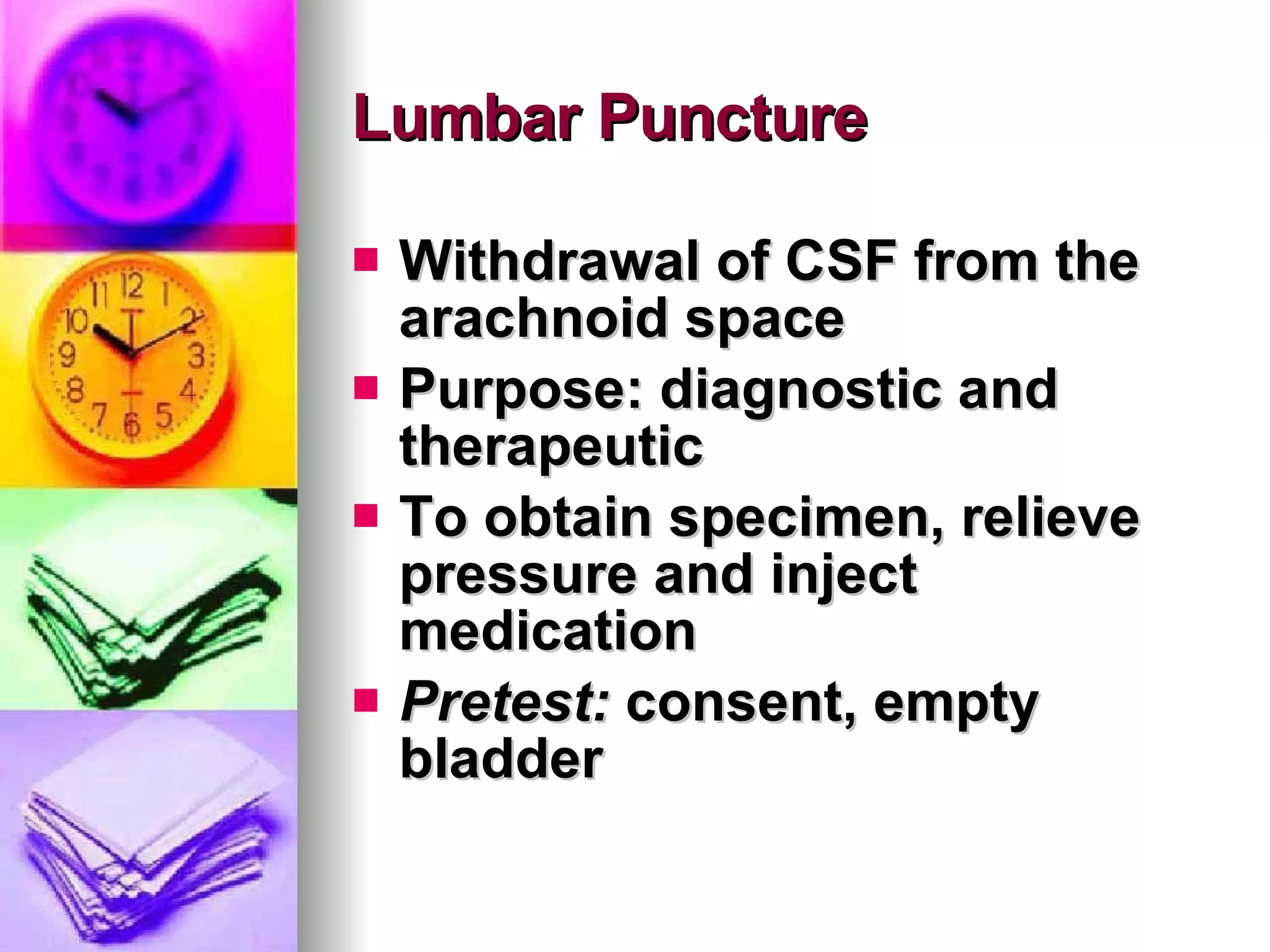
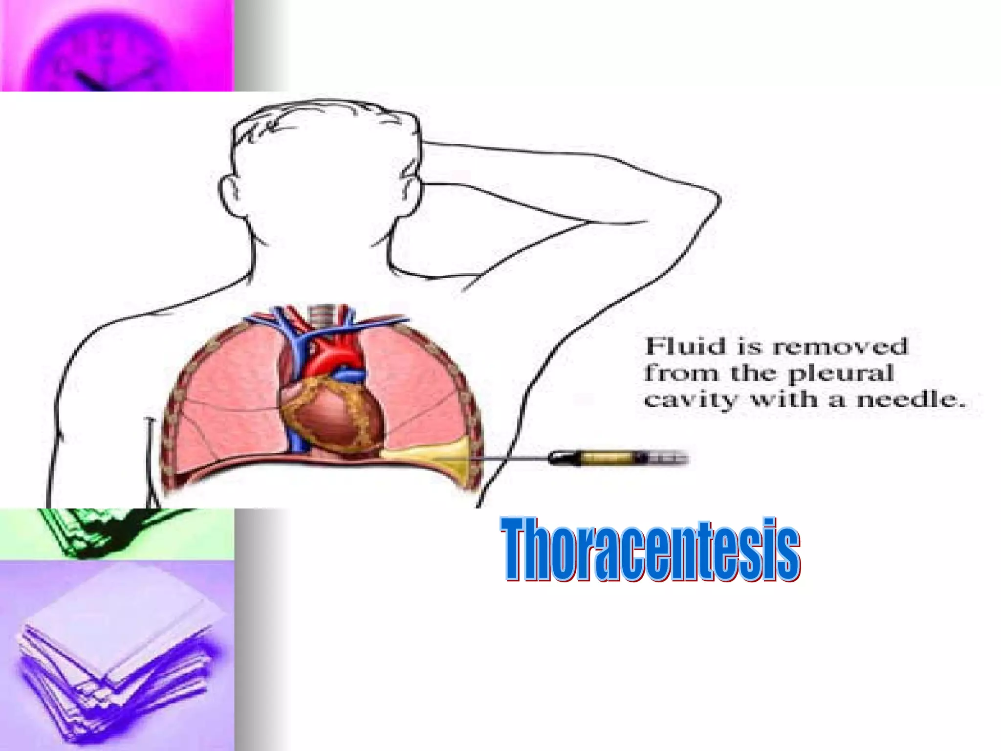
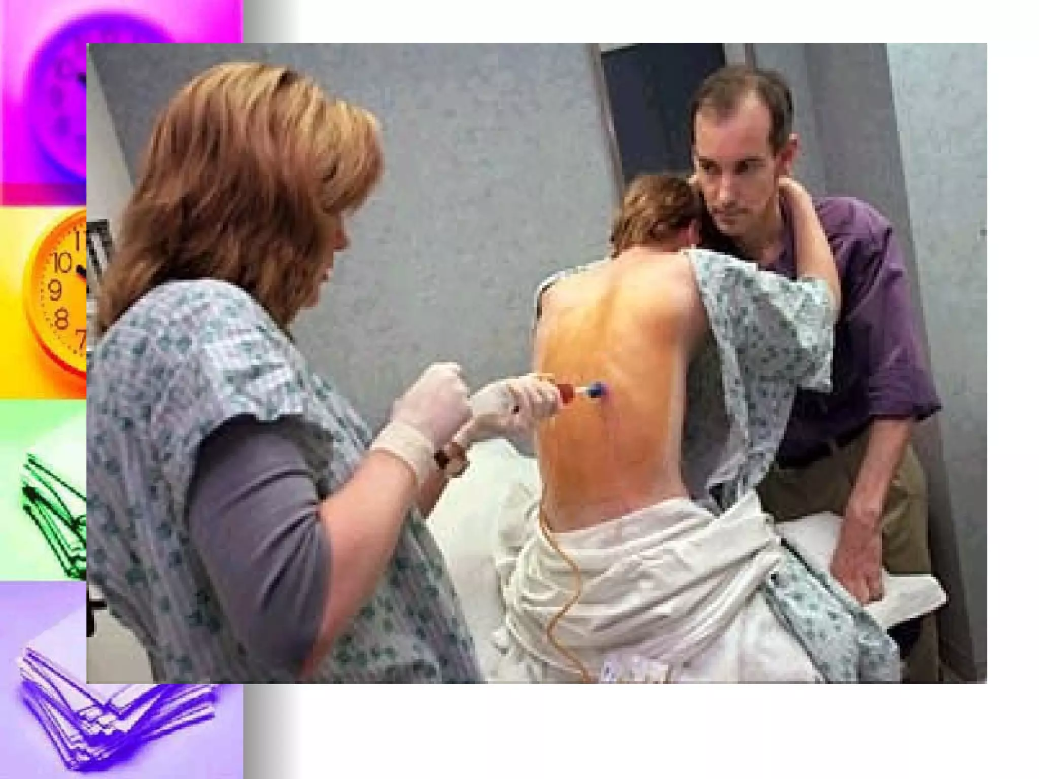
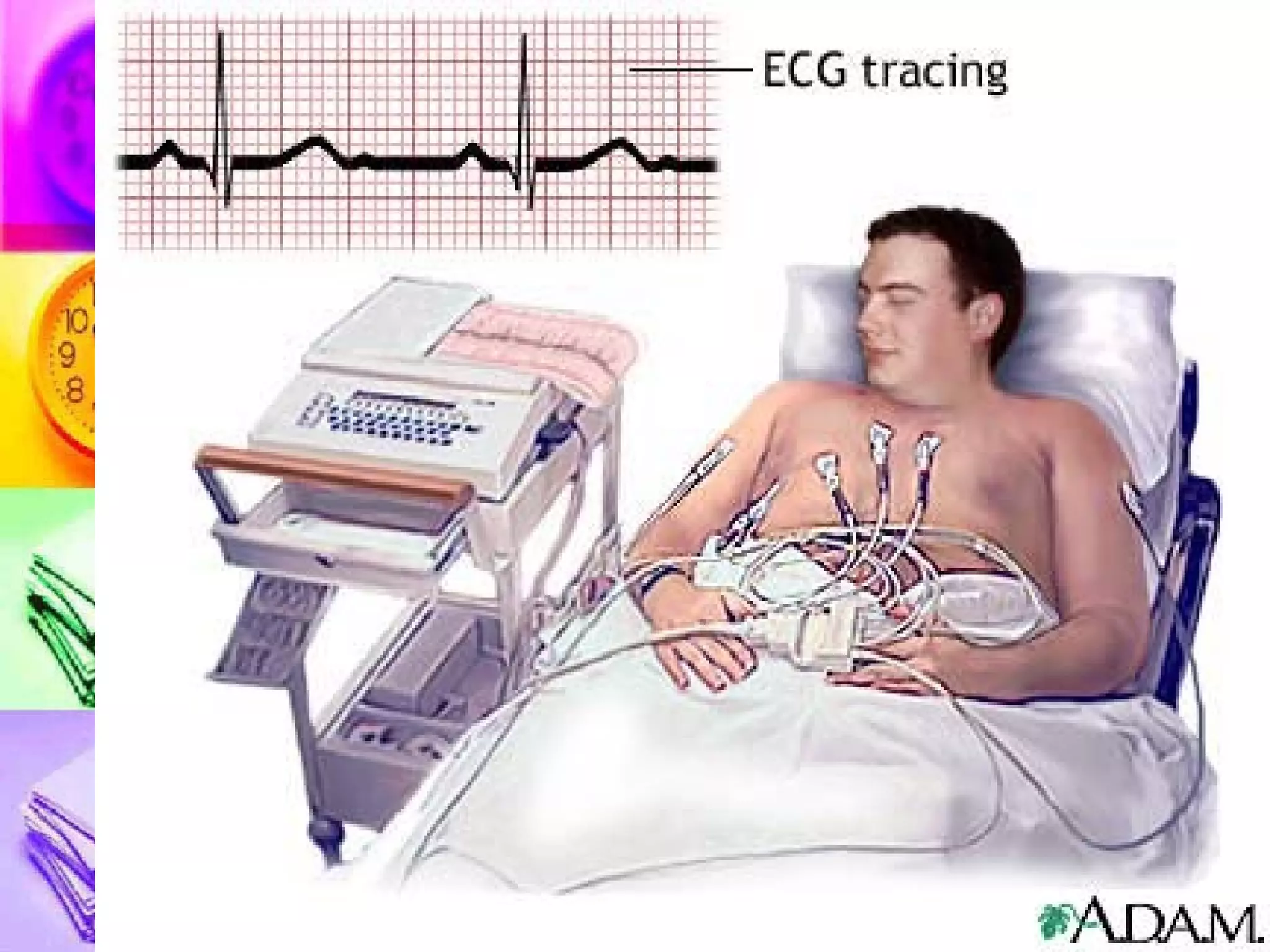
Common Laboratory procedures: Nursing Responsibilities and Implications pinoynursing.webkotoh.com
3 Phases of Diagnostic testing Pretest Client preparation Intra-test specimen collection and VS monitoring Post-test Monitoring and follow-up nursing care
Related Nursing Diagnoses Anxiety Fear Impaired physical mobility Deficient knowledge
Blood tests
BLOOD TESTS CBC Hemoglobin, Hematocrit, WBC, RBC and platelet Serum Electrolytes Arterial blood gases Blood Chemistry Drug and Hormone Assay
Complete Blood Count Specimen: Venous blood Pretest : obtain syringe, tourniquet, vial with appropriate anticoagulant Intratest:  Cubital vein commonly used for venipuncture Post-test : direct pressure and observe for bleeding, label vial
Normal values for CBC RBC  (M) 4.7-6.1/ (F) 4.2-5.4 Hgb  (M) 14-18/ (F) 12-16 mg/dL Hct  (M) 42-52/ (F) 33-47 % WBC  5-10,000 cells/cubic cm Differential count Neutrophils- 55- 70% Lymphocytes- 20-40% Monocytes- 2-5% Eosinophils- 1-4% Platelets  150,000-400,000
 
Table. 11.2
 
CBC PARASITIC infection  Increased Eosinophils CHRONIC bacterial infection VIRAL infection Increased Lymphocytes ACUTE bacterial infection Increased Neutrophils More than 10, 000 Increased WBC (Leukocytosis) 5-10,000 cell/cm3 Normal WBC count
Serum Electrolytes Specimen: venous blood Pretest/Intratest/Post-test- same Commonly ordered: Sodium- 135-145 mEq/L Potassium- 3.5-5.0 mEq/L Chloride- 95-105 mEq/L Magnesium-  1.3 to 2.1 mEq/L   Calcium-  8 to 10 mg/dL
Serum Electrolytes Problems can be Hyper   if increased Hypo   if decreased
Blood Chemistry Specimen: Venous blood, serum Pretest/Intratrest/Post-test-same Examined are enzymes, hormones, lipid profile BUN , Creatinine, etc… Place  patient on NPO for 8 h * Creatinine  is produced relatively constant by muscles, excreted by the kidneys and is the  RELIABLE  Reflection of Renal Status
Blood Chemistry Normal values for : Creatinine: 0.7 to 1.4 mg/dL BUN: 10-20 mg/dL Creatinine clearance: 1.67 to 2.5 mL/s Serum uric acid: 2.5 to 8 mg/dL Blood osmolality= 250 to 290 mOsm/L
Blood Chemistry SLE diagnosis Anti-DNA antibody For Rheumatoid arthritis Rheumatoid factor Gout detection Uric acid Identifies Cardiac damage or muscle damage CK-MB, LDH and Troponin Liver function test SGOT/SGPT Purpose Enzymes/acids
Blood Chemistry Measures Platelet function Bleeding time 1-9 minutes Same as PTT, measures effectiveness of HEPARIN (more specific than PTT) aPTT 30-40 seconds The BEST single screening test for coagulation disorders PTT 60-70 seconds Measures the effectiveness of Warfarin PT 12-16 seconds Purpose Coagulation studies
Blood Chemistry To detect hyperlipidemia Blood lipids Cholesterol= 150-200 mg/dL Triglycerides= 140-200 mg/dL Measures the rate at which the RBCs settle out of the anti-coagulated blood Elevates in inflammation auto immune diseases  ESR (erythrocyte sedimentation rate) 10-20 mm/hour Purpose Others
Diabetes Mellitus DIAGNOSTIC CRITERIA FBS equal to or greater than 126 mg/dL (7.0mmol/L)  (Normal 8 hour FBS- 80-109 mg/dL)
Diabetes Mellitus DIAGNOSTIC CRITERIA OGTT value 1 and 2 hours post-prandial equal to or greater than 200 mg/dL Normal OGTT 1 and 2 hours post-prandial- is 140 mg/dL
Diabetes Mellitus DIAGNOSTIC CRITERIA RBS of equal to or greater than 200 mg/dL  PLUS the 3 P’s
Diabetes Mellitus DIAGNOSTIC CRITERIA Glycosylated hemoglobin (HbA1c) is a monitoring test to assess the adherence to diabetic medication
Arterial Blood Gases Specimen: arterial blood Pretest : obtain syringe with heparin, rubber stopper, container with ice Intratest :  usual site-radial artery, perform  Allen’s test Post-test : Apply direct pressure on site for 5-10 minutes, send specimen with occluded needle on ice
Normal ABG values pH  7.35-7.45 pCO2  35-45 mmHg paO2  80-100 mmHg HCO3  22-26 mEq/L Base excess  -2 to +2 O2 sat   95-98%
ABG interpretation Metabolic >26 Metabolic <22 22-26 mEq/L HCO3 Respiratory <35 Respiratory >45 35-45 mmHg paCO2 95-98% SaO2 95-100 mmHg paO2 Above 7.45 Below 7.35 7.35-7.45 pH Alkalosis Acidosis Normal Value
Urine exams
Urine Analysis Specimens Clean-voided urine for routine urinalysis Clean-catch or midstream urine for urine culture Suprapubic and catheterized urine for urine culture
Routine Urinalysis Specimen: Clean voided Pretest : give clean vial and instruct to void directly into the specimen bottle Intratest:  Allow a 10 ml collection Post-test : prompt delivery to laboratory * First voided urine in a.m. is highly concentrated, more uniform concentration and with more acidic pH
Urine Culture: Normal is <100,000  Specimen: clean catch, midstream or catheterized urine Pretest : Instruct to wash and dry genitalia/perineum with soap and water. (M)- circular motion, (F)-front to back direction Intratest:   Midstream urine , 30-60 ml Post-test:  Cap and label, prompt delivery and documentation
Special Urine Collection Infants Special urine bag Or cut a hole of the diaper  (front for the boy, middle for the girl) pulling out through the hole the special bag Children May use potty chair or bedpan Give another vial to play with, allow parent to assist Elderly Assistance may be required
Timed-urine collection Collection of  ALL urine  voided over a specified time Refrigerated  or with preservative Pretest:  Specimen container with preservative, receptacle for collection, a  post sign Intratest:  A t the start of collection, have patient void and  discard the urine At the end of collection period, instruct to completely void and  save the urine Post test:  Documentation
Catheter specimen  Sterile urine Insert needle of the syringe through a drainage port Only done with the rubber catheter not the plastic, silastic or silicone catheter. Intratest : Clamp catheter x 30 mins if no urine Wipe area where needle will be inserted 30-45 ° angle, 3 ml for culture Post-test  : Unclamp catheter after collection
Stool Exams
Stool Analysis Occult Blood   GUAIAC test Steatorrhea Ova/Parasites Bacteria Viruses
General Nursing consideration for stool collection Pretest : Determine purpose/s, obtain gloves, container and tongue blade Intratest:   Instruct to defecate in clean bed pan Void before collection Do not discard tissue in bedpan Obtain 2.5 (1 inch) formed stool 15-30 ml of liquid stool Post-test : prompt delivery
Occult Blood: Guaiac Test Detect the presence of enzyme:  Peroxidase (+) blue color   positive guaiac Restrict intake of red meats, some medications and Vitamin C for 3-7 days FALSE (+):   red meat , raw fruits and vegetables especially radish, turnip, melon and horseradish; meds like aspirin, NSAIDS, iron and anticoagulants FALSE (-): Vitamin C, ingested 250 mg per day from any source
Sputum Exams
Sputum Analysis For Culture and sensitivity For sputum cytology For sputum AFB For monitoring of the effectiveness of therapy
Sputum examination Pretest:  Morning specimen is collected Intratest: Mouthwash with  plain water Deeply inhale x 2 then cough Wear gloves in collecting specimen Expectorate needed- 1-2 Tbsp or 15-30 ml Post-test : oral care and prompt delivery to lab
Visualization:  DIRECT  and INDIRECT
VISUALIZATION PROCEDURES Invasive  procedures are  direct  methods and need CONSENT Non-invasive  procedures are  indirect  methods and may need written consent in some instances
Visualization procedures They can be: Radiographic procedures “ Scopic”  procedures
GI tract Visualization
GIT Visualization Barium Swallow- UGIS Pretest: written consent, NPO the night Intratest: administer barium orally, then  followed by X-ray Post-test: Laxative for constipation, increased fluids, assess for intestinal obstruction , warn that stool is light colored!
 
 
GIT Visualization Barium Enema- LGIS Pretest: Informed consent, NPO the night, Enema the morning Intratest: Position on  LEFT side, administer enema, then X-ray follow Post-test: Cleansing enema , Laxative for constipation, assess for intestinal obstruction
 
GIT Visualization Esophagogastroscopy Pretest: Informed consent, NPO for 8 hours, warn that gag reflex is abolished Intratest: Position on  LEFT side during scope insertion Post-test: NPO until gag returns. Monitor for complications
 
GIT Visualization Anoscopy, proctoscopy, proctosigmoidoscopy, colonoscopy Pretest: Consent, NPO, and enema administration the morning Intratest: Position on the  LEFT  side during scope insertion Post-test: Monitor for complications
 
 
Gallbladder Oral cholescystogram PTC ERCP Ultrasound
IV Cholecystogram X-ray visualization of the gallbladder after administration of contrast media intravenously  Pre-test: Allergy to iodine and  sea-foods Intra-test: ensure patent IV line Post-test: increase fluid intake to flush out the dye, Assess for delayed hypersensitivity reaction to the dye like chills and N/V
 
Oral Cholecystogram X-ray visualization of the gallbladder after administration of contrast media Done 10 hours after ingestion of contrast tablets Done to determine the patency of biliary duct
 
 
Endoscopic retrograde cholangiopancreatography Examination where a flexible endoscope is inserted into the mouth and via the common bile duct and pancreatic duct to visualize the structures Iodinated dye can also be injected after for the x-ray procedure
Endoscopic retrograde cholangiopancreatography Pre-test: consent, NPO for 12 hours, Allergy to sea-foods, Atropine sulfate Intra-test: Gag reflex is abolished, Position on LEFT side Post-test: NPO until gag reflex returns, Position side lying and monitor for perforation and hemorrhage


Percutaneous Transhepatic Cholangiogram Under fluoroscopy, the bile duct is entered percutaneously and injected with a dye to observe filling of hepatic and biliary ducts
 
Ultrasound of the liver, gallbladder and pancreas Consent MAY be needed Place patient on NPO!!! Laxative may be given to  decrease the bowel gas
urinary visualization
Urinary Visualization Non-invasive: KUB, IVP, Ultrasound Pretest: Elicit allergy to iodine and seafood, NPO after midnight Intra-test: IV iodinated  Dye is administered then X-ray is taken Post-test: Increase fluids to flush the dye. Documentation, VS monitoring
 
 
Urinary Visualization Invasive: retrograde cystourethrogram Pretest: Elicit allergy to iodine and seafood Intra-test: catheter is inserted with dye is administered then X-ray is taken as patient  voids Post-test: Increase fluids to flush the dye. Documentation, VS monitoring
 
Pulmonary test
Pulmonary visualization Invasive: Bronchoscopy, laryngoscopy Non-invasive: CXR and Scan
Bronchoscopy Purpose: Diagnostic and therapeutic Pretest:  Consent, NPO, client teaching, anti-anxiety drugs Intratest : gag reflex is abolished, instruct to remain still during procedure, FOWLER or  SUPINE Post-test : NPO until gag reflex returns, monitor patient for complication like perforation/bleed
 
 
Pulmonary function test Test to determine lung volumes and capacities
 
 
LUNG VOLUMES 1. Tidal volume – TV 2. Inspiratory Reserve Volume- IRV 3. Expiratory Reserve Volume- ERV 4. Residual volume- RV
LUNG CAPACITIES Lung volume + another lung volume 1. Inspiratory Capacity- IC 2. Functional Residual Capacity- FRC 3. Vital capacity- VC 4. Total Lung capacity- TLC
Pulmonary &quot;Volumes” 1.  Tidal Volume: -volume of air inspired or expired with each normal breath, about 500ml 2.  Inspiratory Reserve Volume -extra volume of air than can be inspired over & beyond the normal tidal volume, about 3000ml
Pulmonary &quot;Volumes” 3.  Expiratory Reserve Volume -amount of air that can still be expired by forceful expiration after the end of a normal tidal expiration -about 1100ml 4. Residual Volume -volume of air still remaining in the lungs after the most forceful expiration, averages about 1200ml
Pulmonary &quot;Capacities:&quot; 1. Inspiratory Capacity -equals TV + IRV, about 3500ml -amount of air that a person can breathe beginning at the normal expiratory level & distending his lungs to maximum amount 2. Functional Residual Capacity -equals ERV + RV -about amount of air remaining in the lungs at the end of normal expiration, about 2300ml
Pulmonary &quot;Capacities:&quot; 3.  Vital Capacity -equals IRV + TV + ERV or 1C + ERV, about 4600ml -maximum amount of air that a person can expel from the lungs after filling the lungs to their maximum extent & expiring to the maximum extent 4. Total Lung Capacity -maximum volume to which the lungs can be expanded with the greatest possible effort -volume of air in the lungs at this level is equal to FRC (2300ml) in young adult
Heart exams
Cardiac Visualization Invasive: angiography. Cardiac catheterization Non-invasive: ECG, Echocardiography, Stress ECG
The Cardiovascular System LABORATORY PROCEDURES ECHOCARDIOGRAM Non-invasive test that studies the structural and functional changes of the heart with the use of ultrasound No special preparation is needed
2 D-echocardiogram
Angiography Pretest: informed consent, allergy to dyes, seafood and iodine Intratest: Monitor VS Post-test: maintain pressure dressing over puncture site Immobilize for 6 hours
 
arteriography
Cardiac Catheterization Introduction of catheter into heart chambers Pretest: informed consent, allergy to dyes, seafood and iodine, NPO 8-12 hours Intra-test: Empty bladder, Monitor VS, explain palpitations Post-test: maintain pressure dressing over puncture site Immobilize for 6-8 hours with extremity straight
Spinal column
Myelography Radiographic examination of the spinal column and sub-arachnoid space to help diagnose back pain causes Pre-test: Consent, NPO, allergy to seafoods Intra-test: like LT Post-test: supine for 12 hours
Myelography
Arthroscopy Insertion of fiber optic scope into the joint to visualize it, perform biopsy  Performed under OR condition After care: Dressing over the puncture site for 24 hours to prevent bleeding Limit activity for several days (7 usually)
 
Arthrogram X-ray visualization of the joint after introduction of contrast medium Pre-test: consent, allergy to seafoods Post-test: Dressing over puncture site and limit joint activity
 
Electromyelography Records the electrical activity in muscles at rest and during involuntary and  electrical  stimulation Detects disorders such as MG, MS and Parkinson’s Explain the use of electrode inserted into the muscles Mild discomfort may be experienced About 45 minutes for one muscle
 
Scans
CT scan Painless, non-invasive, x-ray procedure Mechanism: distinguish density of tissues
 
MRI Painless, non-invasive, no radiation Creates a magnetic field Contraindications: (+) pacemaker (+) metal prosthesis Client teaching: Lie still during the procedure for 60-90 minutes Earplugs to reduce noise discomfort Claustrophobia No radiation
MRI
PET scan
Bone Scan
Aspiration and Biopsy
ASPIRATION AND BIOSPY Aspiration : withdrawal of fluid Biopsy : removal and exam of tissue Invasive procedure needs INFORMED CONSENT
Lumbar Puncture Withdrawal of CSF from the arachnoid space Purpose: diagnostic and therapeutic To obtain specimen, relieve pressure and inject medication  Pretest:  consent, empty bladder
Lumbar Puncture Intra-test:  Site used-between L4/L5 Position- flexion of the trunk Post-test:  Flat on bed (8-12 hours) Offer fluids to 3 Liters Oral analgesic for headache Monitor bleeding, swelling and changes in neurologic status
 
 
Abdominal Paracentesis Withdrawal of fluid from the peritoneal space Purpose: diagnostic and therapeutic Pretest:  consent, empty bladder Position: sitting Site: midway between the umbilicus and symphysis
Abdominal Paracentesis Intratest:  1,500 ml maximum amount collected at one time, Monitor VS Post-test : monitor VS, bleeding complication Measure abdominal girth and weight
 
Thoracentesis Removal of fluid from the pleural space Purpose: Diagnostic and therapeutic Pretest:  Consent, teach to avoid coughing Position: sitting with arms above head
Thoracentesis Intra-test:  Support and observation Post-test:  Assess VS Position Post-procedure: lie on the  UNAFFECTED SIDE  with head elevated 30 ° x 30 minutes to facilitate expansion of the affected lungs
Thoracentesis
 
Bone marrow Biopsy Removal of specimen of bone marrow Purpose: diagnostic Pretest : consent, teach that procedure is painful Site: POSTERIOR SUPERIOR ILIAC CREST (adult); PROXIMAL TIBIA (pedia) Position: prone or lateral
Bone marrow Biopsy Intratest:  Monitor, maintain pressure dressing over punctured site X 10 mins  Post-test:  Asses for discomfort, administer prescribed pain meds
Liver Biopsy Liver tissue obtained for diagnostic purpose Pretest:  consent, administer Vitamin K, monitor bleeding parameters, NPO 2 hours before procedure Position: Supine or semi-fowlers with upper right quadrant of abdomen exposed
Liver Biopsy Intra-test : Monitor VS Take few deep inhalation and exhalation and hold final breath in exhalation x 10 seconds as needle is injected Post-test:  monitor VS, bleeding Position post-procedure: RIGHT side-lying with folded towel/pillow under biopsy site for 4-6 hours
Papanicolau Smear Done as screening test for cervical cancer, for culture Pre-test: no coitus for 2-3 days, no menstrual bleeding Intra-test: Lithotomy, speculum with  water for lubrication,  specimen obtained for cervix and vagina Post-test: monitor for bleeding
Others
The Cardiovascular System LABORATORY PROCEDURES ELECTROCARDIOGRAM (ECG) A non-invasive procedure that evaluates the electrical activity of the heart Electrodes and wires are attached to the patient


What the waves represent? P wave= Atrial Depolarization QRS= Ventricular Depolarization  T wave= Ventricular REPOLARIZATION
 
LABORATORY PROCEDURES CVP The CVP is the pressure within the SVC Reflects the pressure under which blood is returned to the SVC and right atrium
LABORATORY PROCEDURES CVP Normal CVP is 0 to 8 mmHg/  4-10 cm H2O
LABORATORY PROCEDURES Measuring CVP 1. Position the client supine with bed elevated at  45 degrees (CBQ) 2. Position the zero point of the CVP line at the level of the right atrium. Usually this is at the  MAL, 4 th  ICS  3. Instruct the client to be relaxed and avoid coughing and straining.
 
Tubes and drainage
Tubes Levine Salem Sump tube Gastrostomy tube Jejunostomy tube
Salem Sump tube
 
T-tube
Drainage Penrose Drain Hemovac Pleuravac Jackson-Pratt
Hemovac
Jackson-Pratt
Penrose
Asked in the local boards DRE Snellen’s chart Weber’s test Rinnes’ test
DRE Position: Left Lateral or Sim’s position with upper leg acutely flexed. Females can also be examined in lithotomy Ask client to  BEAR DOWN   To accentuate rectal fissure, prolapse ,polyps To relax the anal sphincter
Snellen’s Chart: test for visual acuity  20 ft or 6 m distance 3 readings: L, R and Both eyes Report: 20/ xxx Numerator: denotes the distance from the chart
 
Snellen’s Chart: test for visual acuity  Denominator denotes the distance from which the normal eye can read the chart 20/60: the person can see at 20 feet, what a normal person can see at 60 feet.
Weber’s test Test for lateralization and bone conduction Tuning fork is placed on top of head NORMAL: sound is heard in BOTH ears, localized at the center of the head:  WEBER NEGATIVE
Weber’s test Sound is heard BETTER in the affected ear: Bone conductive hearing loss Sound is heard only or better on the NORMAL ear: Sensorineural heating loss ABNORMAL:  WEBER POSITIVE
Rinne’s Test Test for  AIR and BONE conduction Tuning fork is initially placed on the mastoid process until no vibration is heard Tuning fork is now placed in front of the ear until sound disappears
Rinne’s Test Air conduction is LONGER than bone conduction Normal is POSITIVE Rinne’s
Rinne’s Test CONDUCTIVE HEARING LOSS: Bone conduction is  GREATER  than or equal to the AIR conduction Abnormal is NEGATIVE RINNEs
Rinne’s Test SENSORINEURAL HEARING LOSS: No bone conduction and air conduction vibration can be assessed NEGATIVE RINNEs
Weber
Rinne’s
Rinne’s
FAILING TO PREPARE  IS  PREPARING TO FAIL…
END

Common Laboratory Procedures

  • 1.
    Common Laboratory procedures:Nursing Responsibilities and Implications pinoynursing.webkotoh.com
  • 2.
    3 Phases ofDiagnostic testing Pretest Client preparation Intra-test specimen collection and VS monitoring Post-test Monitoring and follow-up nursing care
  • 3.
    Related Nursing DiagnosesAnxiety Fear Impaired physical mobility Deficient knowledge
  • 4.
  • 5.
    BLOOD TESTS CBCHemoglobin, Hematocrit, WBC, RBC and platelet Serum Electrolytes Arterial blood gases Blood Chemistry Drug and Hormone Assay
  • 6.
    Complete Blood CountSpecimen: Venous blood Pretest : obtain syringe, tourniquet, vial with appropriate anticoagulant Intratest: Cubital vein commonly used for venipuncture Post-test : direct pressure and observe for bleeding, label vial
  • 7.
    Normal values forCBC RBC (M) 4.7-6.1/ (F) 4.2-5.4 Hgb (M) 14-18/ (F) 12-16 mg/dL Hct (M) 42-52/ (F) 33-47 % WBC 5-10,000 cells/cubic cm Differential count Neutrophils- 55- 70% Lymphocytes- 20-40% Monocytes- 2-5% Eosinophils- 1-4% Platelets 150,000-400,000
  • 8.
  • 9.
  • 10.
  • 11.
    CBC PARASITIC infection Increased Eosinophils CHRONIC bacterial infection VIRAL infection Increased Lymphocytes ACUTE bacterial infection Increased Neutrophils More than 10, 000 Increased WBC (Leukocytosis) 5-10,000 cell/cm3 Normal WBC count
  • 12.
    Serum Electrolytes Specimen:venous blood Pretest/Intratest/Post-test- same Commonly ordered: Sodium- 135-145 mEq/L Potassium- 3.5-5.0 mEq/L Chloride- 95-105 mEq/L Magnesium- 1.3 to 2.1 mEq/L Calcium- 8 to 10 mg/dL
  • 13.
    Serum Electrolytes Problemscan be Hyper  if increased Hypo  if decreased
  • 14.
    Blood Chemistry Specimen:Venous blood, serum Pretest/Intratrest/Post-test-same Examined are enzymes, hormones, lipid profile BUN , Creatinine, etc… Place patient on NPO for 8 h * Creatinine is produced relatively constant by muscles, excreted by the kidneys and is the RELIABLE Reflection of Renal Status
  • 15.
    Blood Chemistry Normalvalues for : Creatinine: 0.7 to 1.4 mg/dL BUN: 10-20 mg/dL Creatinine clearance: 1.67 to 2.5 mL/s Serum uric acid: 2.5 to 8 mg/dL Blood osmolality= 250 to 290 mOsm/L
  • 16.
    Blood Chemistry SLEdiagnosis Anti-DNA antibody For Rheumatoid arthritis Rheumatoid factor Gout detection Uric acid Identifies Cardiac damage or muscle damage CK-MB, LDH and Troponin Liver function test SGOT/SGPT Purpose Enzymes/acids
  • 17.
    Blood Chemistry MeasuresPlatelet function Bleeding time 1-9 minutes Same as PTT, measures effectiveness of HEPARIN (more specific than PTT) aPTT 30-40 seconds The BEST single screening test for coagulation disorders PTT 60-70 seconds Measures the effectiveness of Warfarin PT 12-16 seconds Purpose Coagulation studies
  • 18.
    Blood Chemistry Todetect hyperlipidemia Blood lipids Cholesterol= 150-200 mg/dL Triglycerides= 140-200 mg/dL Measures the rate at which the RBCs settle out of the anti-coagulated blood Elevates in inflammation auto immune diseases ESR (erythrocyte sedimentation rate) 10-20 mm/hour Purpose Others
  • 19.
    Diabetes Mellitus DIAGNOSTICCRITERIA FBS equal to or greater than 126 mg/dL (7.0mmol/L) (Normal 8 hour FBS- 80-109 mg/dL)
  • 20.
    Diabetes Mellitus DIAGNOSTICCRITERIA OGTT value 1 and 2 hours post-prandial equal to or greater than 200 mg/dL Normal OGTT 1 and 2 hours post-prandial- is 140 mg/dL
  • 21.
    Diabetes Mellitus DIAGNOSTICCRITERIA RBS of equal to or greater than 200 mg/dL PLUS the 3 P’s
  • 22.
    Diabetes Mellitus DIAGNOSTICCRITERIA Glycosylated hemoglobin (HbA1c) is a monitoring test to assess the adherence to diabetic medication
  • 23.
    Arterial Blood GasesSpecimen: arterial blood Pretest : obtain syringe with heparin, rubber stopper, container with ice Intratest : usual site-radial artery, perform Allen’s test Post-test : Apply direct pressure on site for 5-10 minutes, send specimen with occluded needle on ice
  • 24.
    Normal ABG valuespH 7.35-7.45 pCO2 35-45 mmHg paO2 80-100 mmHg HCO3 22-26 mEq/L Base excess -2 to +2 O2 sat 95-98%
  • 25.
    ABG interpretation Metabolic>26 Metabolic <22 22-26 mEq/L HCO3 Respiratory <35 Respiratory >45 35-45 mmHg paCO2 95-98% SaO2 95-100 mmHg paO2 Above 7.45 Below 7.35 7.35-7.45 pH Alkalosis Acidosis Normal Value
  • 26.
  • 27.
    Urine Analysis SpecimensClean-voided urine for routine urinalysis Clean-catch or midstream urine for urine culture Suprapubic and catheterized urine for urine culture
  • 28.
    Routine Urinalysis Specimen:Clean voided Pretest : give clean vial and instruct to void directly into the specimen bottle Intratest: Allow a 10 ml collection Post-test : prompt delivery to laboratory * First voided urine in a.m. is highly concentrated, more uniform concentration and with more acidic pH
  • 29.
    Urine Culture: Normalis <100,000 Specimen: clean catch, midstream or catheterized urine Pretest : Instruct to wash and dry genitalia/perineum with soap and water. (M)- circular motion, (F)-front to back direction Intratest: Midstream urine , 30-60 ml Post-test: Cap and label, prompt delivery and documentation
  • 30.
    Special Urine CollectionInfants Special urine bag Or cut a hole of the diaper (front for the boy, middle for the girl) pulling out through the hole the special bag Children May use potty chair or bedpan Give another vial to play with, allow parent to assist Elderly Assistance may be required
  • 31.
    Timed-urine collection Collectionof ALL urine voided over a specified time Refrigerated or with preservative Pretest: Specimen container with preservative, receptacle for collection, a post sign Intratest: A t the start of collection, have patient void and discard the urine At the end of collection period, instruct to completely void and save the urine Post test: Documentation
  • 32.
    Catheter specimen Sterile urine Insert needle of the syringe through a drainage port Only done with the rubber catheter not the plastic, silastic or silicone catheter. Intratest : Clamp catheter x 30 mins if no urine Wipe area where needle will be inserted 30-45 ° angle, 3 ml for culture Post-test : Unclamp catheter after collection
  • 33.
  • 34.
    Stool Analysis OccultBlood  GUAIAC test Steatorrhea Ova/Parasites Bacteria Viruses
  • 35.
    General Nursing considerationfor stool collection Pretest : Determine purpose/s, obtain gloves, container and tongue blade Intratest: Instruct to defecate in clean bed pan Void before collection Do not discard tissue in bedpan Obtain 2.5 (1 inch) formed stool 15-30 ml of liquid stool Post-test : prompt delivery
  • 36.
    Occult Blood: GuaiacTest Detect the presence of enzyme: Peroxidase (+) blue color  positive guaiac Restrict intake of red meats, some medications and Vitamin C for 3-7 days FALSE (+): red meat , raw fruits and vegetables especially radish, turnip, melon and horseradish; meds like aspirin, NSAIDS, iron and anticoagulants FALSE (-): Vitamin C, ingested 250 mg per day from any source
  • 37.
  • 38.
    Sputum Analysis ForCulture and sensitivity For sputum cytology For sputum AFB For monitoring of the effectiveness of therapy
  • 39.
    Sputum examination Pretest: Morning specimen is collected Intratest: Mouthwash with plain water Deeply inhale x 2 then cough Wear gloves in collecting specimen Expectorate needed- 1-2 Tbsp or 15-30 ml Post-test : oral care and prompt delivery to lab
  • 40.
    Visualization: DIRECT and INDIRECT
  • 41.
    VISUALIZATION PROCEDURES Invasive procedures are direct methods and need CONSENT Non-invasive procedures are indirect methods and may need written consent in some instances
  • 42.
    Visualization procedures Theycan be: Radiographic procedures “ Scopic” procedures
  • 43.
  • 44.
    GIT Visualization BariumSwallow- UGIS Pretest: written consent, NPO the night Intratest: administer barium orally, then followed by X-ray Post-test: Laxative for constipation, increased fluids, assess for intestinal obstruction , warn that stool is light colored!
  • 45.
  • 46.
  • 47.
    GIT Visualization BariumEnema- LGIS Pretest: Informed consent, NPO the night, Enema the morning Intratest: Position on LEFT side, administer enema, then X-ray follow Post-test: Cleansing enema , Laxative for constipation, assess for intestinal obstruction
  • 48.
  • 49.
    GIT Visualization EsophagogastroscopyPretest: Informed consent, NPO for 8 hours, warn that gag reflex is abolished Intratest: Position on LEFT side during scope insertion Post-test: NPO until gag returns. Monitor for complications
  • 50.
  • 51.
    GIT Visualization Anoscopy,proctoscopy, proctosigmoidoscopy, colonoscopy Pretest: Consent, NPO, and enema administration the morning Intratest: Position on the LEFT side during scope insertion Post-test: Monitor for complications
  • 52.
  • 53.
  • 54.
  • 55.
    IV Cholecystogram X-rayvisualization of the gallbladder after administration of contrast media intravenously Pre-test: Allergy to iodine and sea-foods Intra-test: ensure patent IV line Post-test: increase fluid intake to flush out the dye, Assess for delayed hypersensitivity reaction to the dye like chills and N/V
  • 56.
  • 57.
    Oral Cholecystogram X-rayvisualization of the gallbladder after administration of contrast media Done 10 hours after ingestion of contrast tablets Done to determine the patency of biliary duct
  • 58.
  • 59.
  • 60.
    Endoscopic retrograde cholangiopancreatographyExamination where a flexible endoscope is inserted into the mouth and via the common bile duct and pancreatic duct to visualize the structures Iodinated dye can also be injected after for the x-ray procedure
  • 61.
    Endoscopic retrograde cholangiopancreatographyPre-test: consent, NPO for 12 hours, Allergy to sea-foods, Atropine sulfate Intra-test: Gag reflex is abolished, Position on LEFT side Post-test: NPO until gag reflex returns, Position side lying and monitor for perforation and hemorrhage
  • 62.
  • 63.
  • 64.
  • 65.
    Percutaneous Transhepatic CholangiogramUnder fluoroscopy, the bile duct is entered percutaneously and injected with a dye to observe filling of hepatic and biliary ducts
  • 66.
  • 67.
    Ultrasound of theliver, gallbladder and pancreas Consent MAY be needed Place patient on NPO!!! Laxative may be given to decrease the bowel gas
  • 68.
  • 69.
    Urinary Visualization Non-invasive:KUB, IVP, Ultrasound Pretest: Elicit allergy to iodine and seafood, NPO after midnight Intra-test: IV iodinated Dye is administered then X-ray is taken Post-test: Increase fluids to flush the dye. Documentation, VS monitoring
  • 70.
  • 71.
  • 72.
    Urinary Visualization Invasive:retrograde cystourethrogram Pretest: Elicit allergy to iodine and seafood Intra-test: catheter is inserted with dye is administered then X-ray is taken as patient voids Post-test: Increase fluids to flush the dye. Documentation, VS monitoring
  • 73.
  • 74.
  • 75.
    Pulmonary visualization Invasive:Bronchoscopy, laryngoscopy Non-invasive: CXR and Scan
  • 76.
    Bronchoscopy Purpose: Diagnosticand therapeutic Pretest: Consent, NPO, client teaching, anti-anxiety drugs Intratest : gag reflex is abolished, instruct to remain still during procedure, FOWLER or SUPINE Post-test : NPO until gag reflex returns, monitor patient for complication like perforation/bleed
  • 77.
  • 78.
  • 79.
    Pulmonary function testTest to determine lung volumes and capacities
  • 80.
  • 81.
  • 82.
    LUNG VOLUMES 1.Tidal volume – TV 2. Inspiratory Reserve Volume- IRV 3. Expiratory Reserve Volume- ERV 4. Residual volume- RV
  • 83.
    LUNG CAPACITIES Lungvolume + another lung volume 1. Inspiratory Capacity- IC 2. Functional Residual Capacity- FRC 3. Vital capacity- VC 4. Total Lung capacity- TLC
  • 84.
    Pulmonary &quot;Volumes” 1. Tidal Volume: -volume of air inspired or expired with each normal breath, about 500ml 2. Inspiratory Reserve Volume -extra volume of air than can be inspired over & beyond the normal tidal volume, about 3000ml
  • 85.
    Pulmonary &quot;Volumes” 3. Expiratory Reserve Volume -amount of air that can still be expired by forceful expiration after the end of a normal tidal expiration -about 1100ml 4. Residual Volume -volume of air still remaining in the lungs after the most forceful expiration, averages about 1200ml
  • 86.
    Pulmonary &quot;Capacities:&quot; 1.Inspiratory Capacity -equals TV + IRV, about 3500ml -amount of air that a person can breathe beginning at the normal expiratory level & distending his lungs to maximum amount 2. Functional Residual Capacity -equals ERV + RV -about amount of air remaining in the lungs at the end of normal expiration, about 2300ml
  • 87.
    Pulmonary &quot;Capacities:&quot; 3. Vital Capacity -equals IRV + TV + ERV or 1C + ERV, about 4600ml -maximum amount of air that a person can expel from the lungs after filling the lungs to their maximum extent & expiring to the maximum extent 4. Total Lung Capacity -maximum volume to which the lungs can be expanded with the greatest possible effort -volume of air in the lungs at this level is equal to FRC (2300ml) in young adult
  • 88.
  • 89.
    Cardiac Visualization Invasive:angiography. Cardiac catheterization Non-invasive: ECG, Echocardiography, Stress ECG
  • 90.
    The Cardiovascular SystemLABORATORY PROCEDURES ECHOCARDIOGRAM Non-invasive test that studies the structural and functional changes of the heart with the use of ultrasound No special preparation is needed
  • 91.
  • 92.
    Angiography Pretest: informedconsent, allergy to dyes, seafood and iodine Intratest: Monitor VS Post-test: maintain pressure dressing over puncture site Immobilize for 6 hours
  • 93.
  • 94.
  • 95.
    Cardiac Catheterization Introductionof catheter into heart chambers Pretest: informed consent, allergy to dyes, seafood and iodine, NPO 8-12 hours Intra-test: Empty bladder, Monitor VS, explain palpitations Post-test: maintain pressure dressing over puncture site Immobilize for 6-8 hours with extremity straight
  • 96.
  • 97.
    Myelography Radiographic examinationof the spinal column and sub-arachnoid space to help diagnose back pain causes Pre-test: Consent, NPO, allergy to seafoods Intra-test: like LT Post-test: supine for 12 hours
  • 98.
  • 99.
    Arthroscopy Insertion offiber optic scope into the joint to visualize it, perform biopsy Performed under OR condition After care: Dressing over the puncture site for 24 hours to prevent bleeding Limit activity for several days (7 usually)
  • 100.
  • 101.
    Arthrogram X-ray visualizationof the joint after introduction of contrast medium Pre-test: consent, allergy to seafoods Post-test: Dressing over puncture site and limit joint activity
  • 102.
  • 103.
    Electromyelography Records theelectrical activity in muscles at rest and during involuntary and electrical stimulation Detects disorders such as MG, MS and Parkinson’s Explain the use of electrode inserted into the muscles Mild discomfort may be experienced About 45 minutes for one muscle
  • 104.
  • 105.
  • 106.
    CT scan Painless,non-invasive, x-ray procedure Mechanism: distinguish density of tissues
  • 107.
  • 108.
    MRI Painless, non-invasive,no radiation Creates a magnetic field Contraindications: (+) pacemaker (+) metal prosthesis Client teaching: Lie still during the procedure for 60-90 minutes Earplugs to reduce noise discomfort Claustrophobia No radiation
  • 109.
  • 110.
  • 111.
  • 112.
  • 113.
    ASPIRATION AND BIOSPYAspiration : withdrawal of fluid Biopsy : removal and exam of tissue Invasive procedure needs INFORMED CONSENT
  • 114.
    Lumbar Puncture Withdrawalof CSF from the arachnoid space Purpose: diagnostic and therapeutic To obtain specimen, relieve pressure and inject medication Pretest: consent, empty bladder
  • 115.
    Lumbar Puncture Intra-test: Site used-between L4/L5 Position- flexion of the trunk Post-test: Flat on bed (8-12 hours) Offer fluids to 3 Liters Oral analgesic for headache Monitor bleeding, swelling and changes in neurologic status
  • 116.
  • 117.
  • 118.
    Abdominal Paracentesis Withdrawalof fluid from the peritoneal space Purpose: diagnostic and therapeutic Pretest: consent, empty bladder Position: sitting Site: midway between the umbilicus and symphysis
  • 119.
    Abdominal Paracentesis Intratest: 1,500 ml maximum amount collected at one time, Monitor VS Post-test : monitor VS, bleeding complication Measure abdominal girth and weight
  • 120.
  • 121.
    Thoracentesis Removal offluid from the pleural space Purpose: Diagnostic and therapeutic Pretest: Consent, teach to avoid coughing Position: sitting with arms above head
  • 122.
    Thoracentesis Intra-test: Support and observation Post-test: Assess VS Position Post-procedure: lie on the UNAFFECTED SIDE with head elevated 30 ° x 30 minutes to facilitate expansion of the affected lungs
  • 123.
  • 124.
  • 125.
    Bone marrow BiopsyRemoval of specimen of bone marrow Purpose: diagnostic Pretest : consent, teach that procedure is painful Site: POSTERIOR SUPERIOR ILIAC CREST (adult); PROXIMAL TIBIA (pedia) Position: prone or lateral
  • 126.
    Bone marrow BiopsyIntratest: Monitor, maintain pressure dressing over punctured site X 10 mins Post-test: Asses for discomfort, administer prescribed pain meds
  • 127.
    Liver Biopsy Livertissue obtained for diagnostic purpose Pretest: consent, administer Vitamin K, monitor bleeding parameters, NPO 2 hours before procedure Position: Supine or semi-fowlers with upper right quadrant of abdomen exposed
  • 128.
    Liver Biopsy Intra-test: Monitor VS Take few deep inhalation and exhalation and hold final breath in exhalation x 10 seconds as needle is injected Post-test: monitor VS, bleeding Position post-procedure: RIGHT side-lying with folded towel/pillow under biopsy site for 4-6 hours
  • 129.
    Papanicolau Smear Doneas screening test for cervical cancer, for culture Pre-test: no coitus for 2-3 days, no menstrual bleeding Intra-test: Lithotomy, speculum with water for lubrication, specimen obtained for cervix and vagina Post-test: monitor for bleeding
  • 130.
  • 131.
    The Cardiovascular SystemLABORATORY PROCEDURES ELECTROCARDIOGRAM (ECG) A non-invasive procedure that evaluates the electrical activity of the heart Electrodes and wires are attached to the patient
  • 132.
  • 133.
  • 134.
  • 135.
    What the wavesrepresent? P wave= Atrial Depolarization QRS= Ventricular Depolarization T wave= Ventricular REPOLARIZATION
  • 136.
  • 137.
    LABORATORY PROCEDURES CVPThe CVP is the pressure within the SVC Reflects the pressure under which blood is returned to the SVC and right atrium
  • 138.
    LABORATORY PROCEDURES CVPNormal CVP is 0 to 8 mmHg/ 4-10 cm H2O
  • 139.
    LABORATORY PROCEDURES MeasuringCVP 1. Position the client supine with bed elevated at 45 degrees (CBQ) 2. Position the zero point of the CVP line at the level of the right atrium. Usually this is at the MAL, 4 th ICS 3. Instruct the client to be relaxed and avoid coughing and straining.
  • 140.
  • 141.
  • 142.
    Tubes Levine SalemSump tube Gastrostomy tube Jejunostomy tube
  • 143.
  • 144.
  • 145.
  • 146.
    Drainage Penrose DrainHemovac Pleuravac Jackson-Pratt
  • 147.
  • 148.
  • 149.
  • 150.
    Asked in thelocal boards DRE Snellen’s chart Weber’s test Rinnes’ test
  • 151.
    DRE Position: LeftLateral or Sim’s position with upper leg acutely flexed. Females can also be examined in lithotomy Ask client to BEAR DOWN To accentuate rectal fissure, prolapse ,polyps To relax the anal sphincter
  • 152.
    Snellen’s Chart: testfor visual acuity 20 ft or 6 m distance 3 readings: L, R and Both eyes Report: 20/ xxx Numerator: denotes the distance from the chart
  • 153.
  • 154.
    Snellen’s Chart: testfor visual acuity Denominator denotes the distance from which the normal eye can read the chart 20/60: the person can see at 20 feet, what a normal person can see at 60 feet.
  • 155.
    Weber’s test Testfor lateralization and bone conduction Tuning fork is placed on top of head NORMAL: sound is heard in BOTH ears, localized at the center of the head: WEBER NEGATIVE
  • 156.
    Weber’s test Soundis heard BETTER in the affected ear: Bone conductive hearing loss Sound is heard only or better on the NORMAL ear: Sensorineural heating loss ABNORMAL: WEBER POSITIVE
  • 157.
    Rinne’s Test Testfor AIR and BONE conduction Tuning fork is initially placed on the mastoid process until no vibration is heard Tuning fork is now placed in front of the ear until sound disappears
  • 158.
    Rinne’s Test Airconduction is LONGER than bone conduction Normal is POSITIVE Rinne’s
  • 159.
    Rinne’s Test CONDUCTIVEHEARING LOSS: Bone conduction is GREATER than or equal to the AIR conduction Abnormal is NEGATIVE RINNEs
  • 160.
    Rinne’s Test SENSORINEURALHEARING LOSS: No bone conduction and air conduction vibration can be assessed NEGATIVE RINNEs
  • 161.
  • 162.
  • 163.
  • 164.
    FAILING TO PREPARE IS PREPARING TO FAIL…
  • 165.