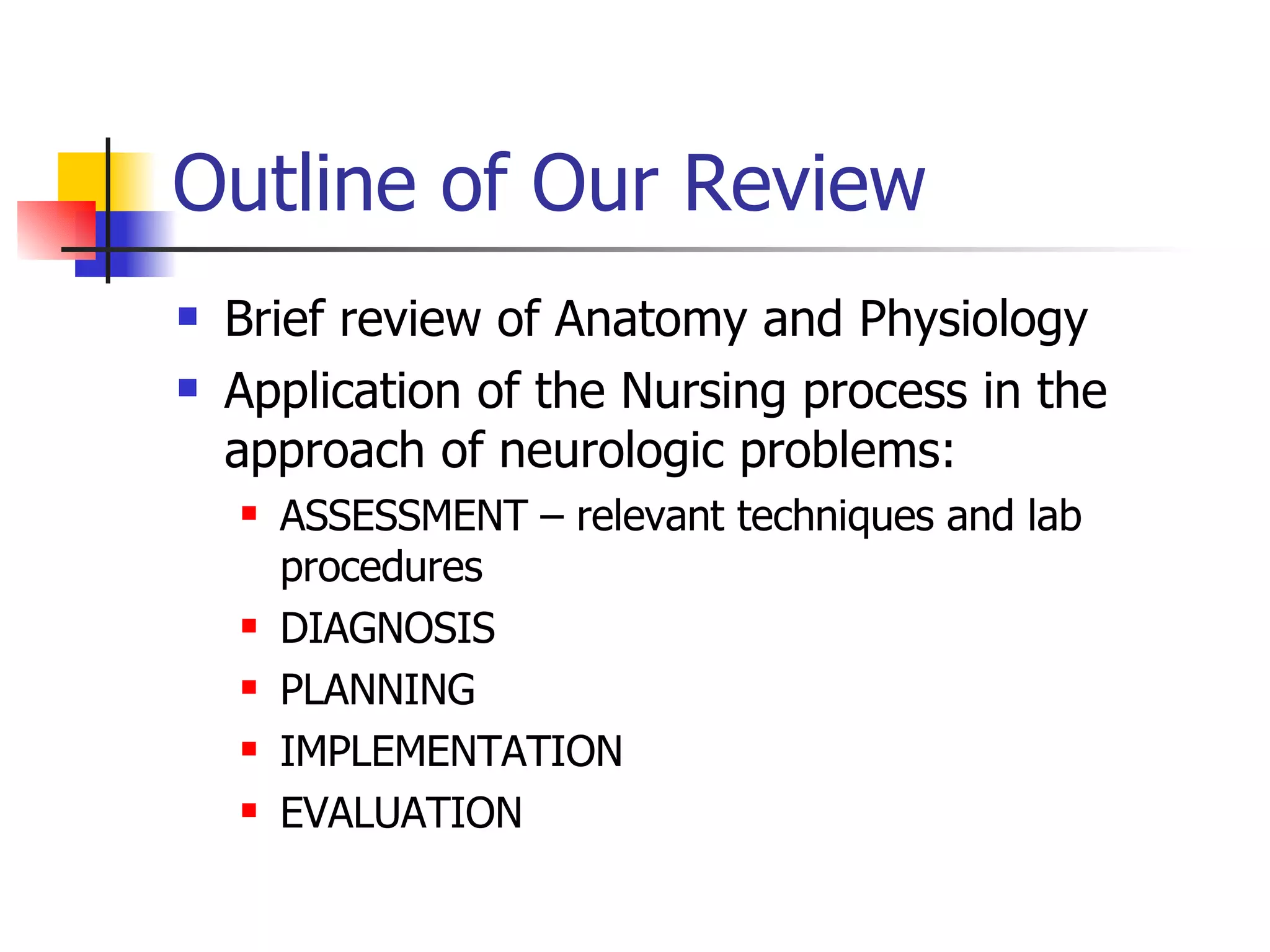
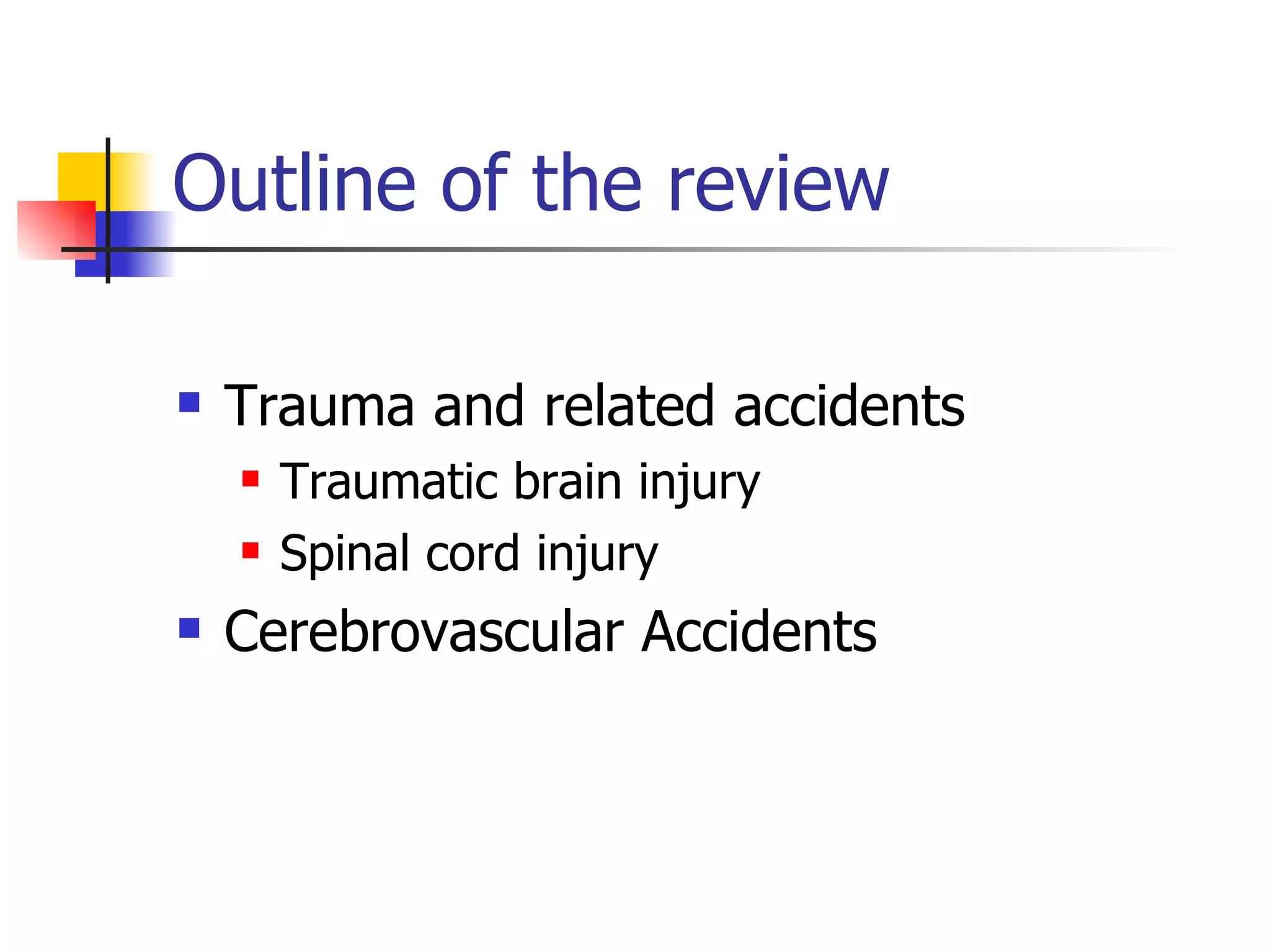
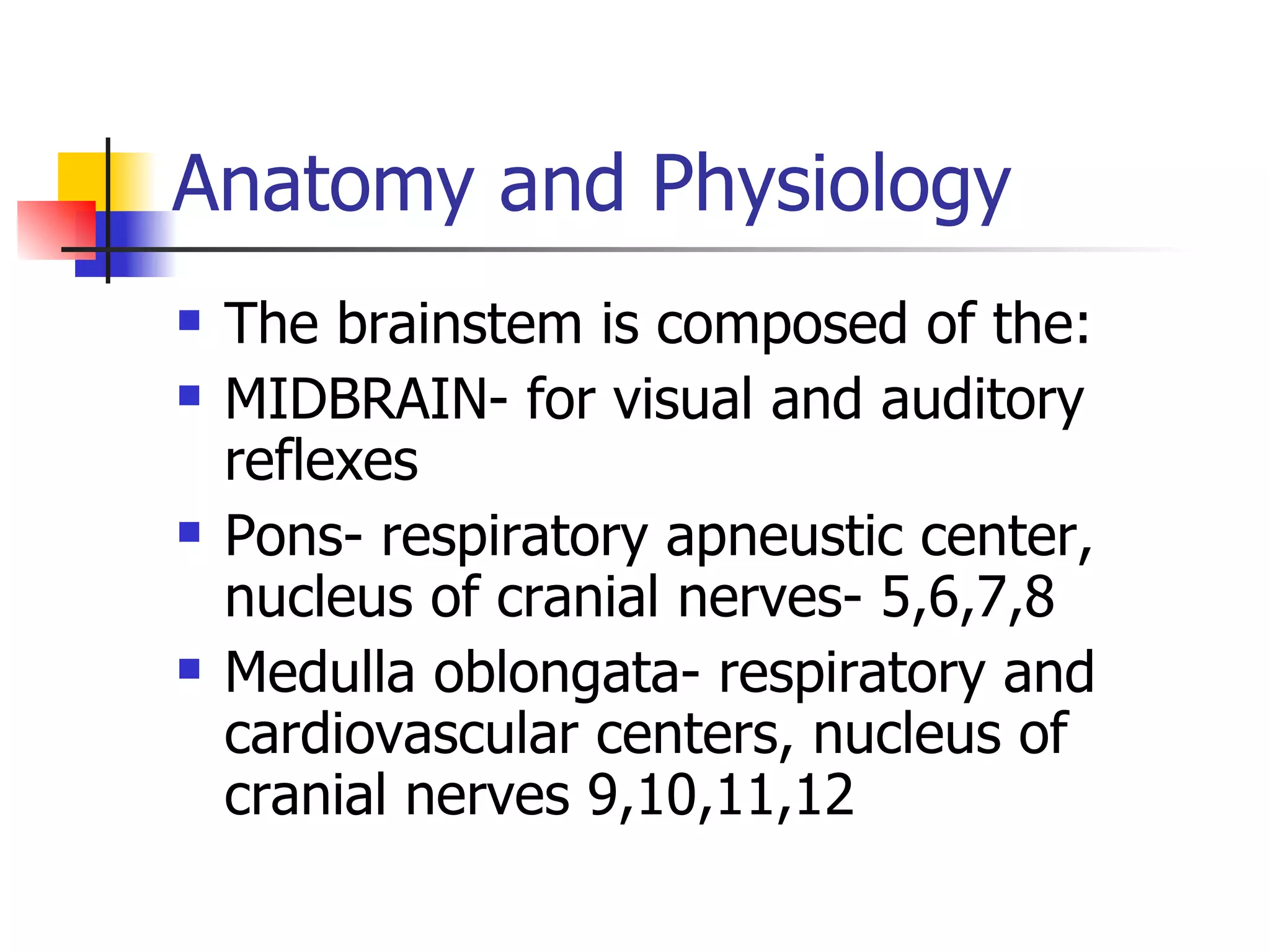
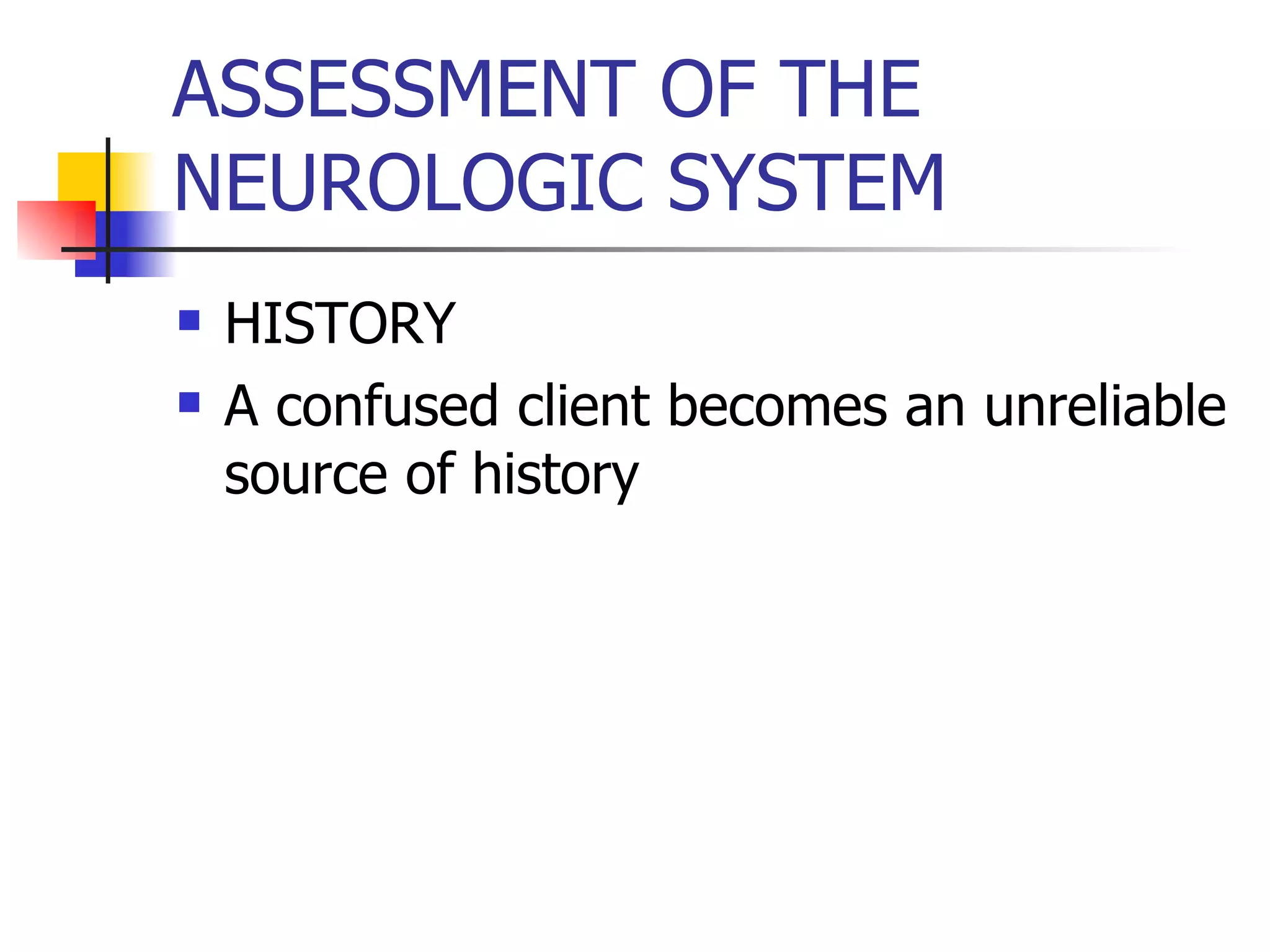
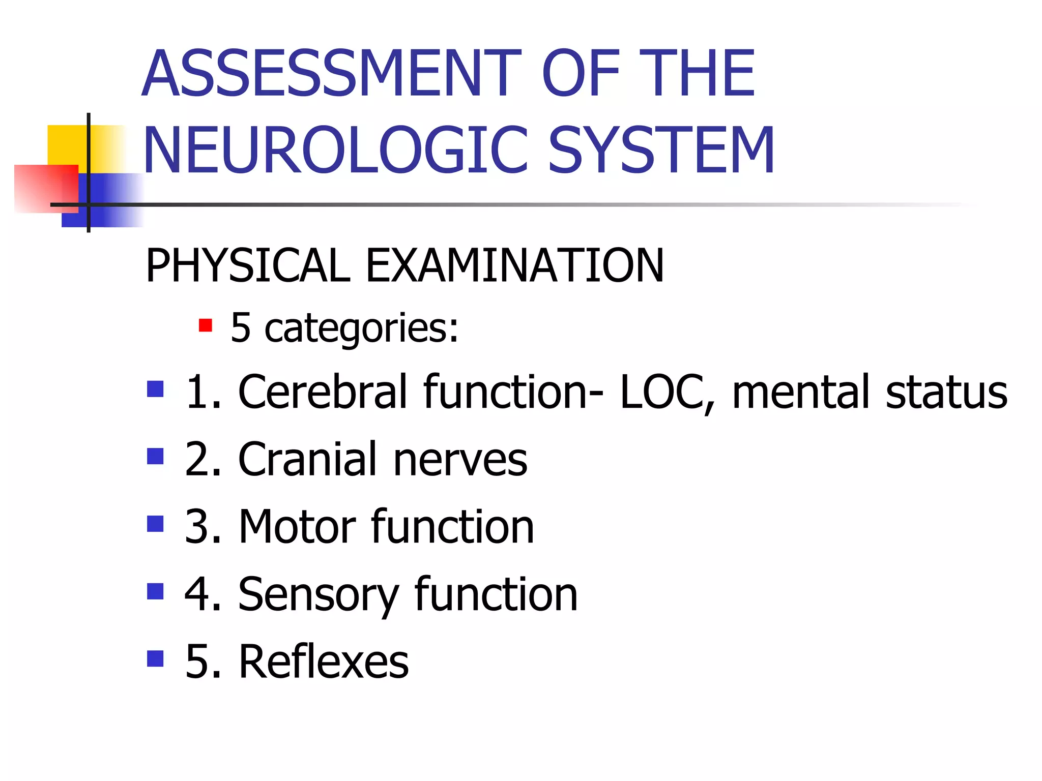
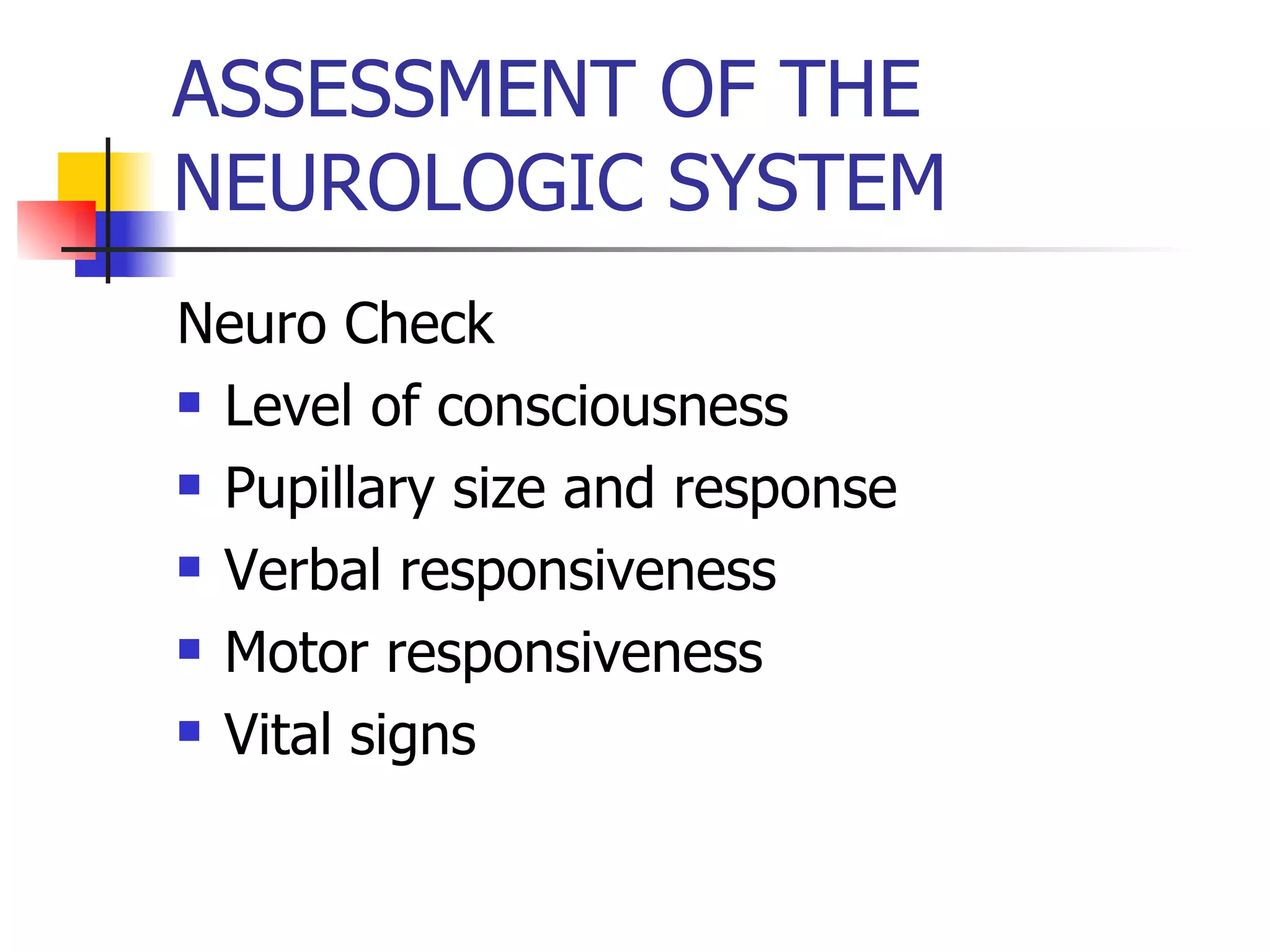
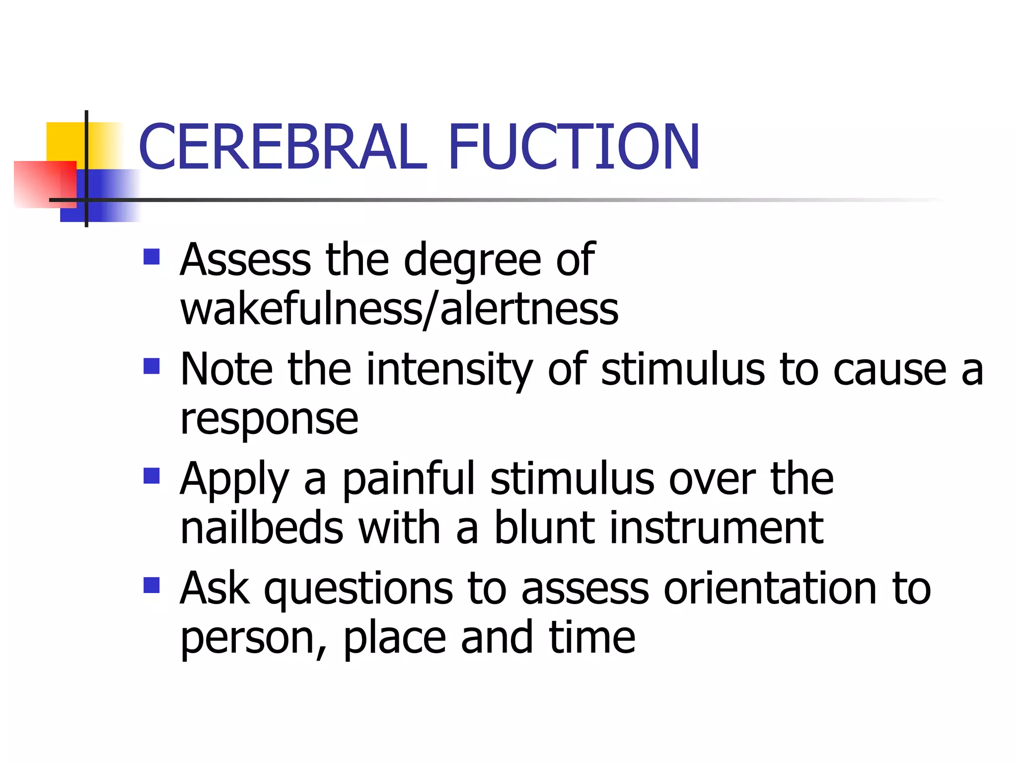
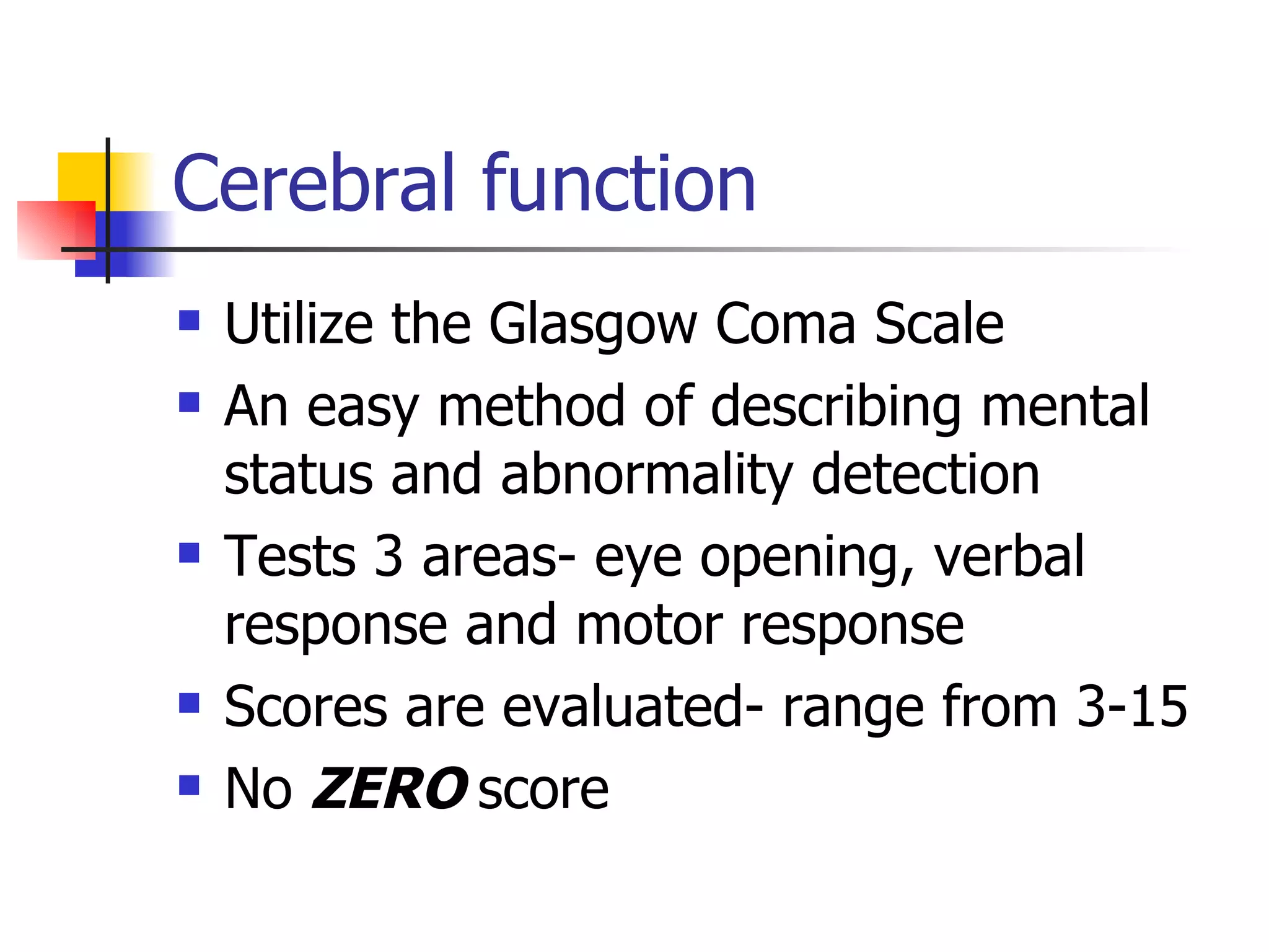
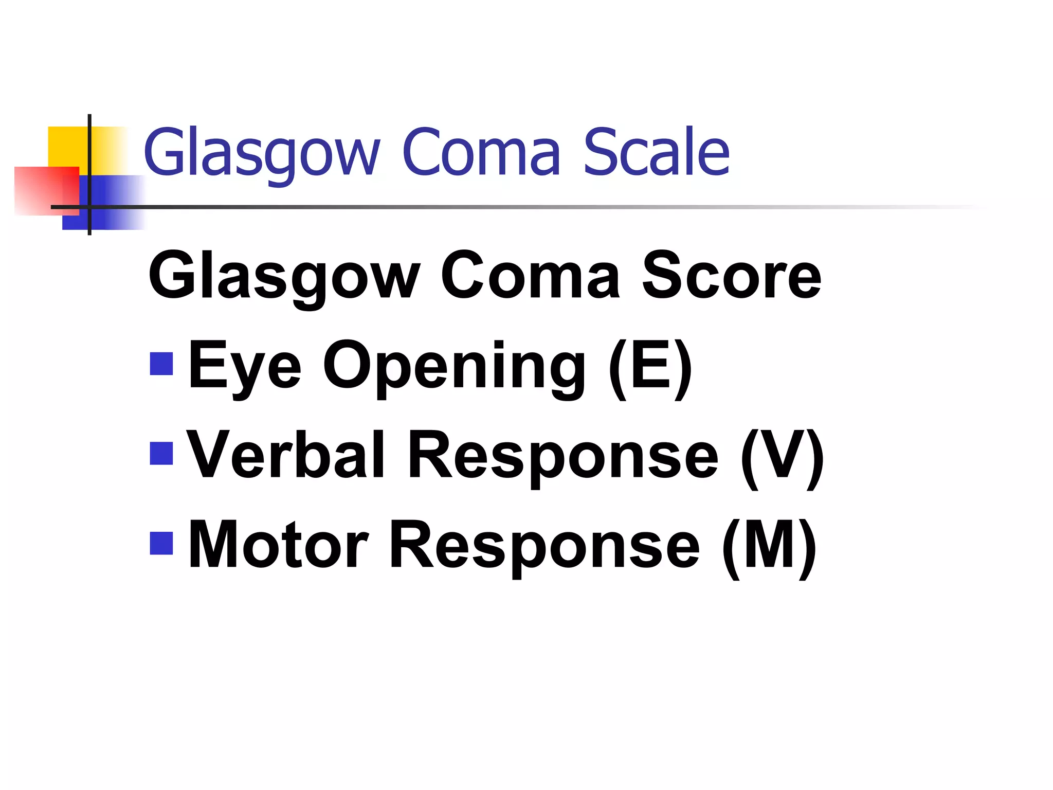
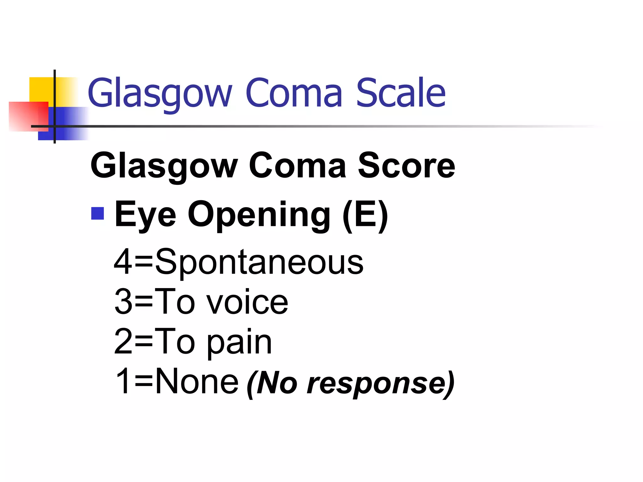
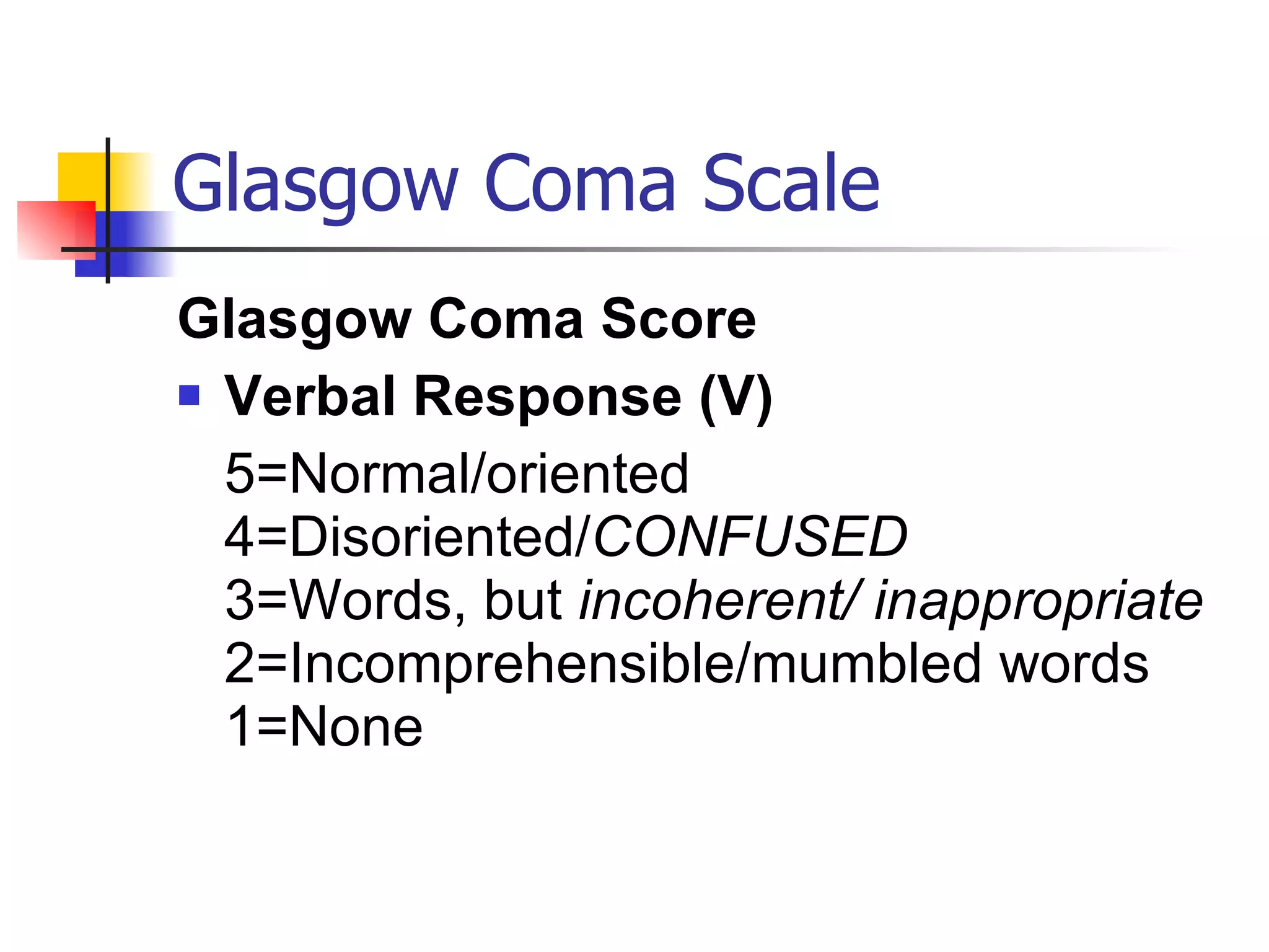
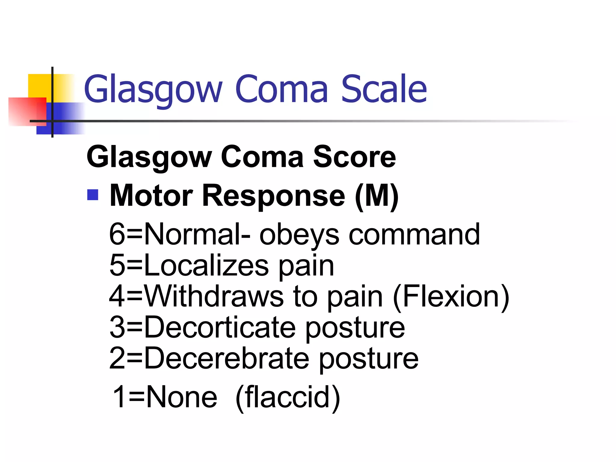
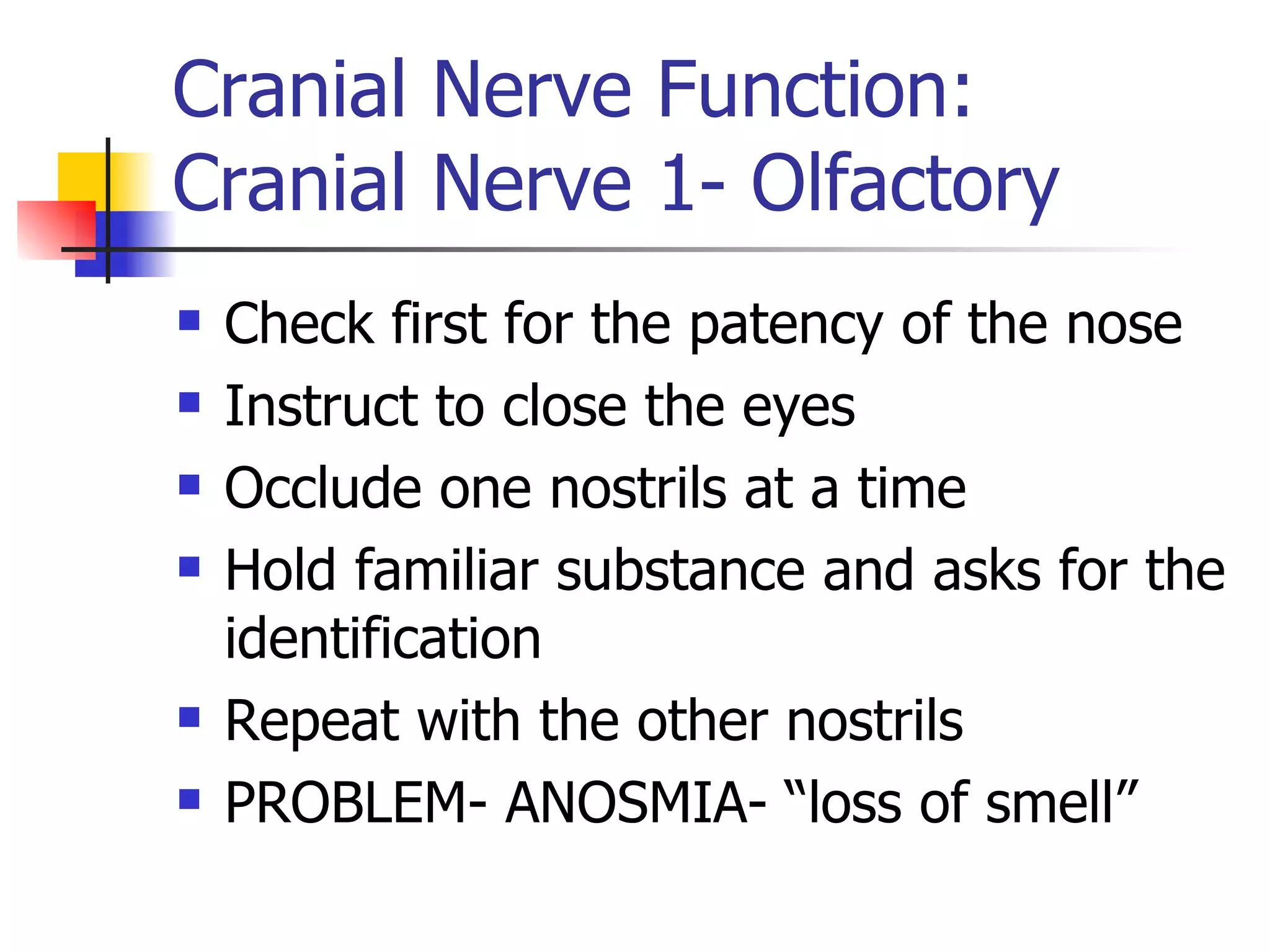
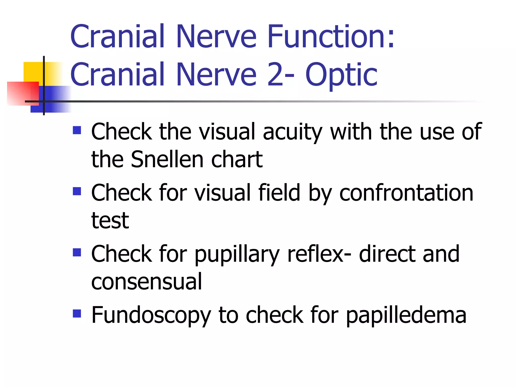
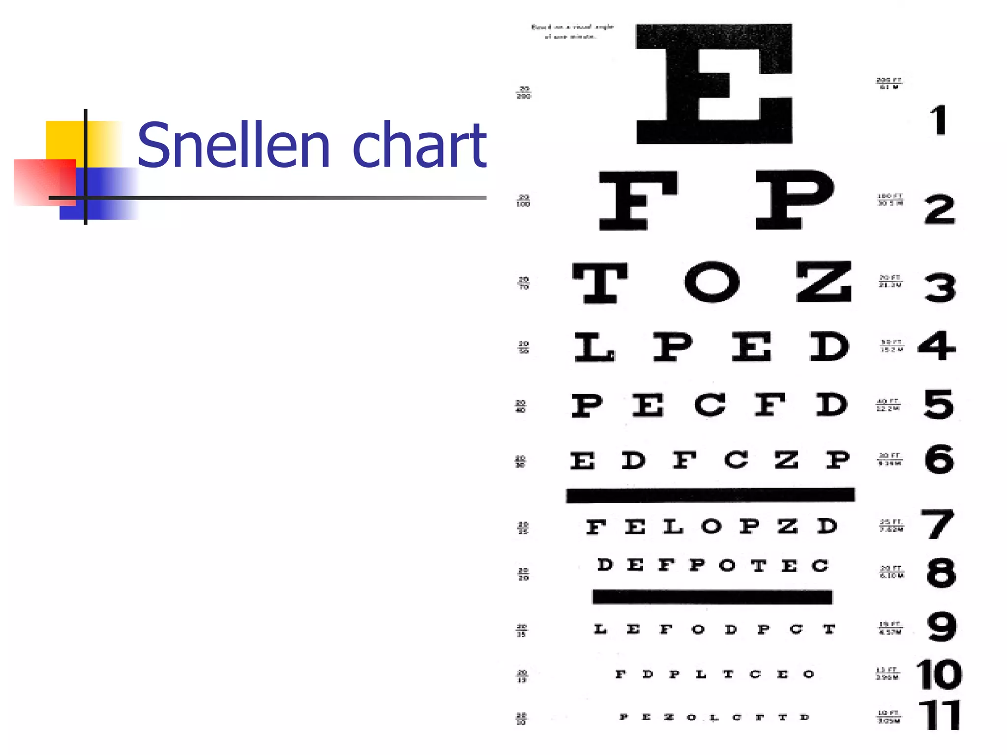
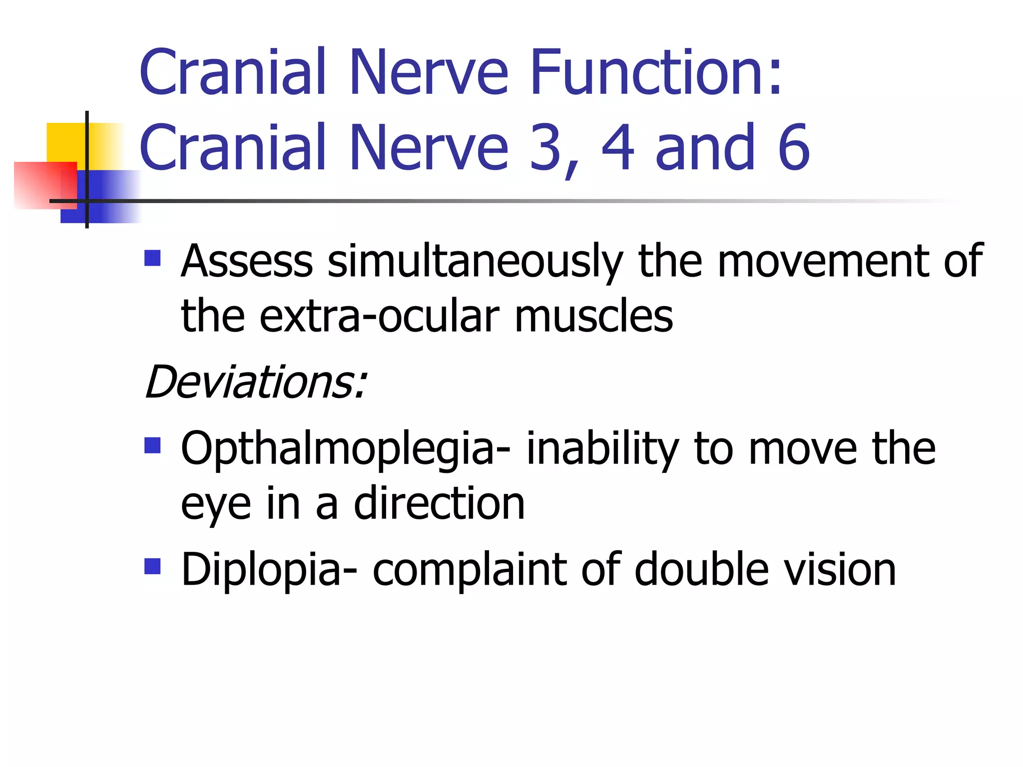
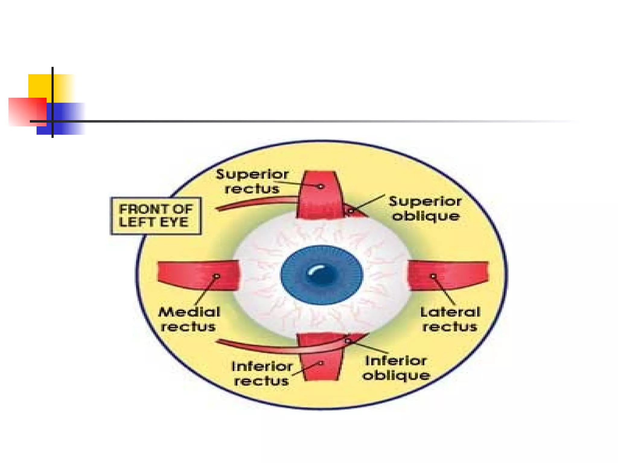
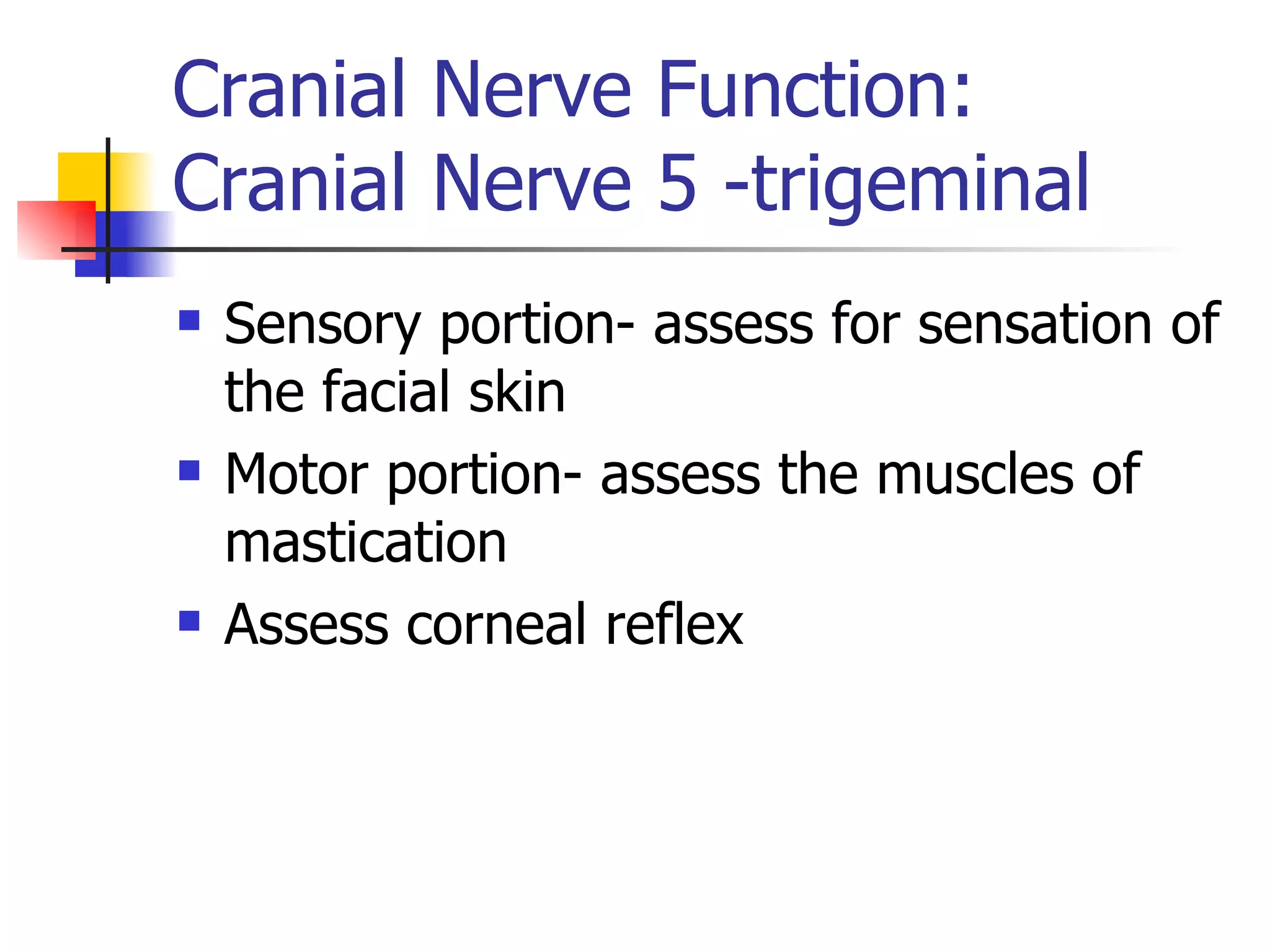
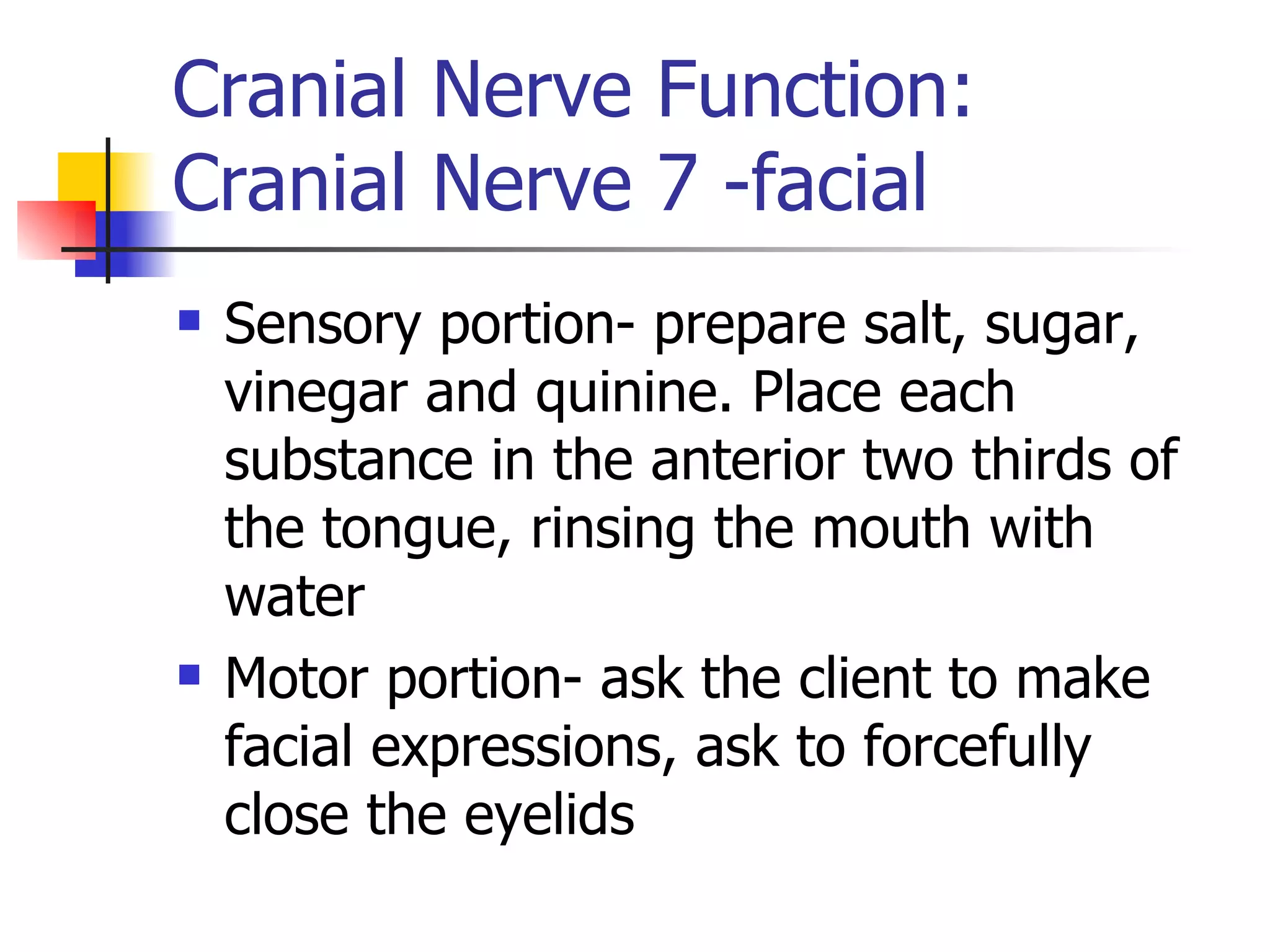
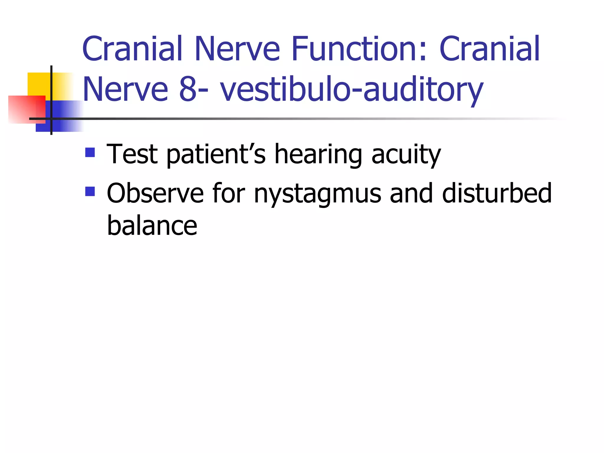
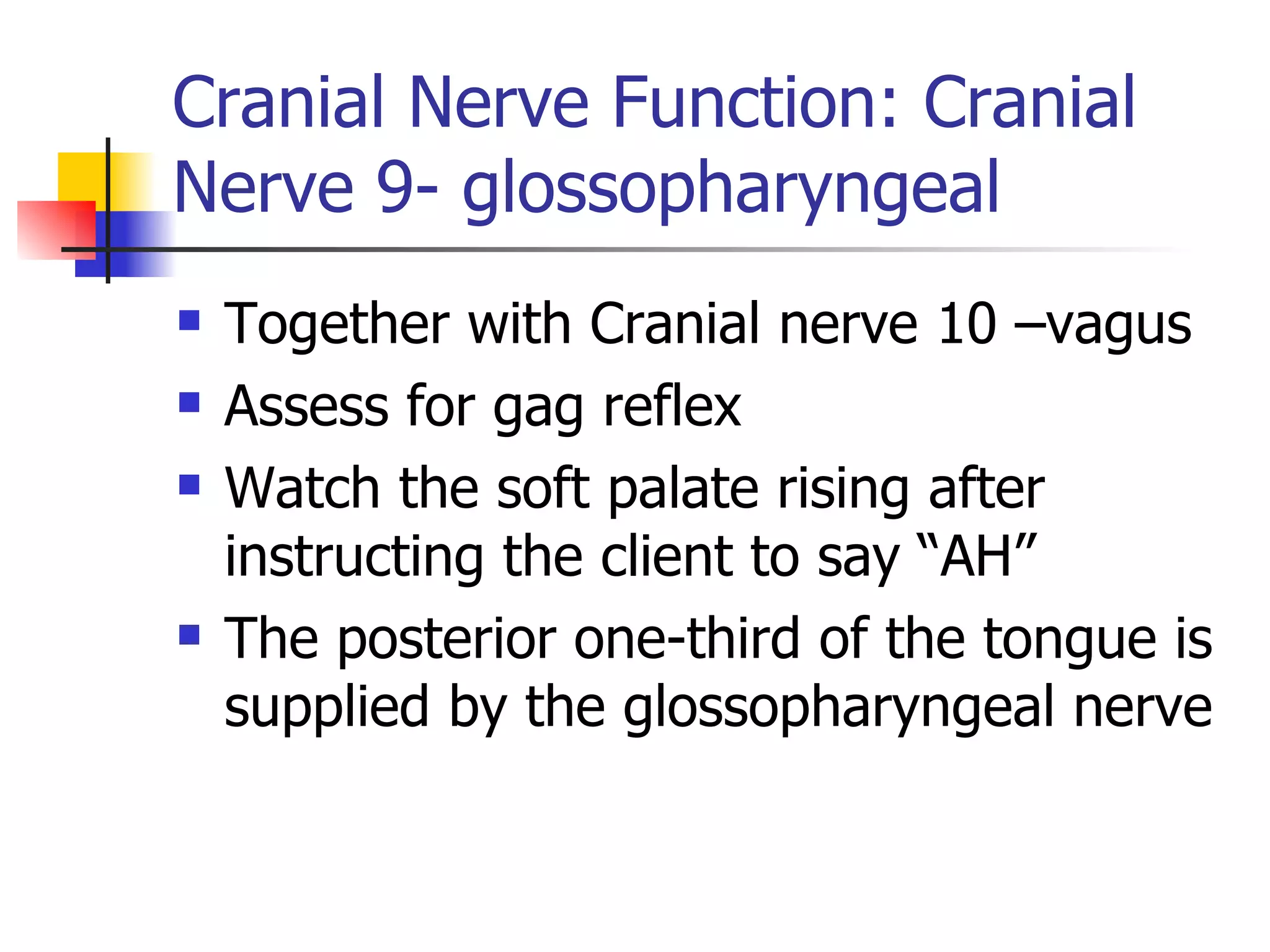
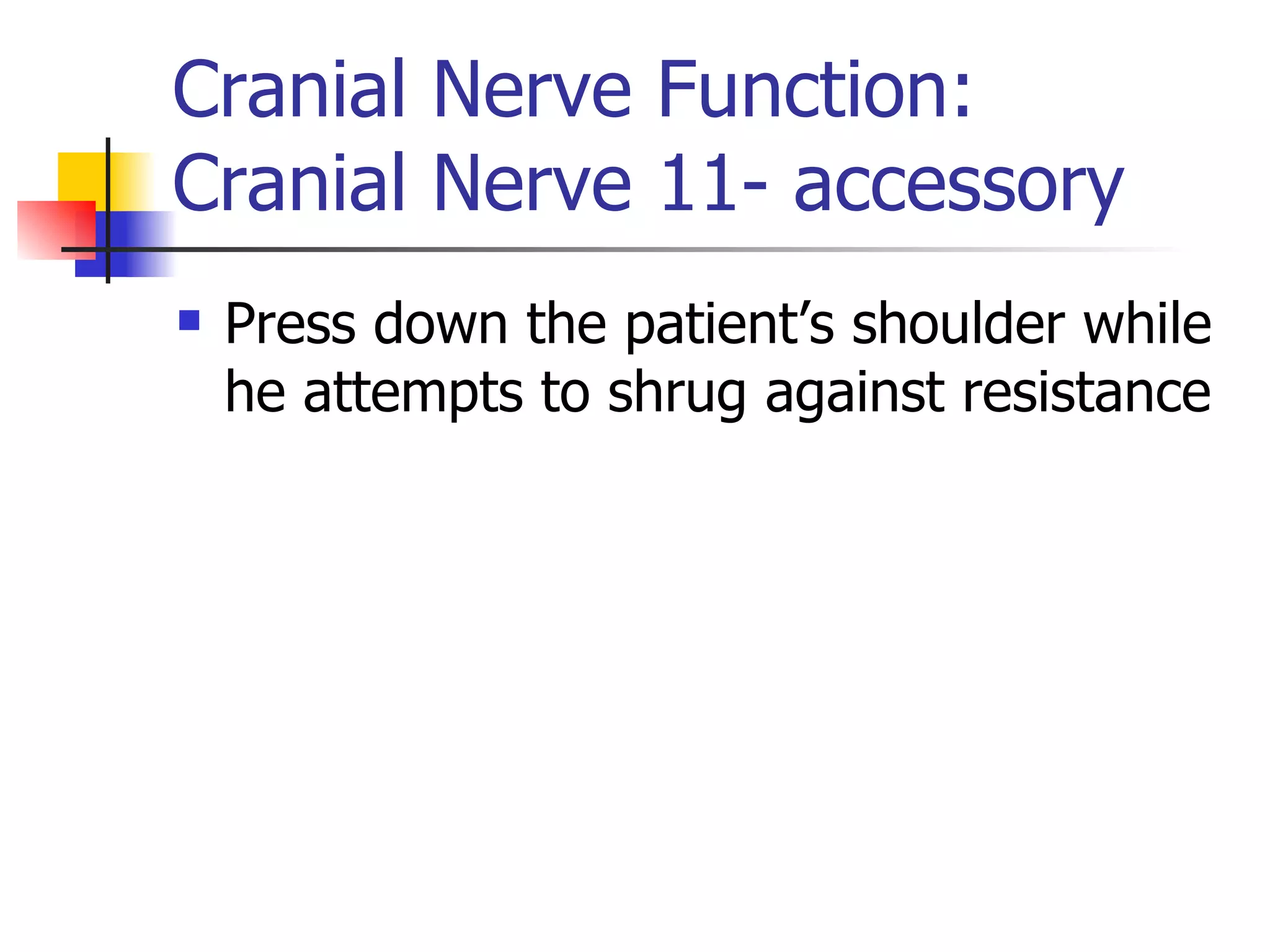
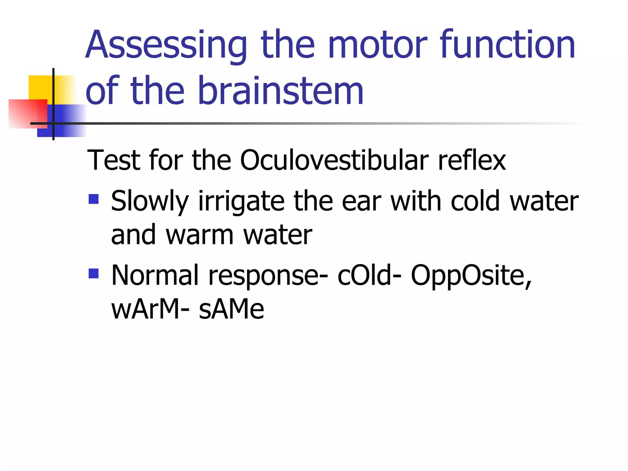
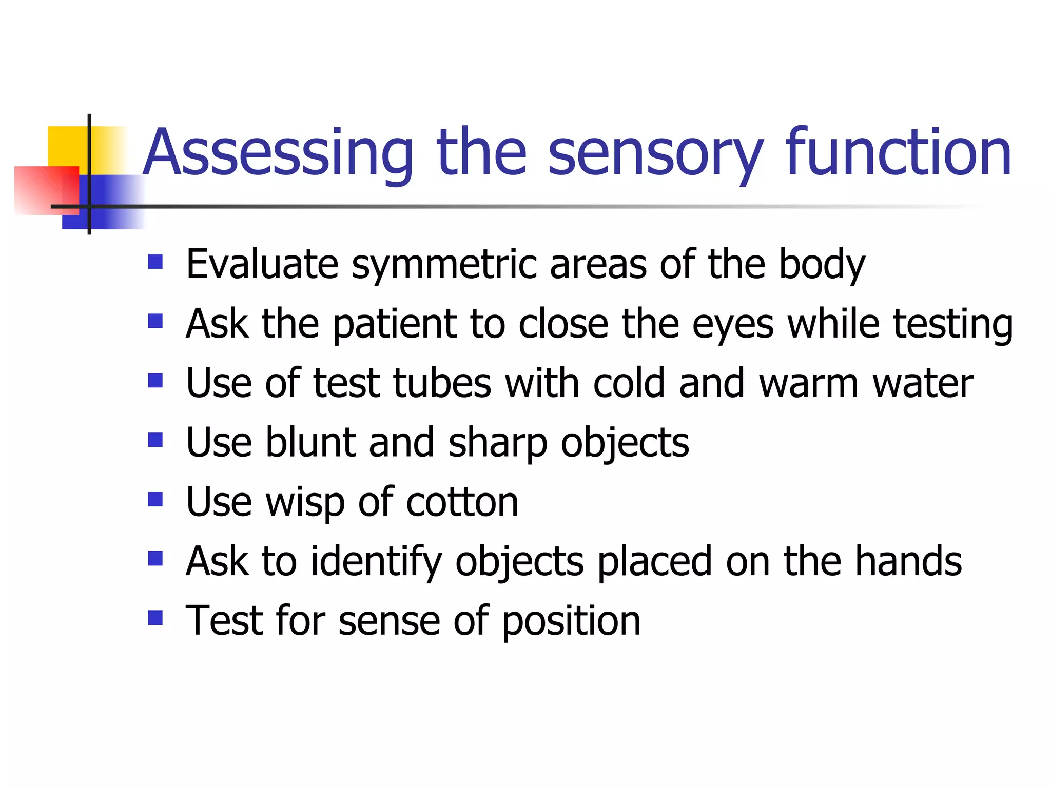
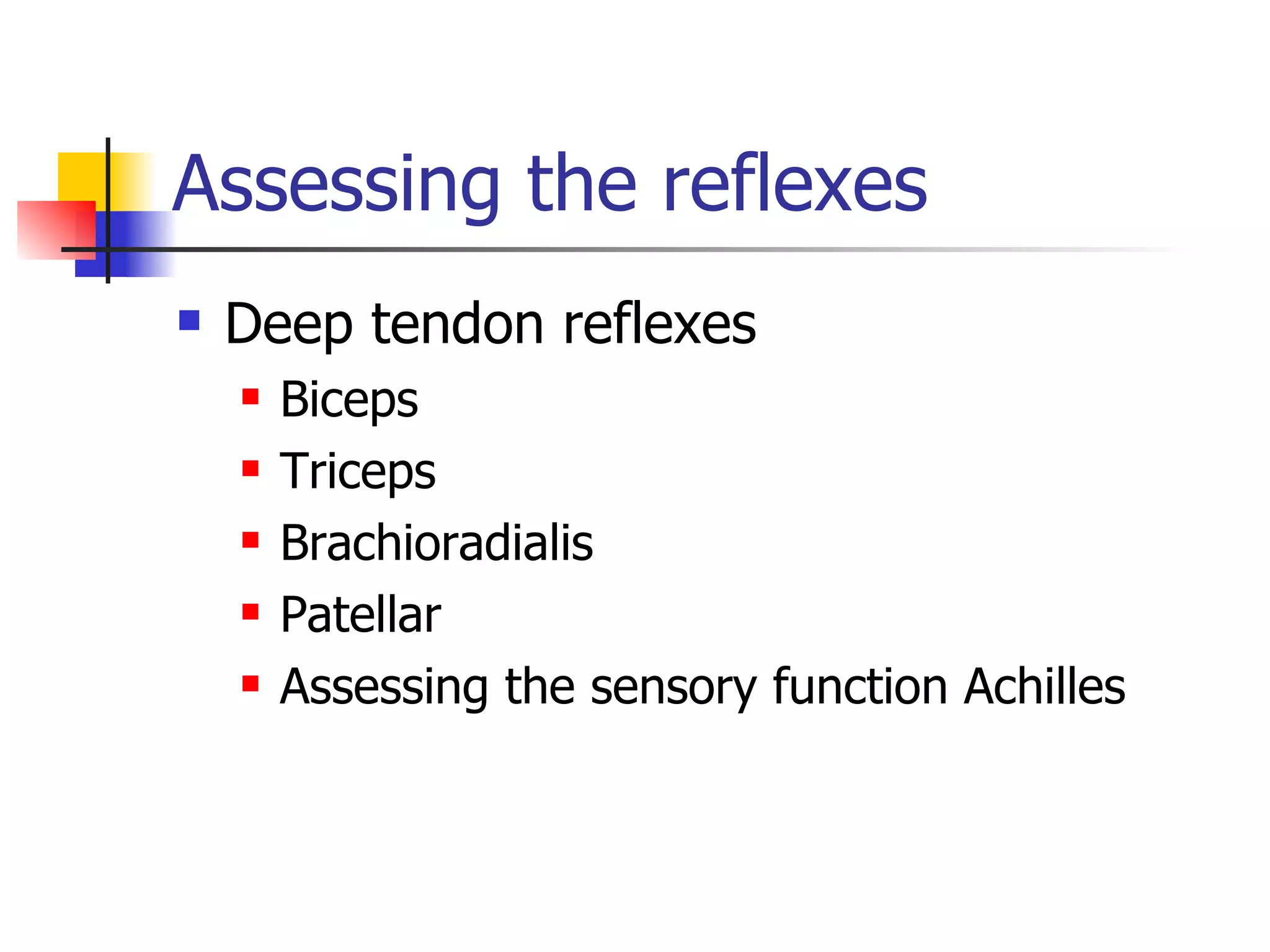
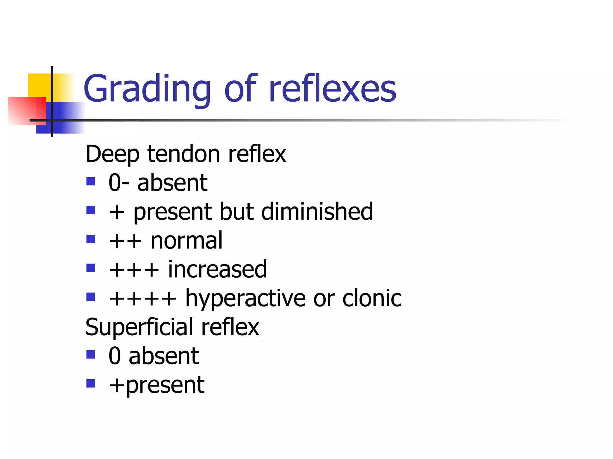
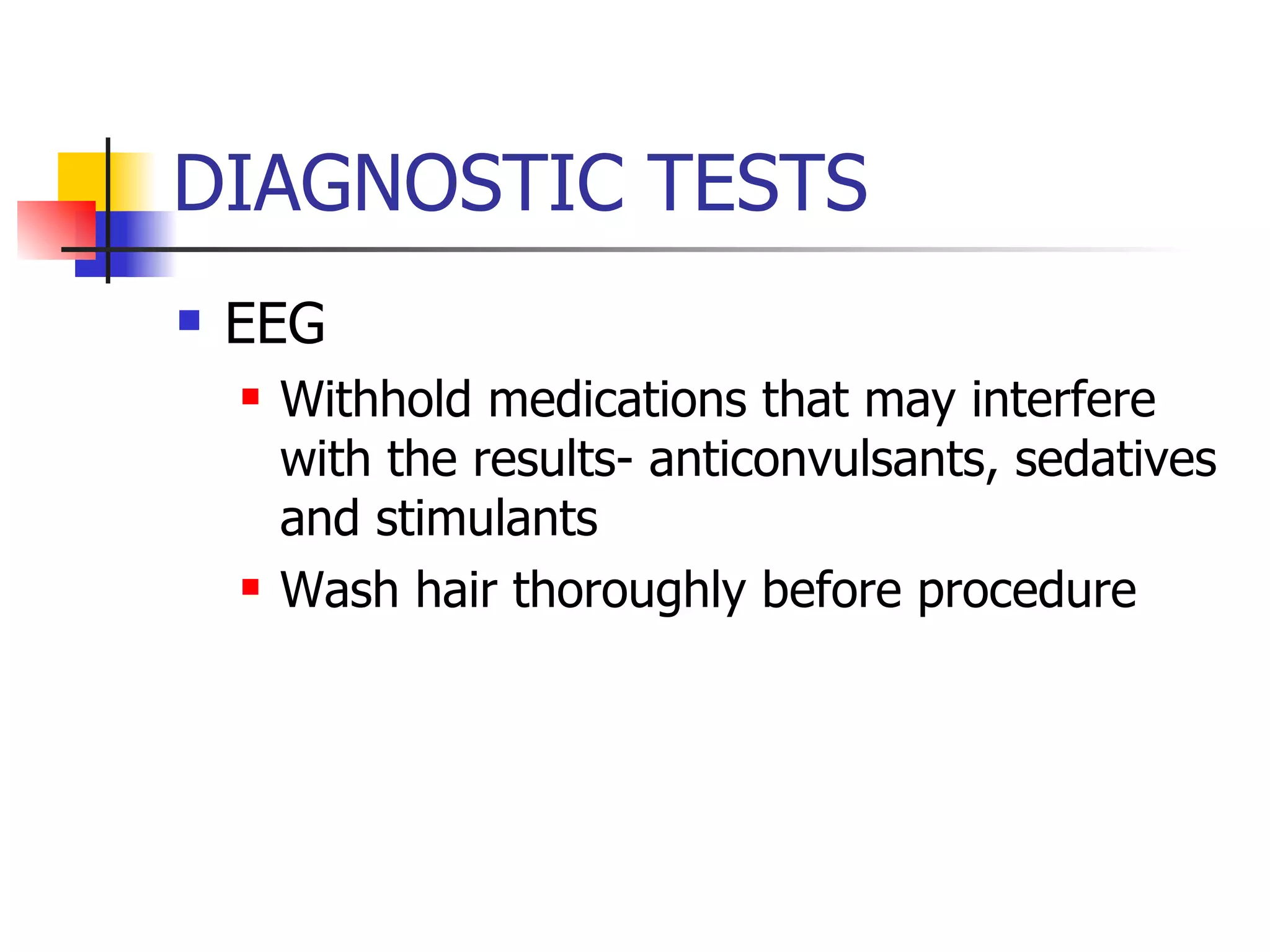
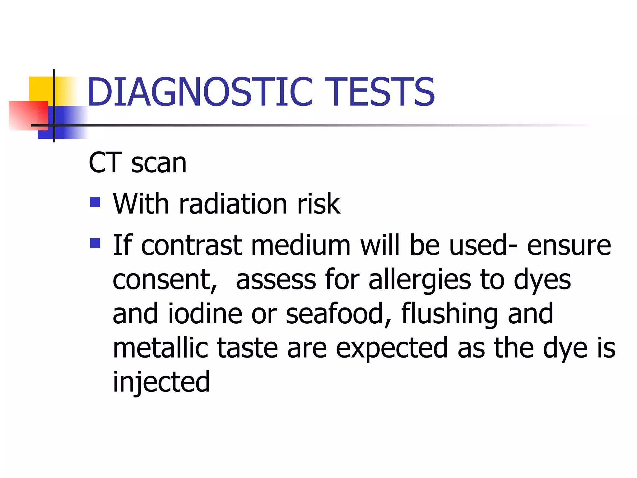
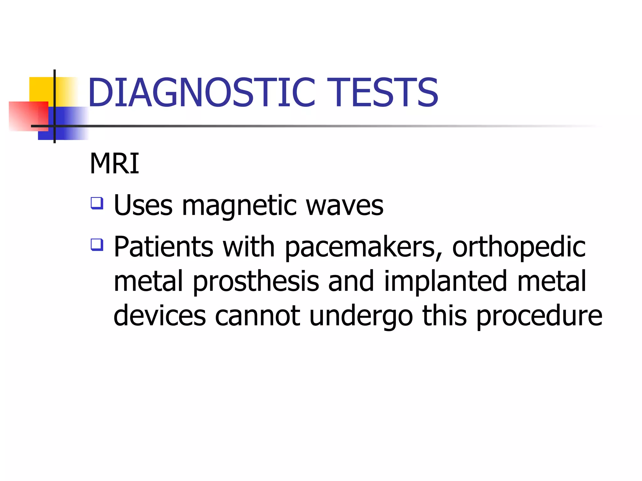
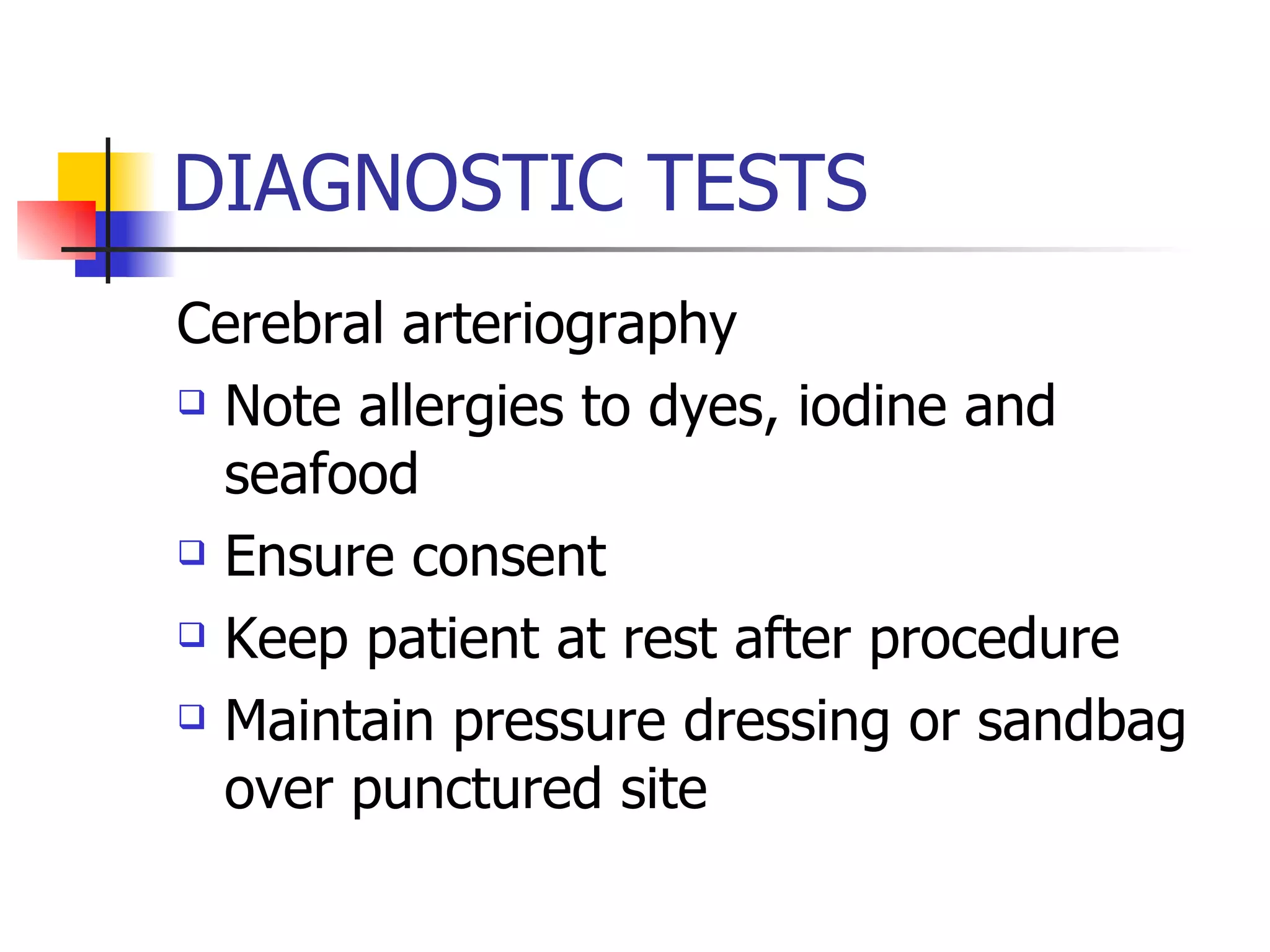
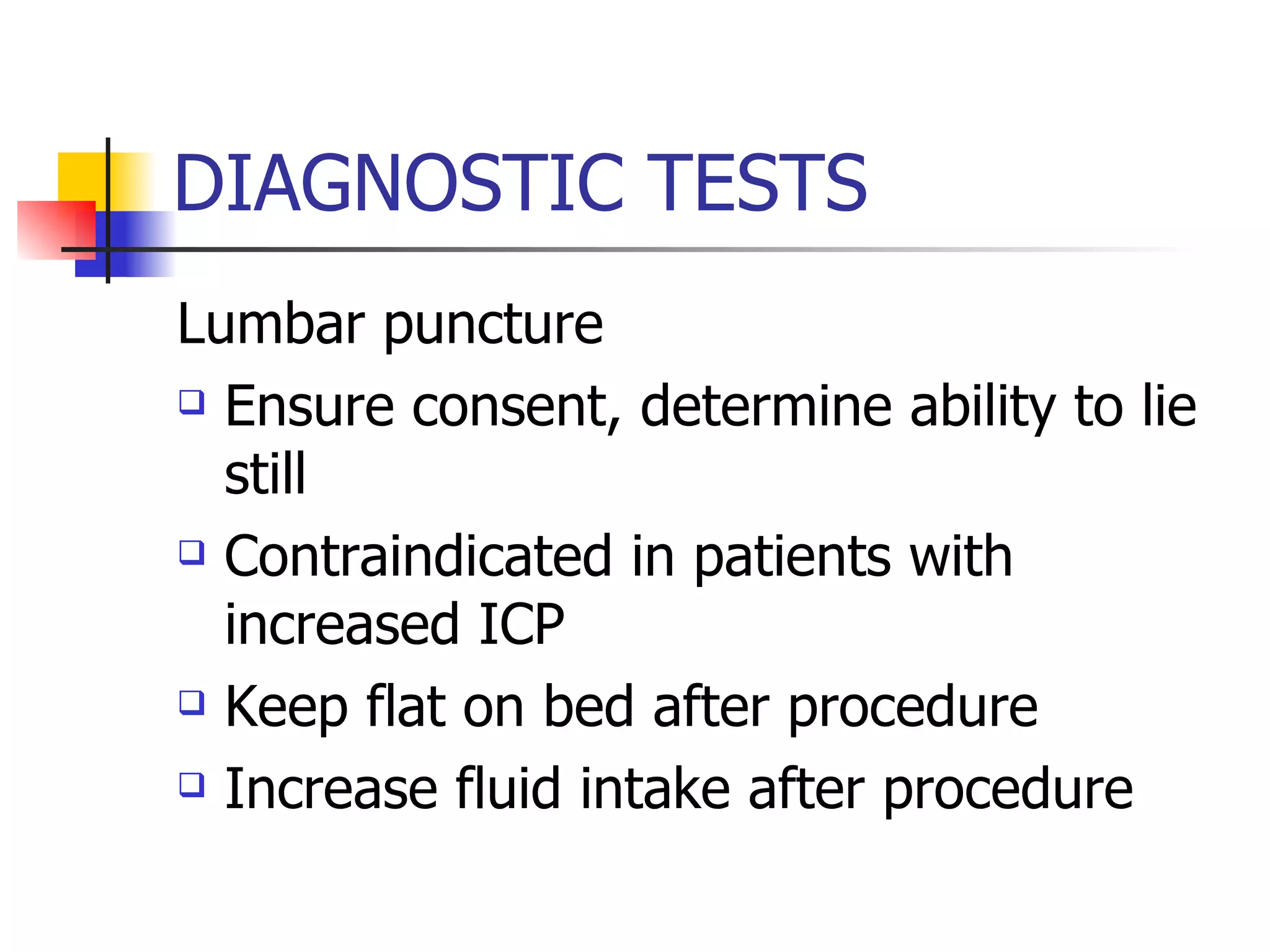
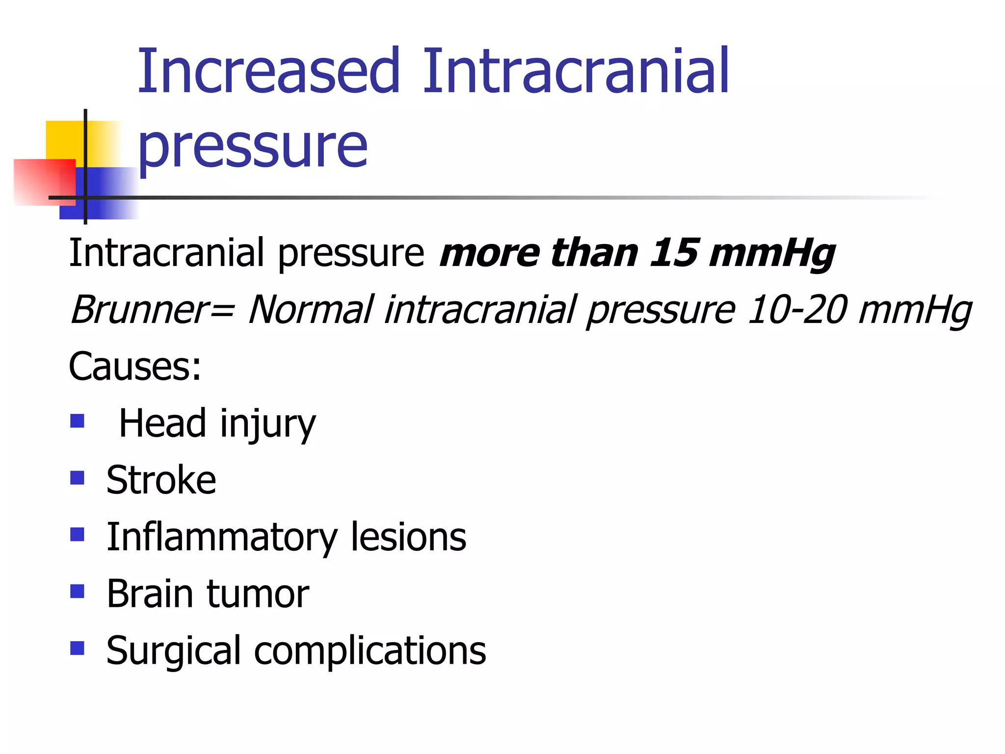
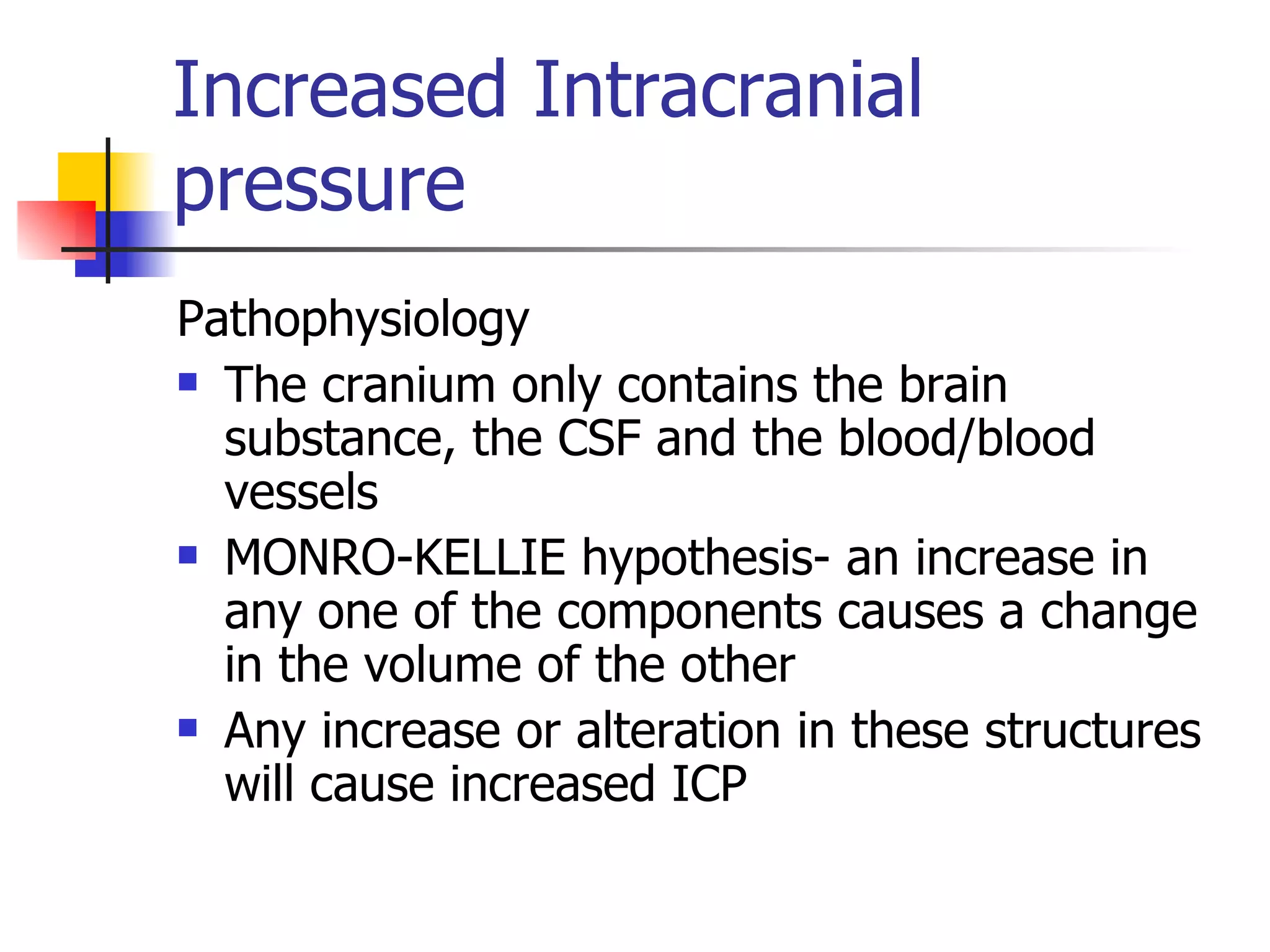
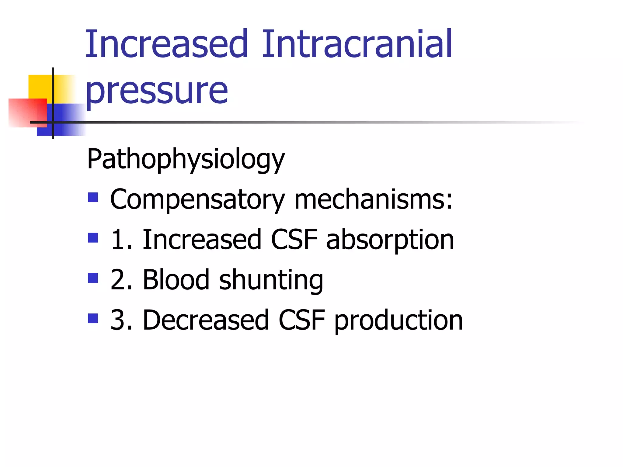
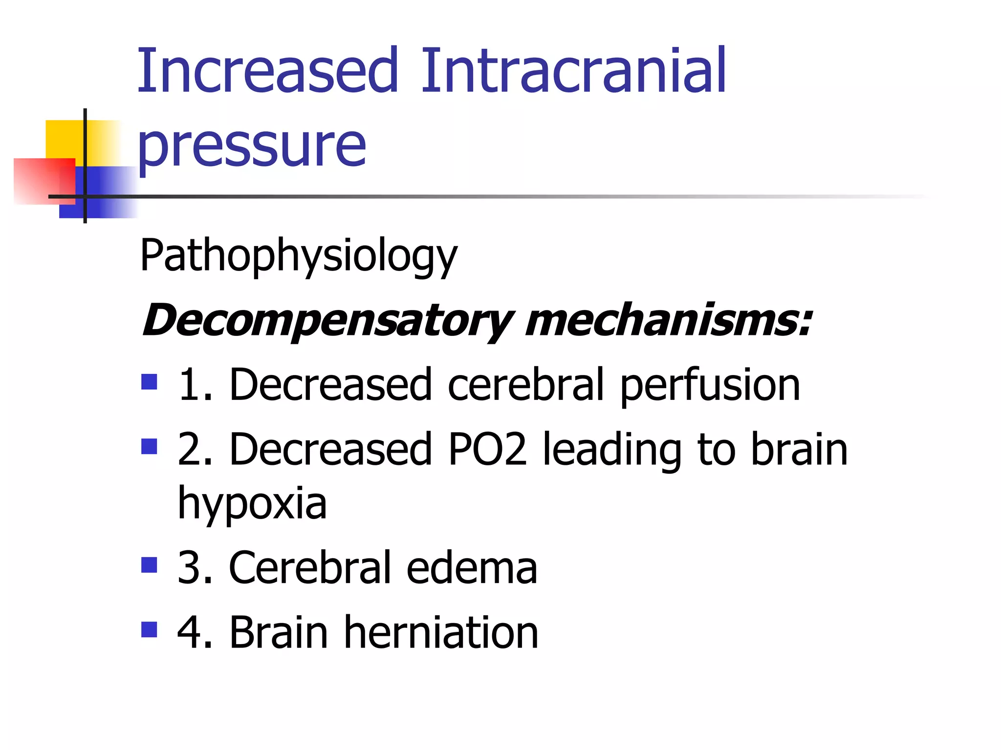
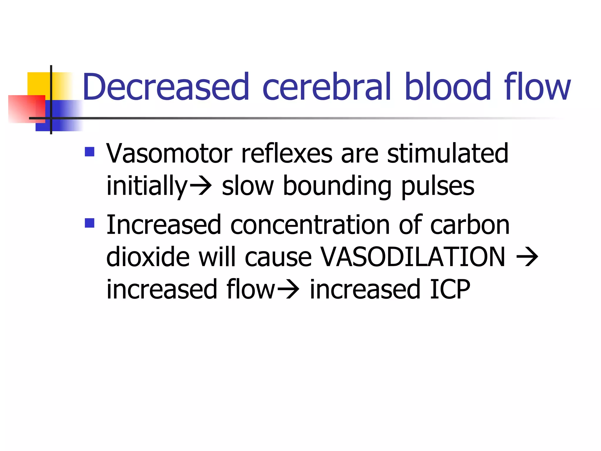
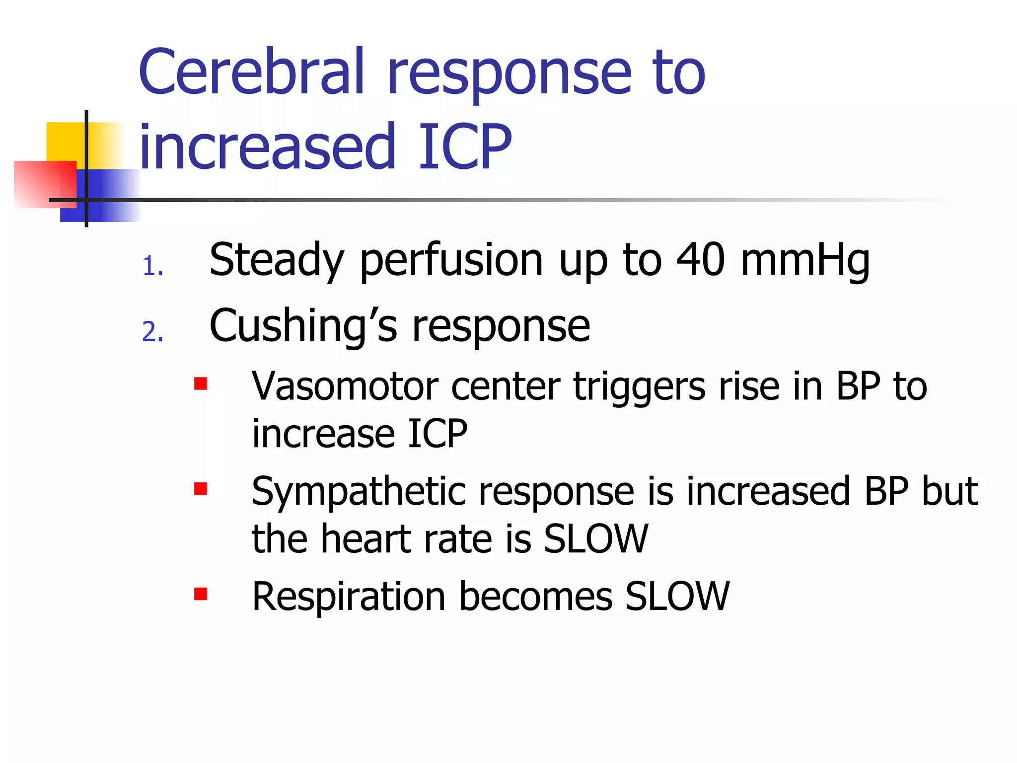
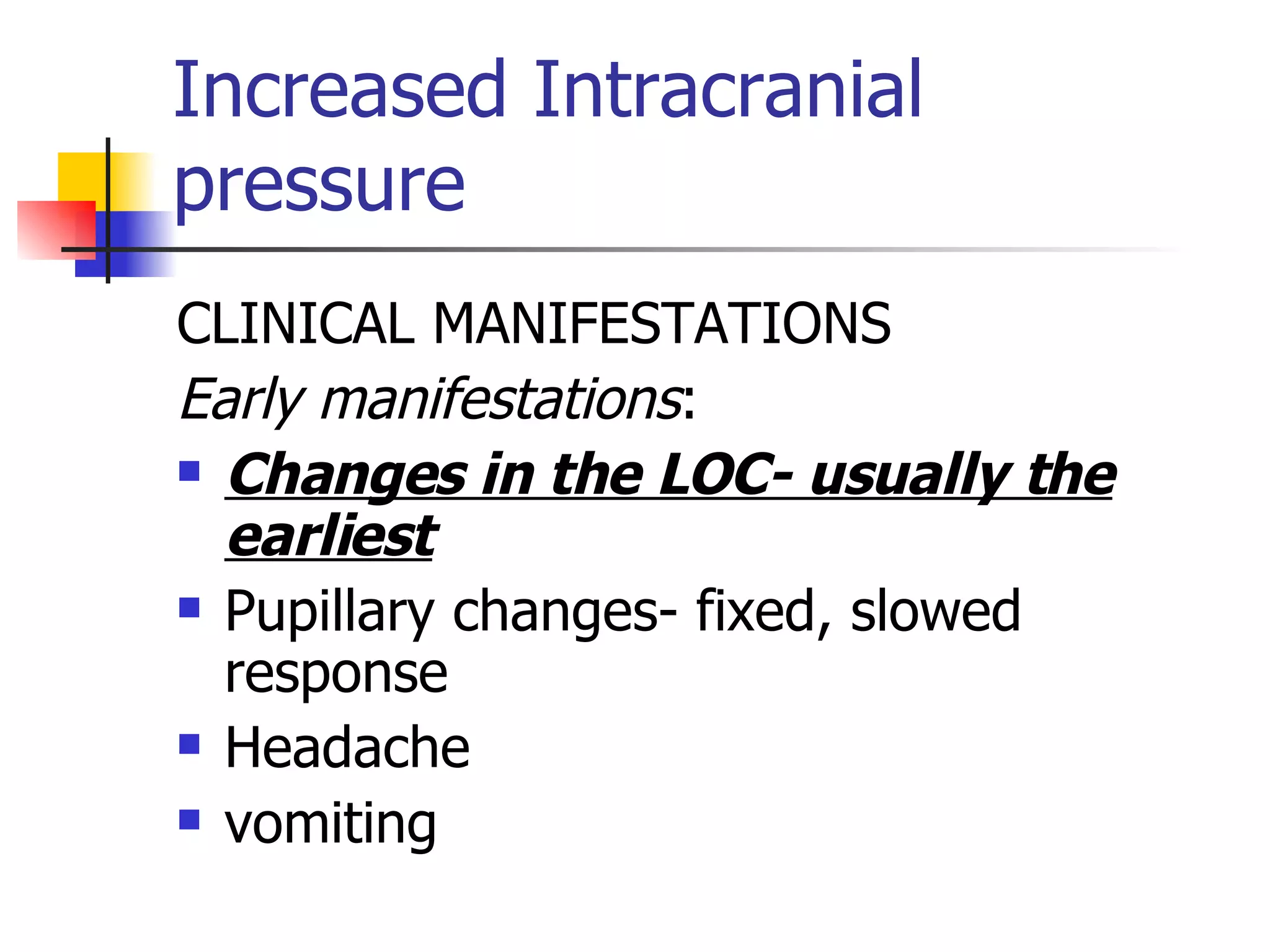
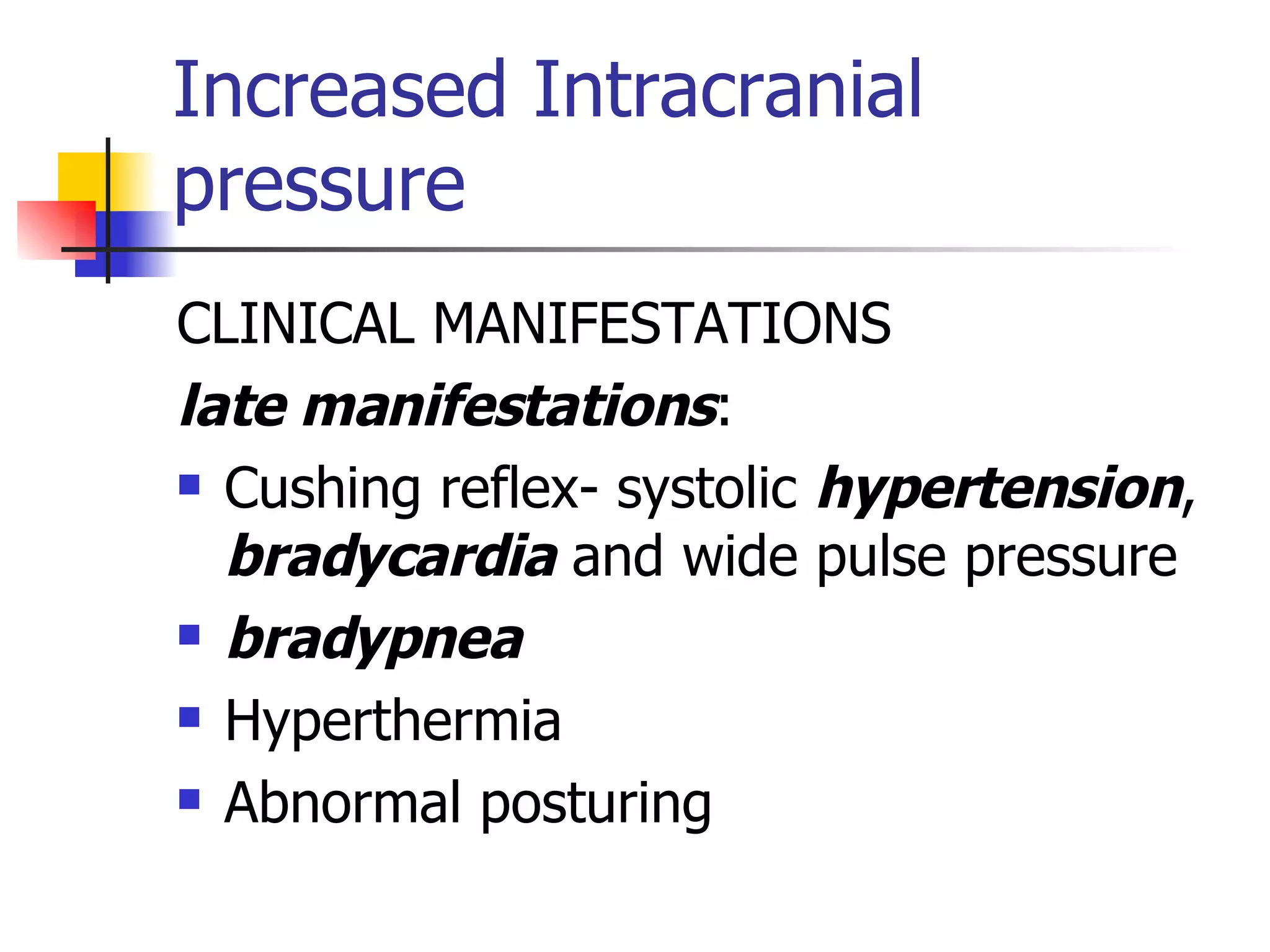
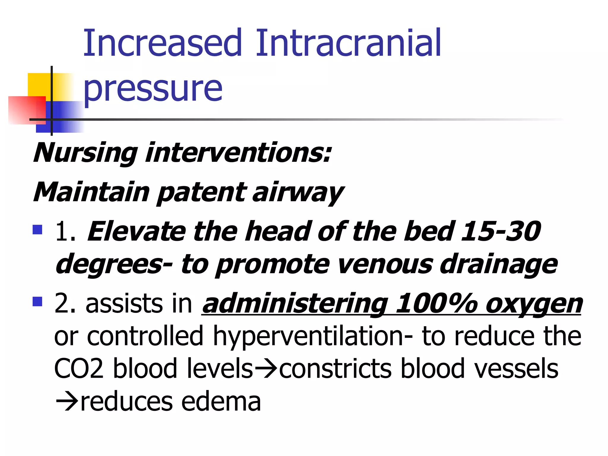
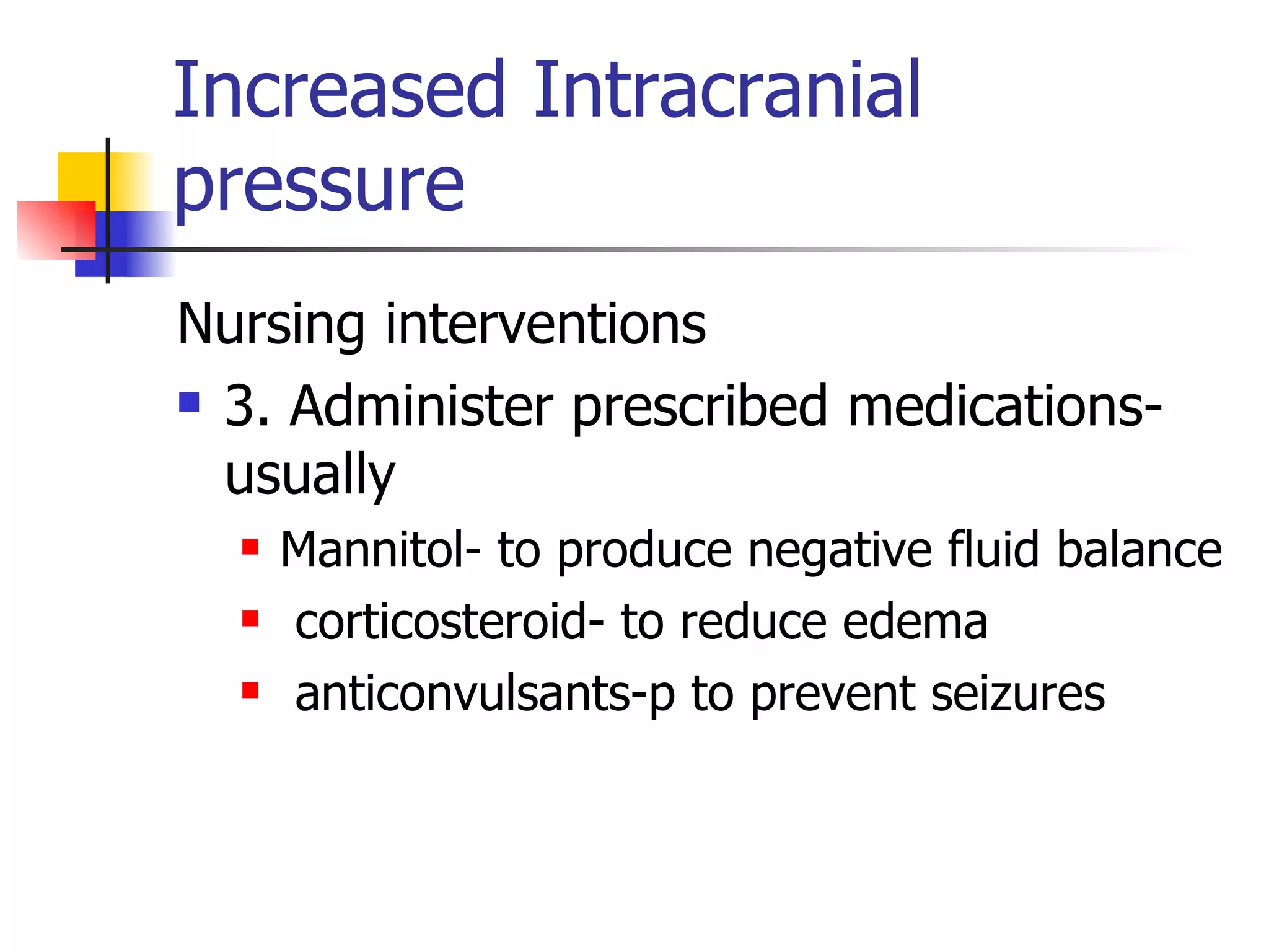
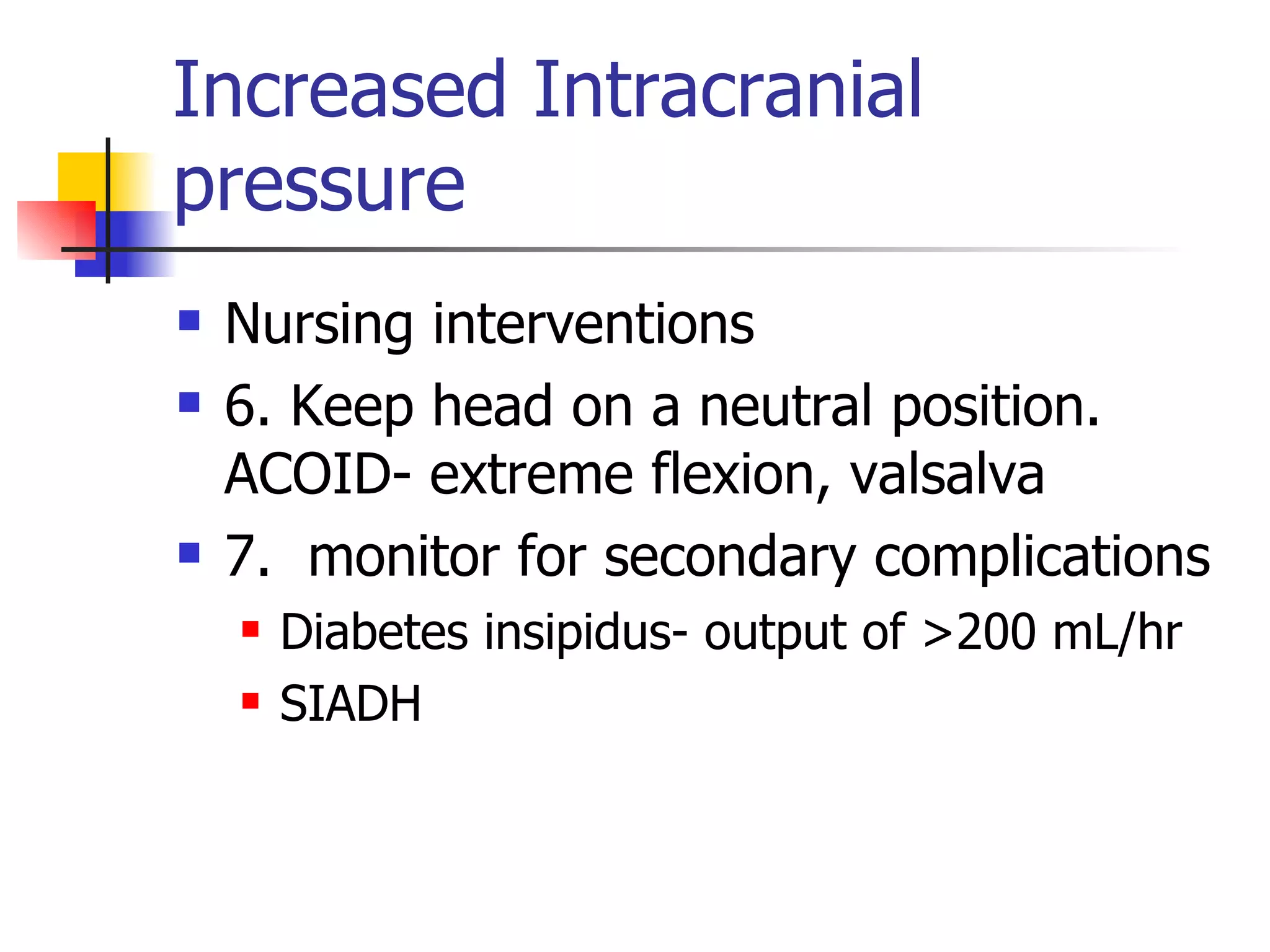
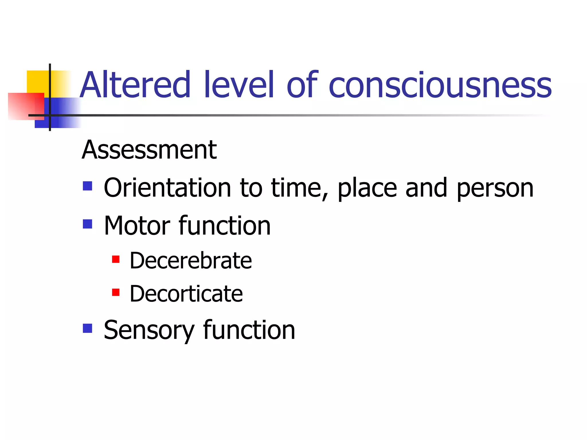
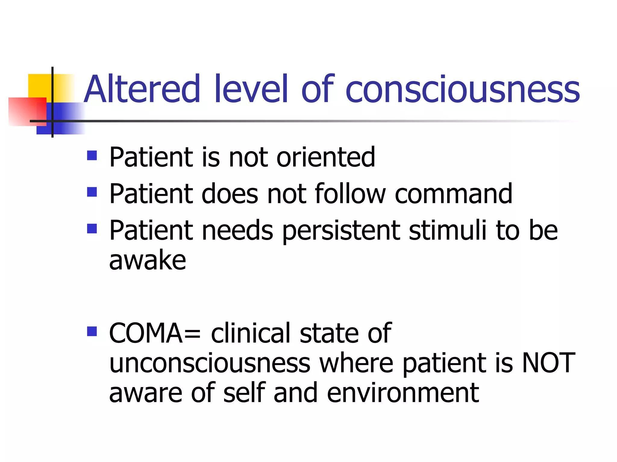
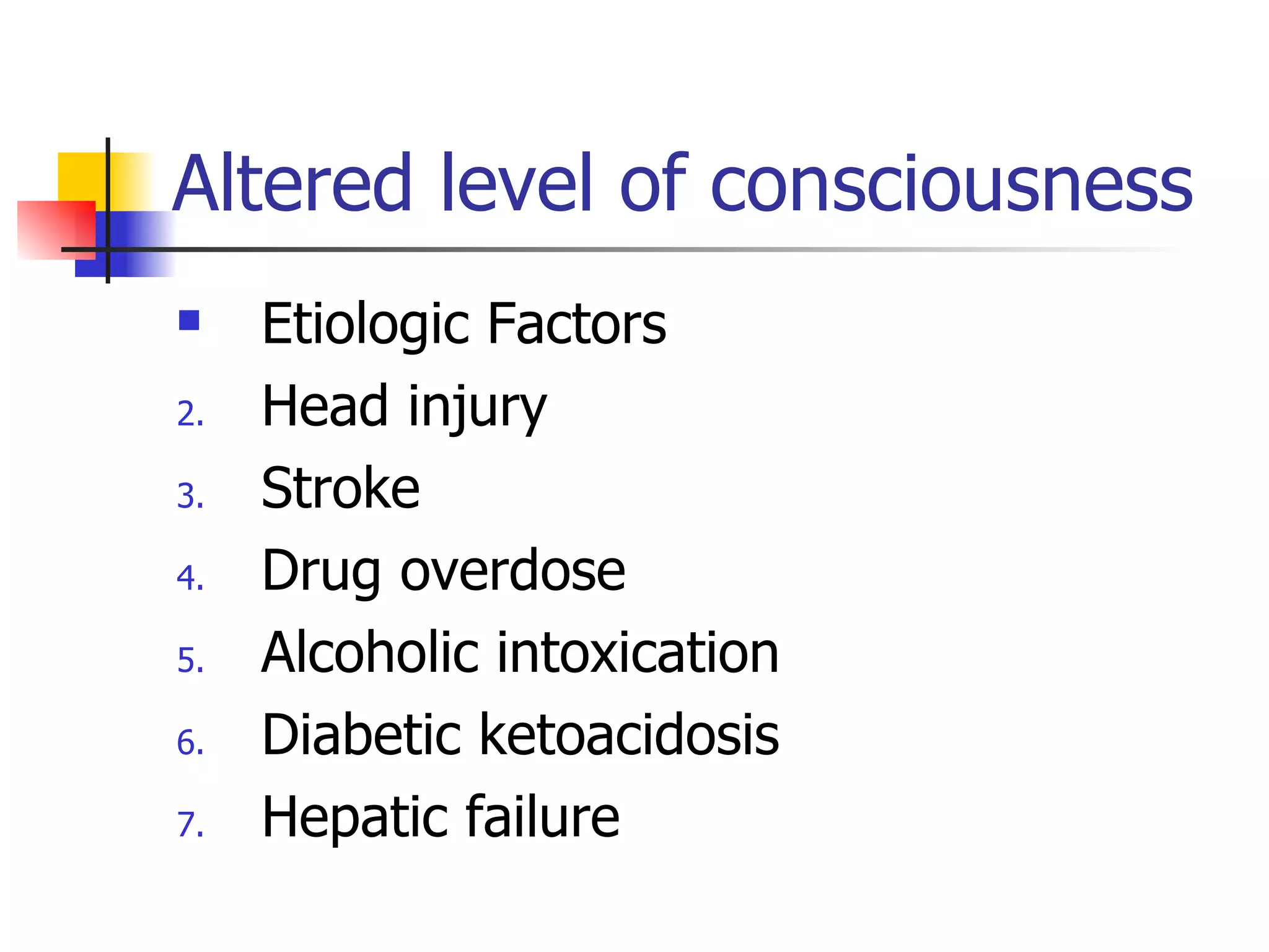
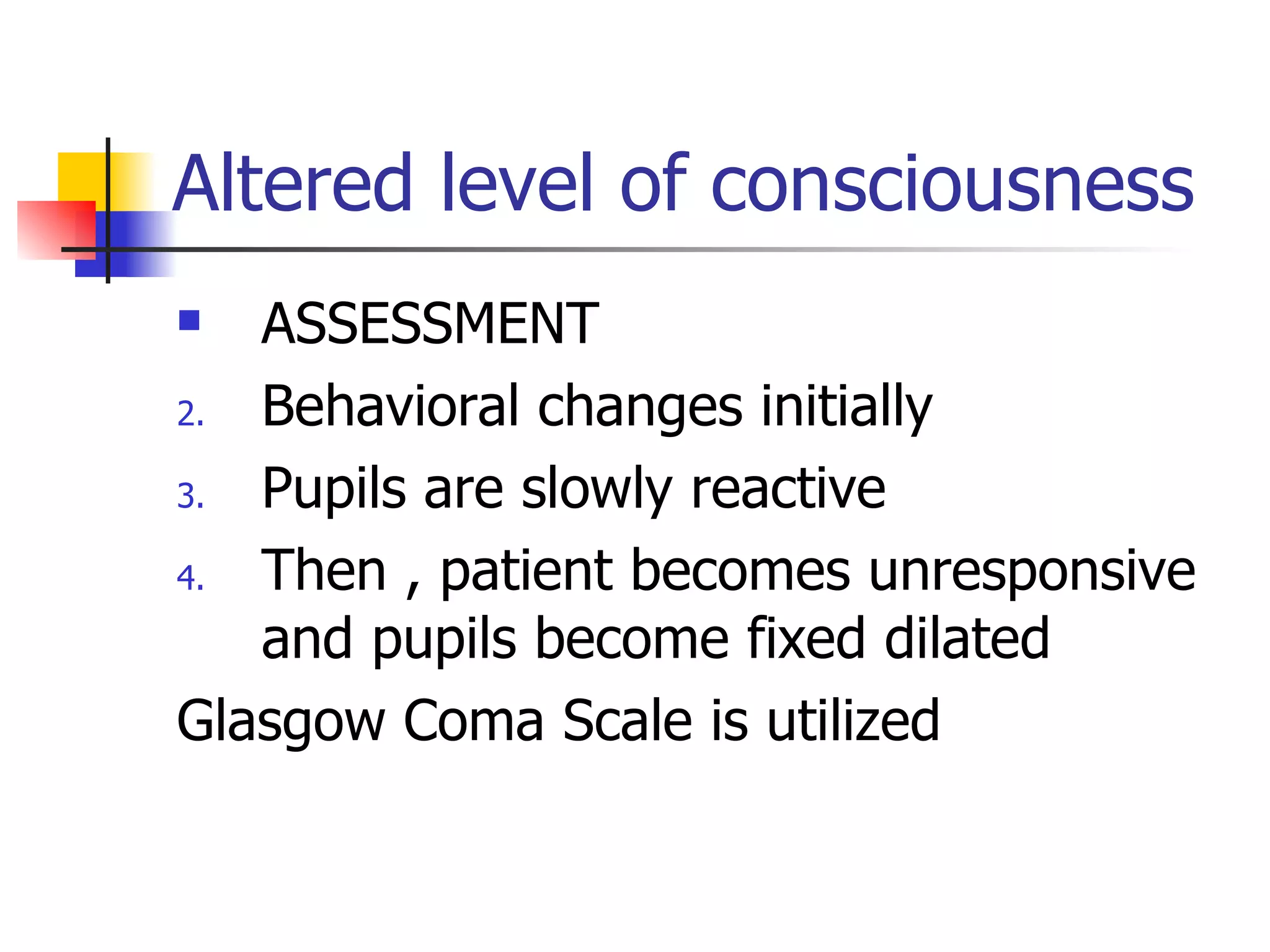
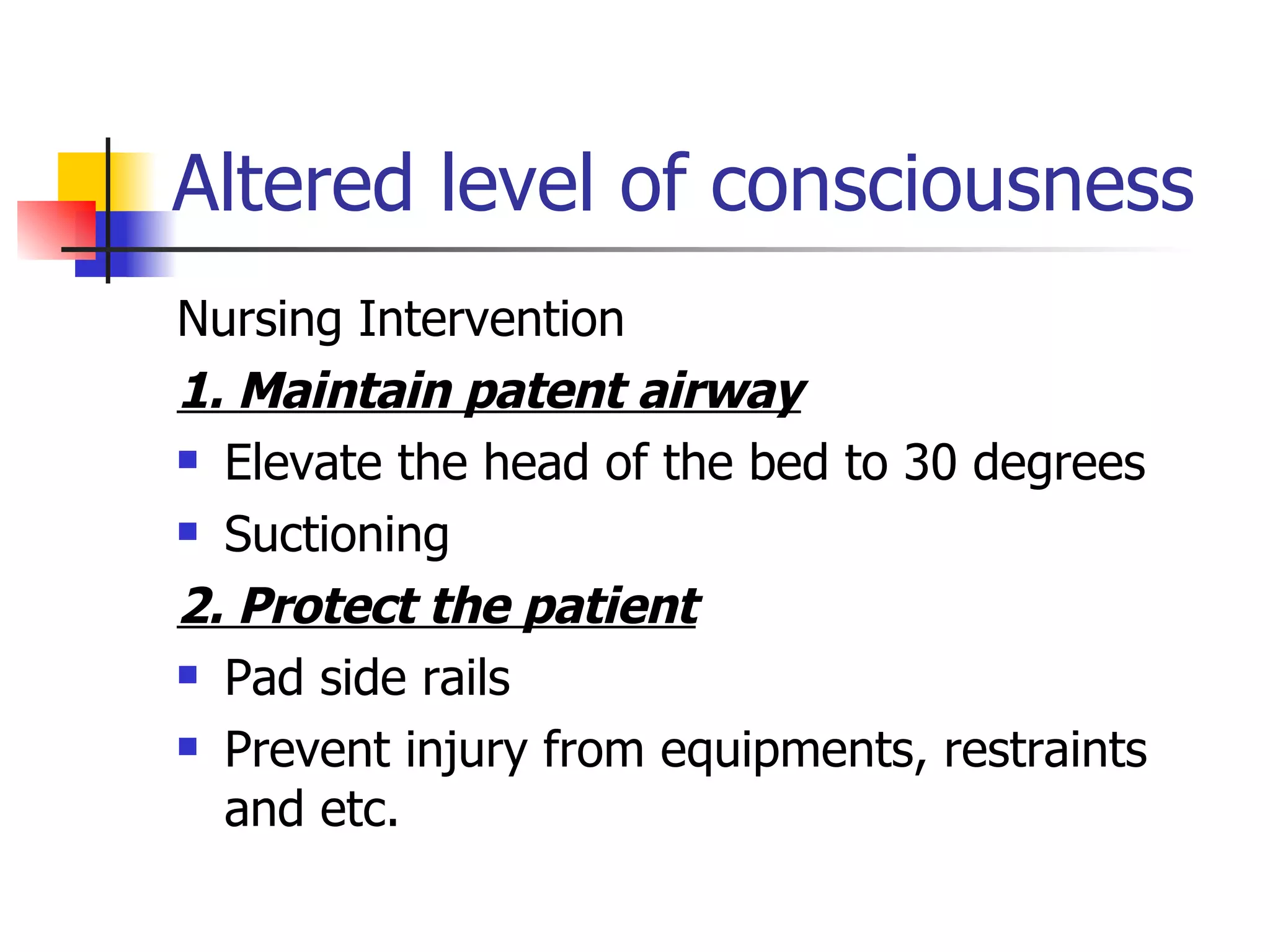
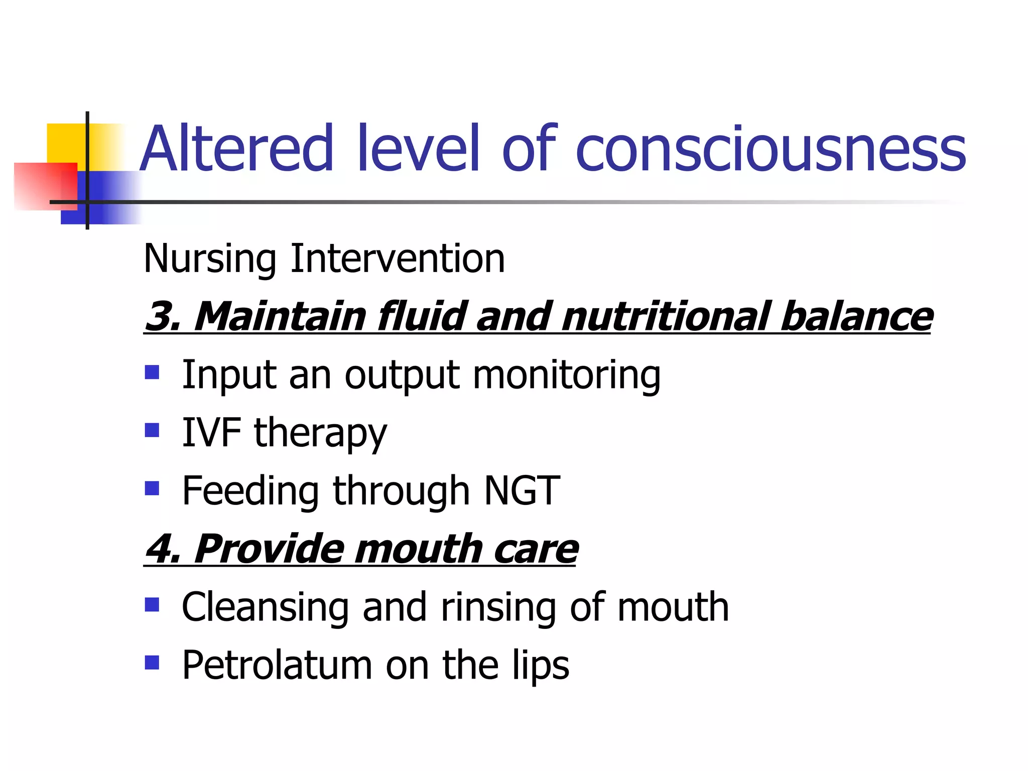
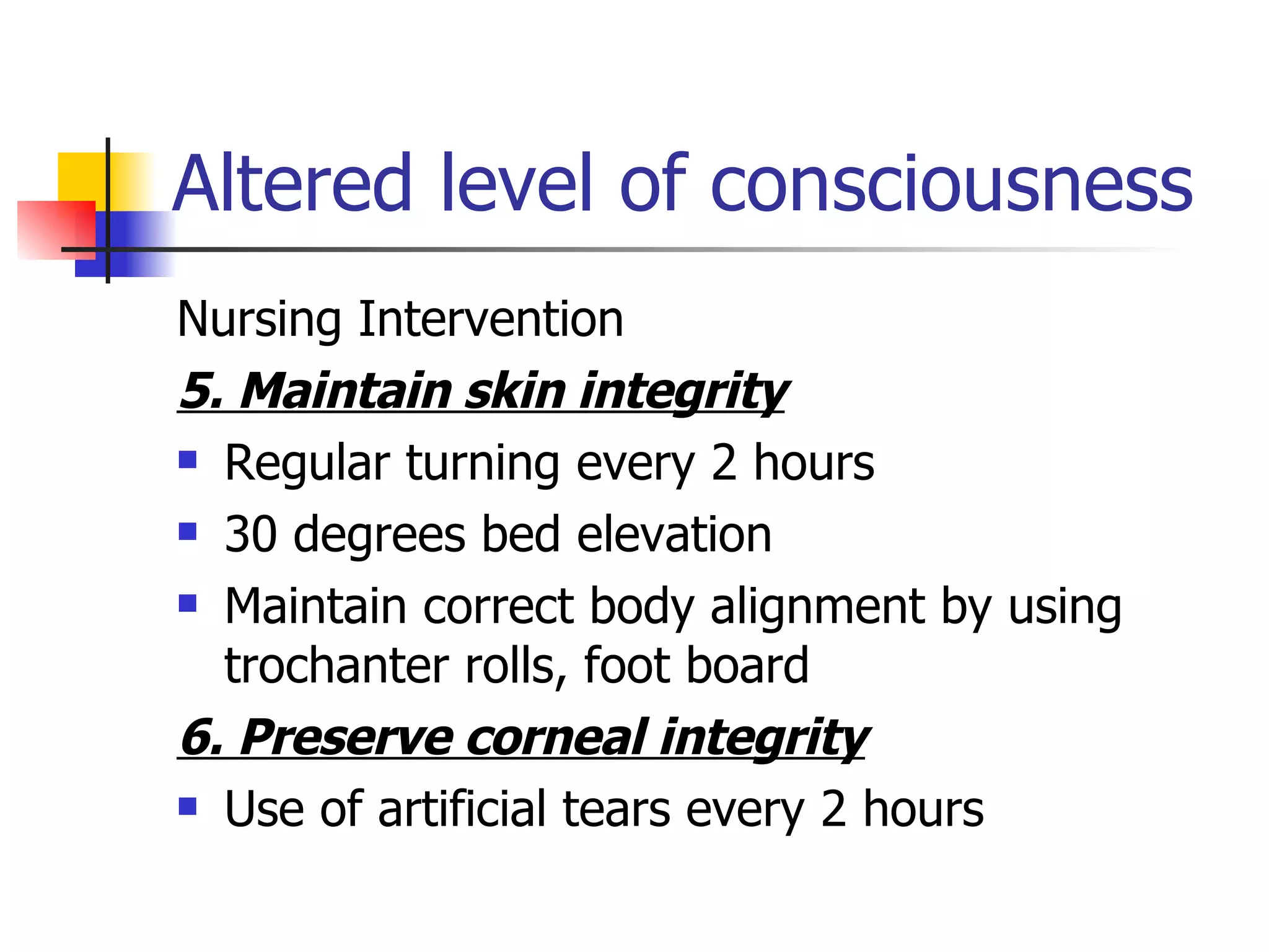
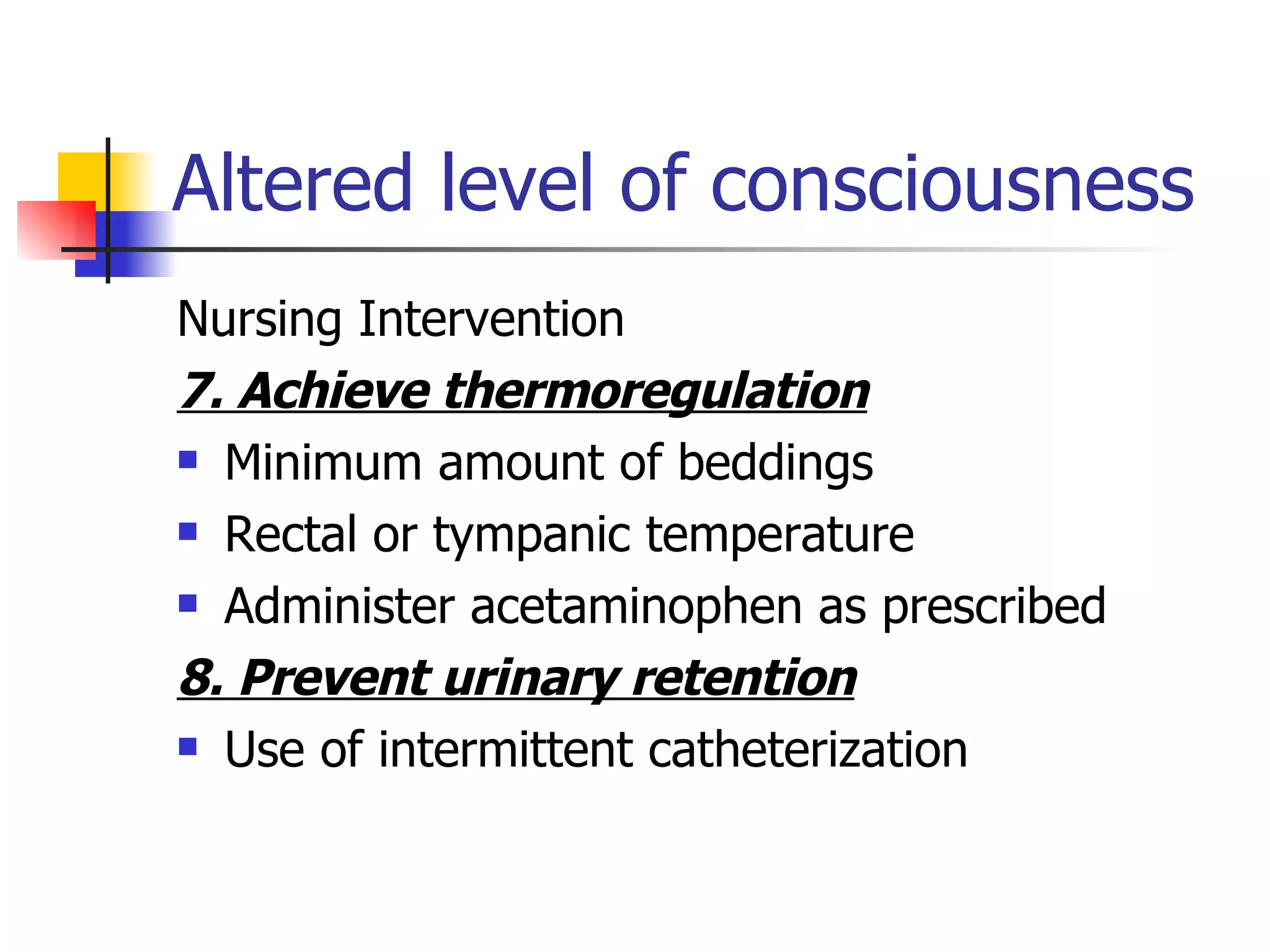
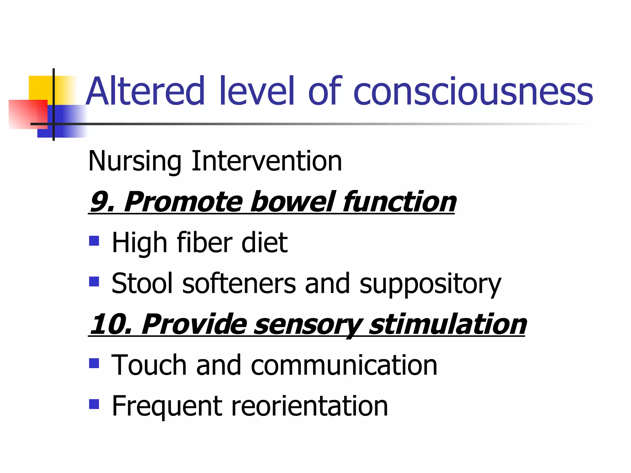
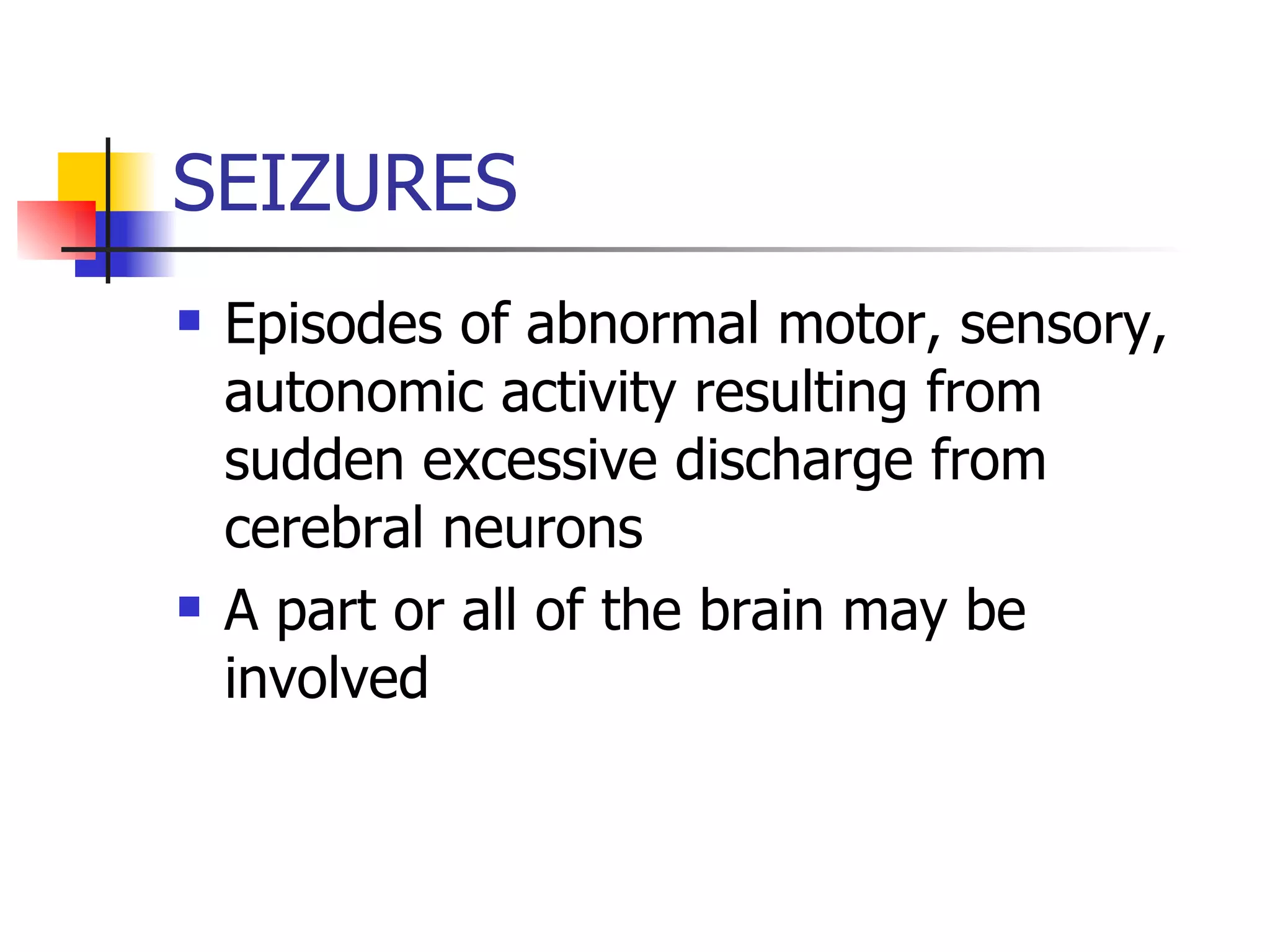
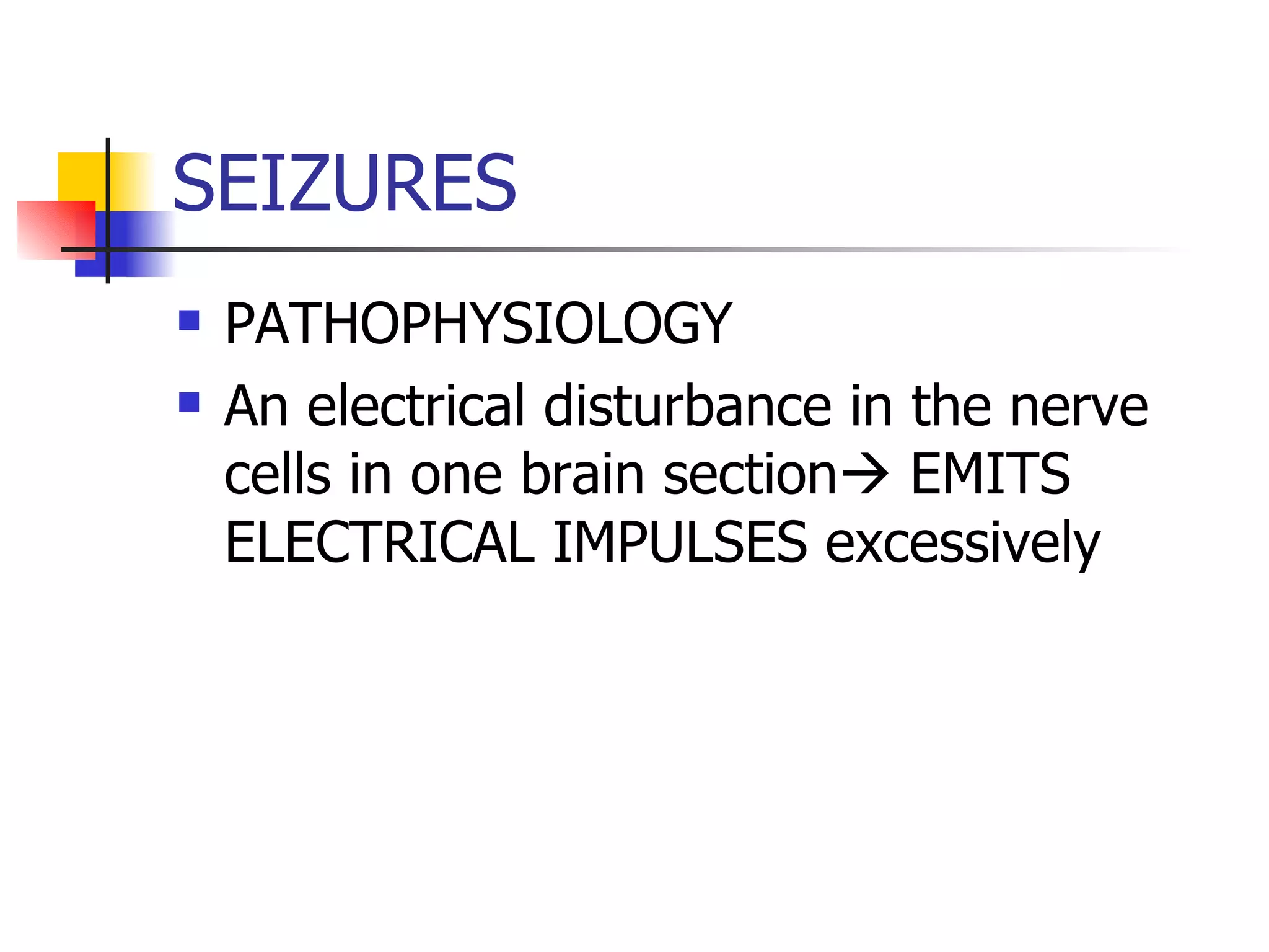
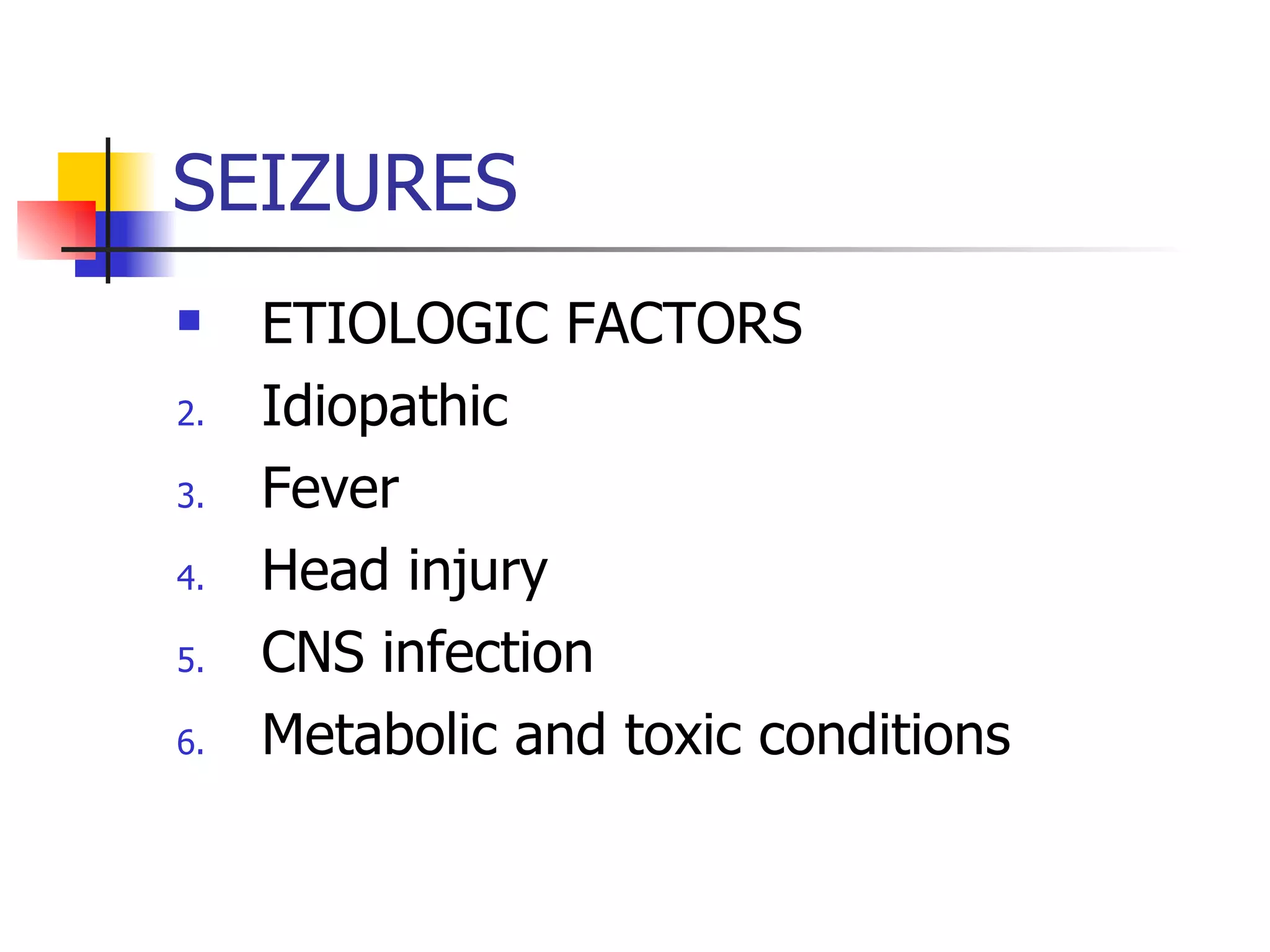
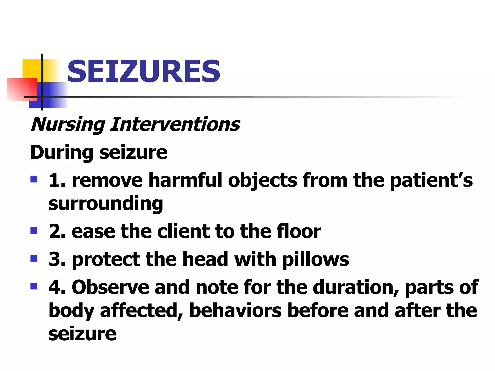
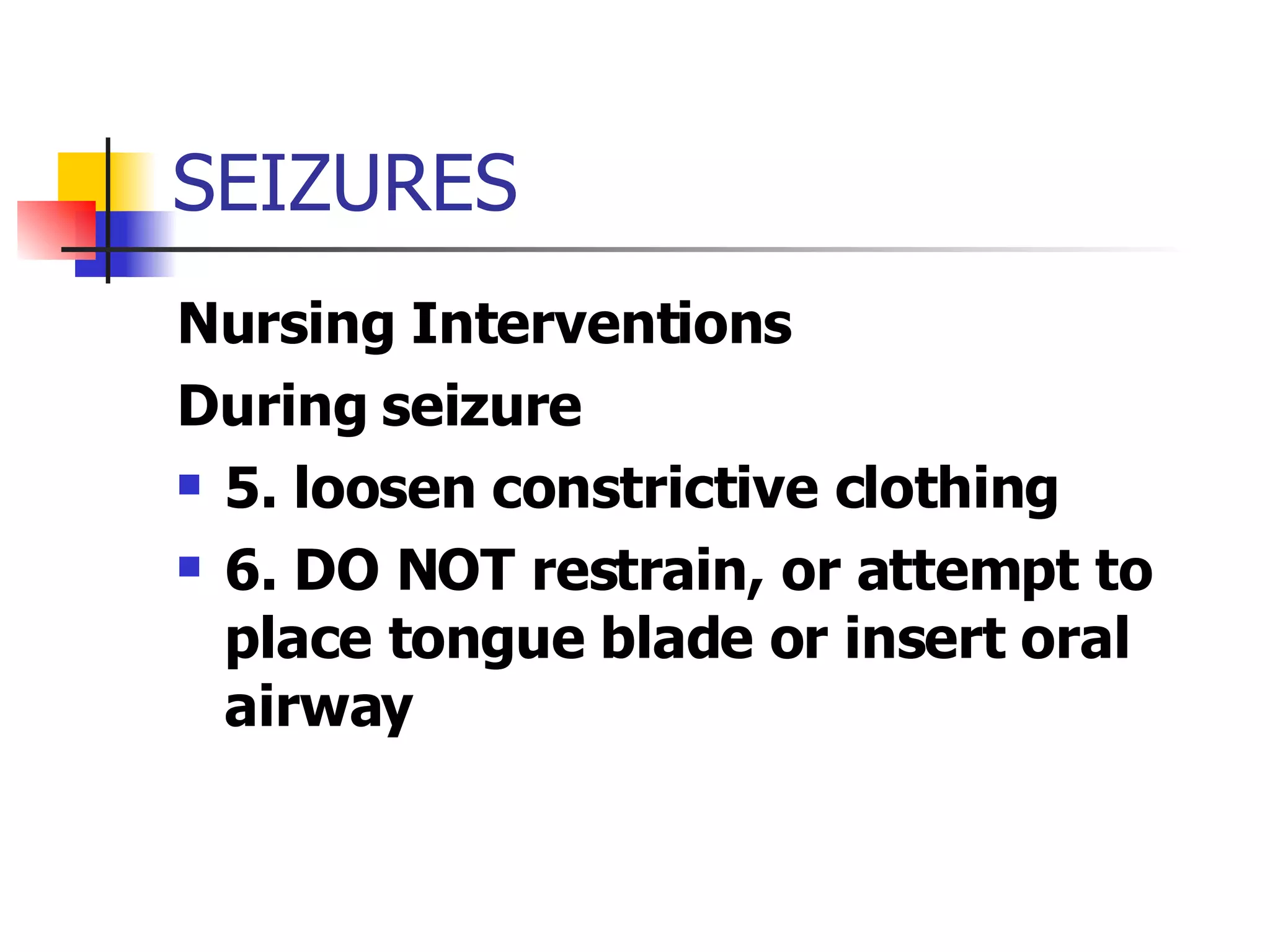
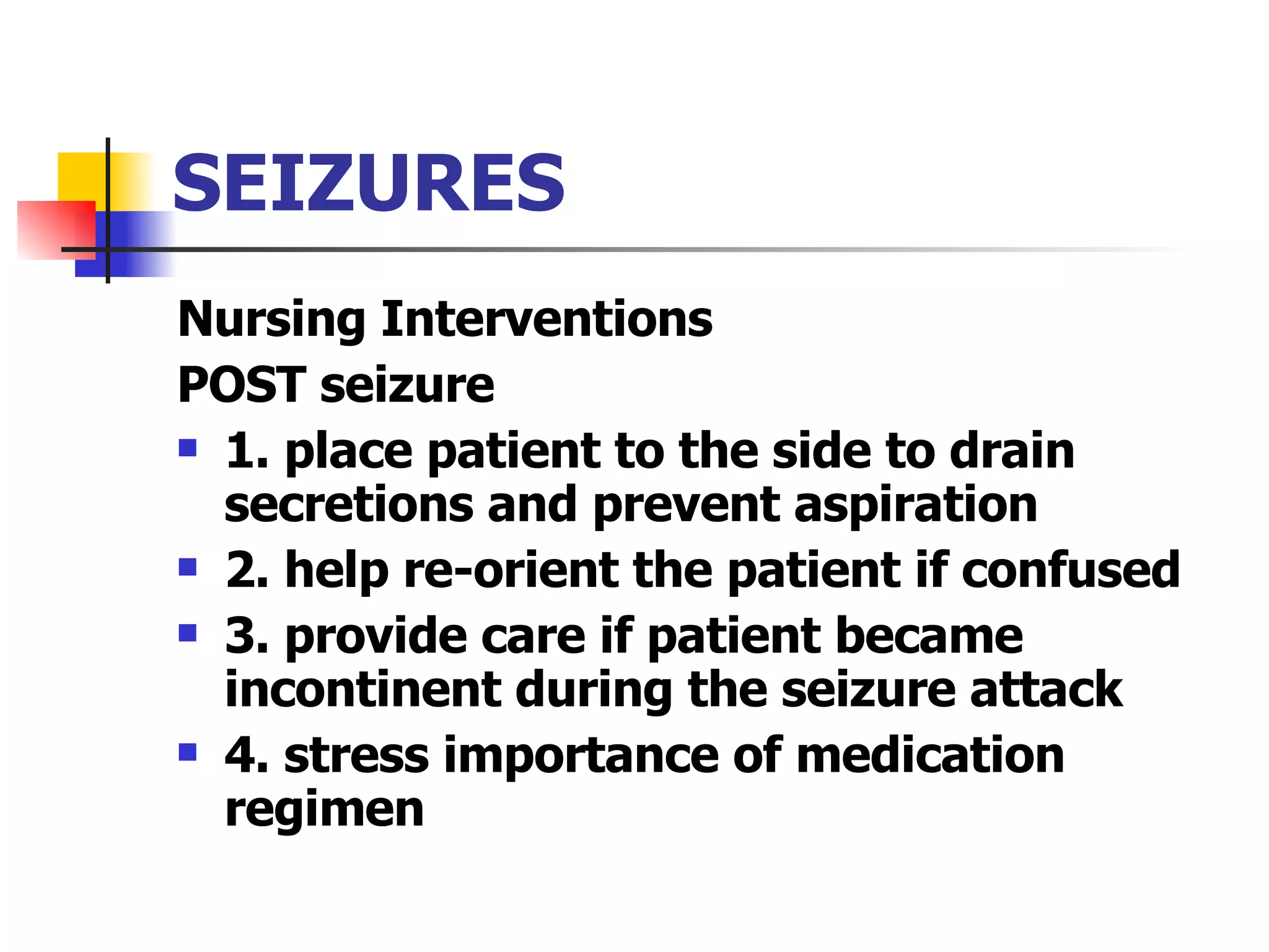
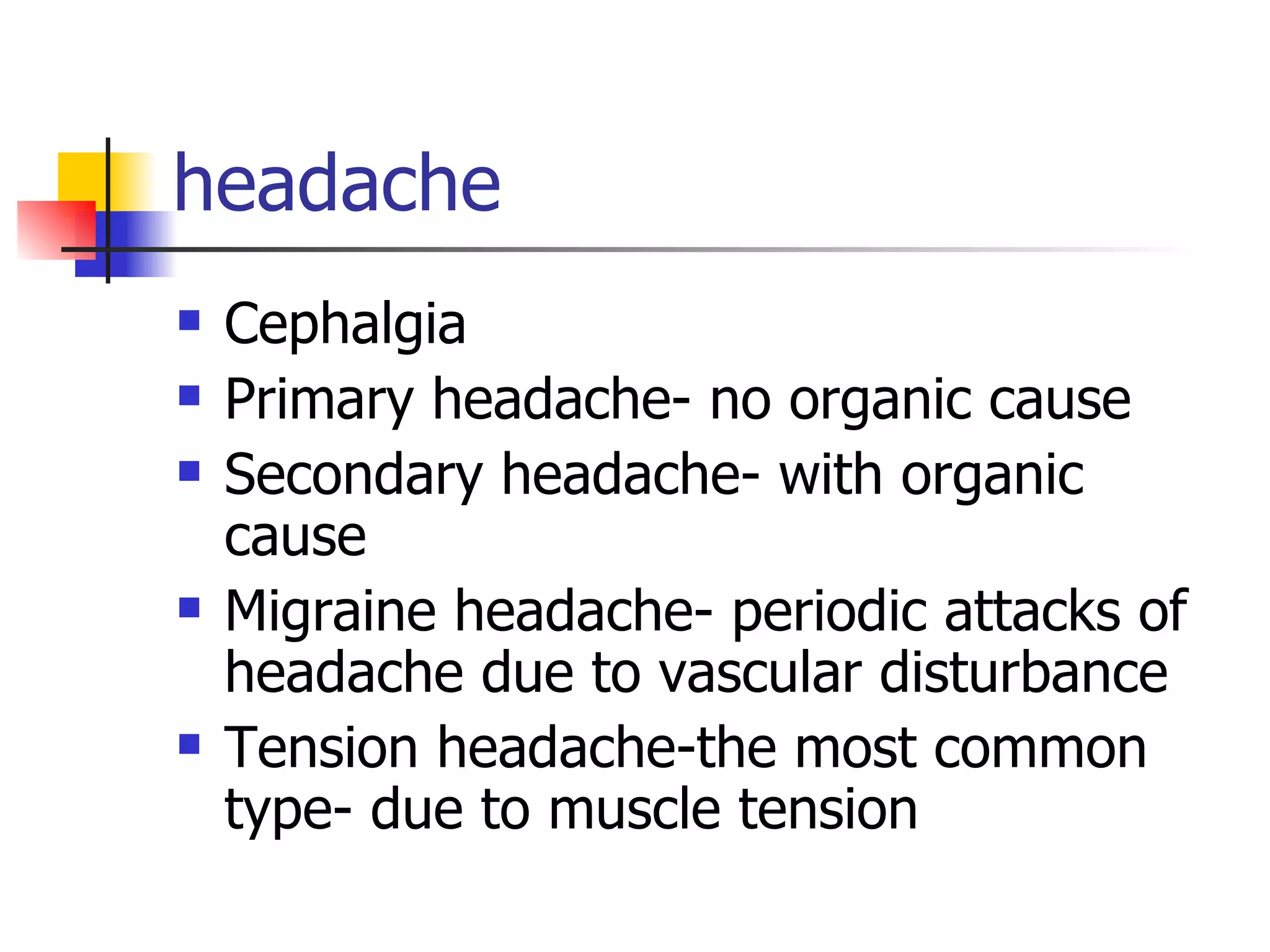
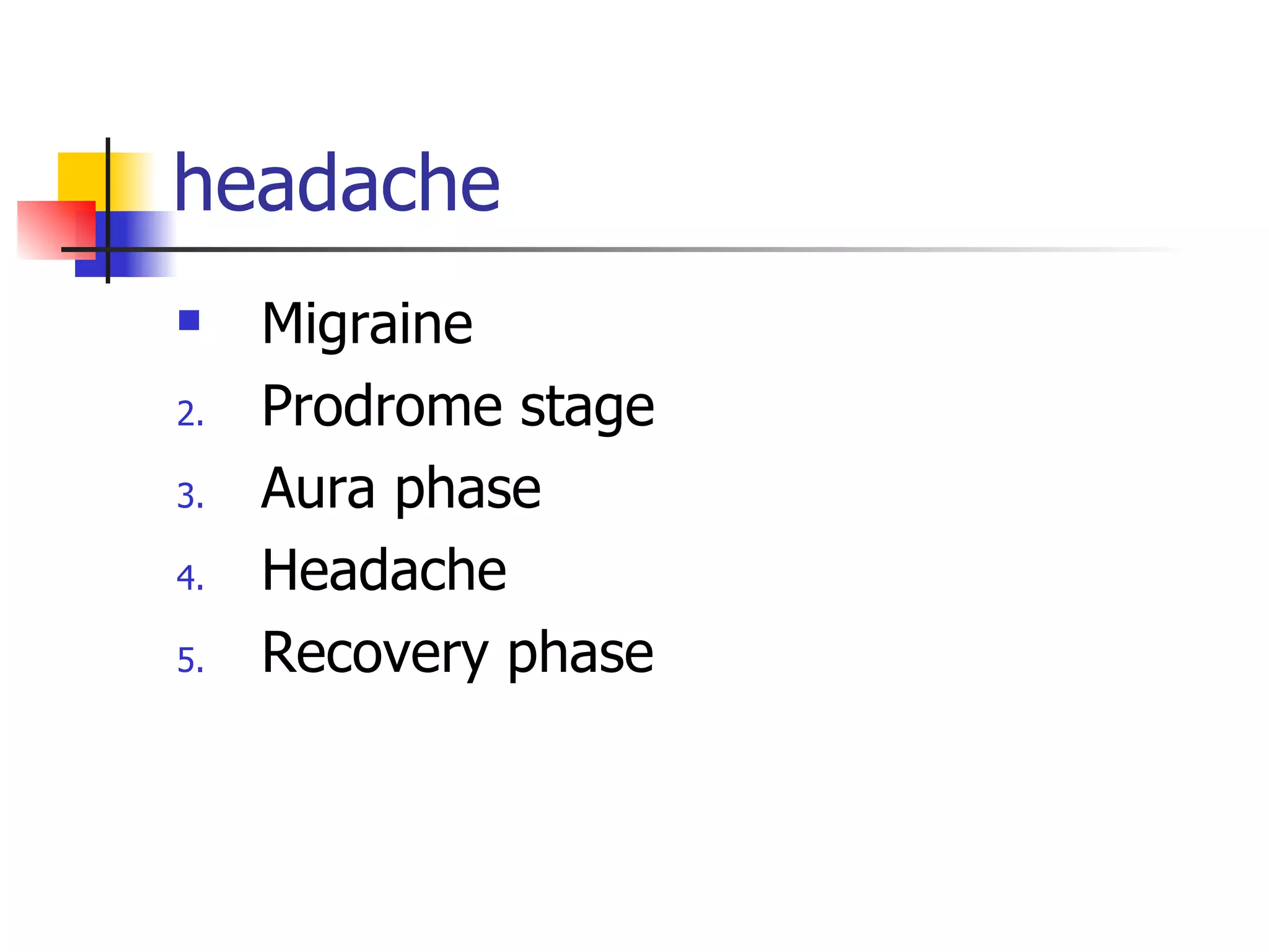
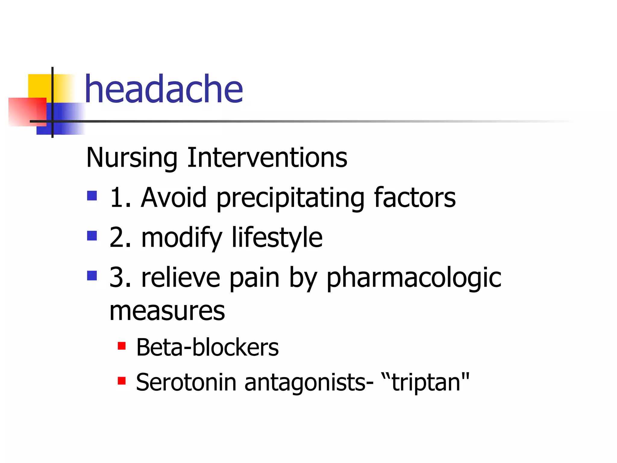
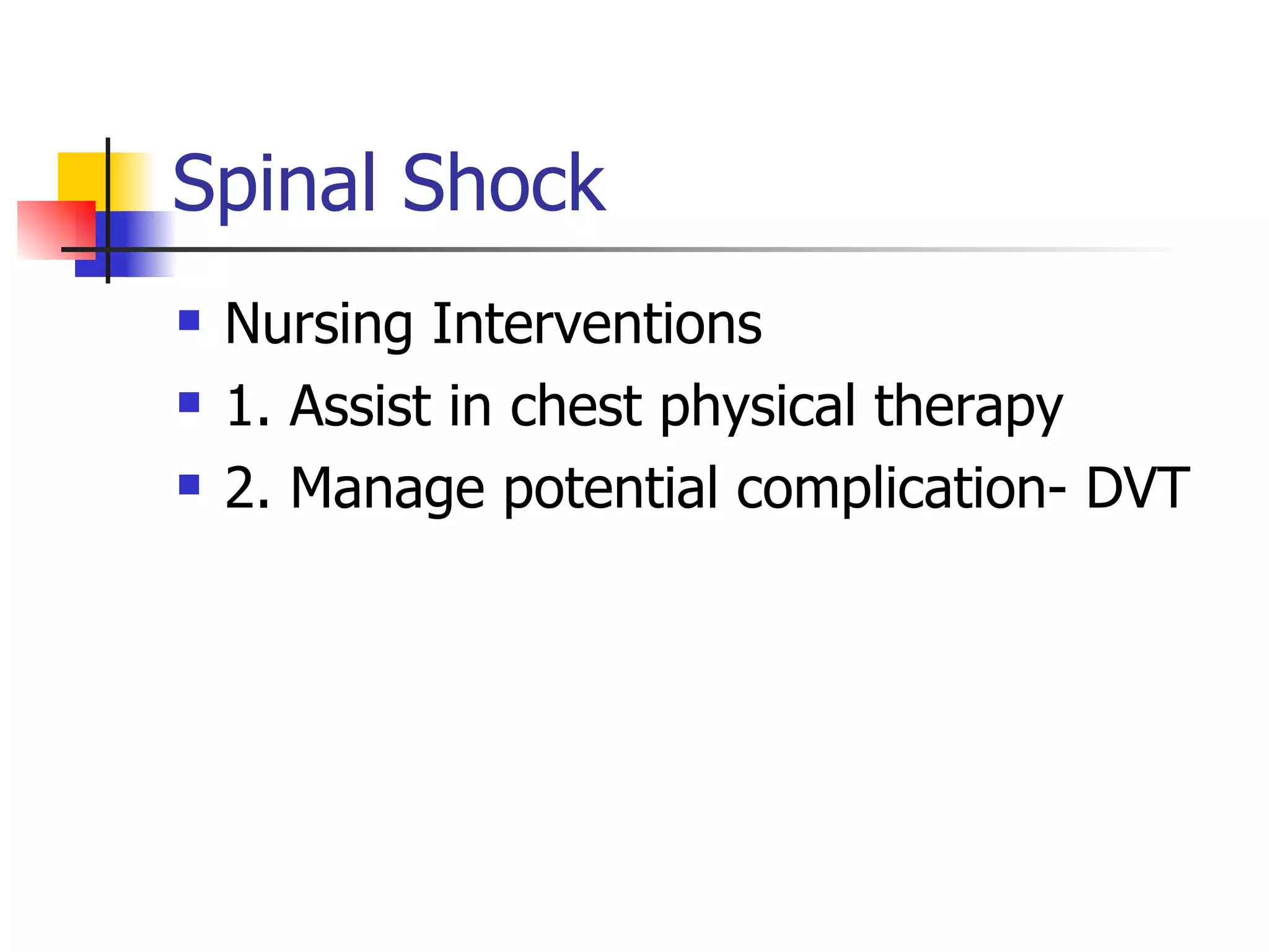
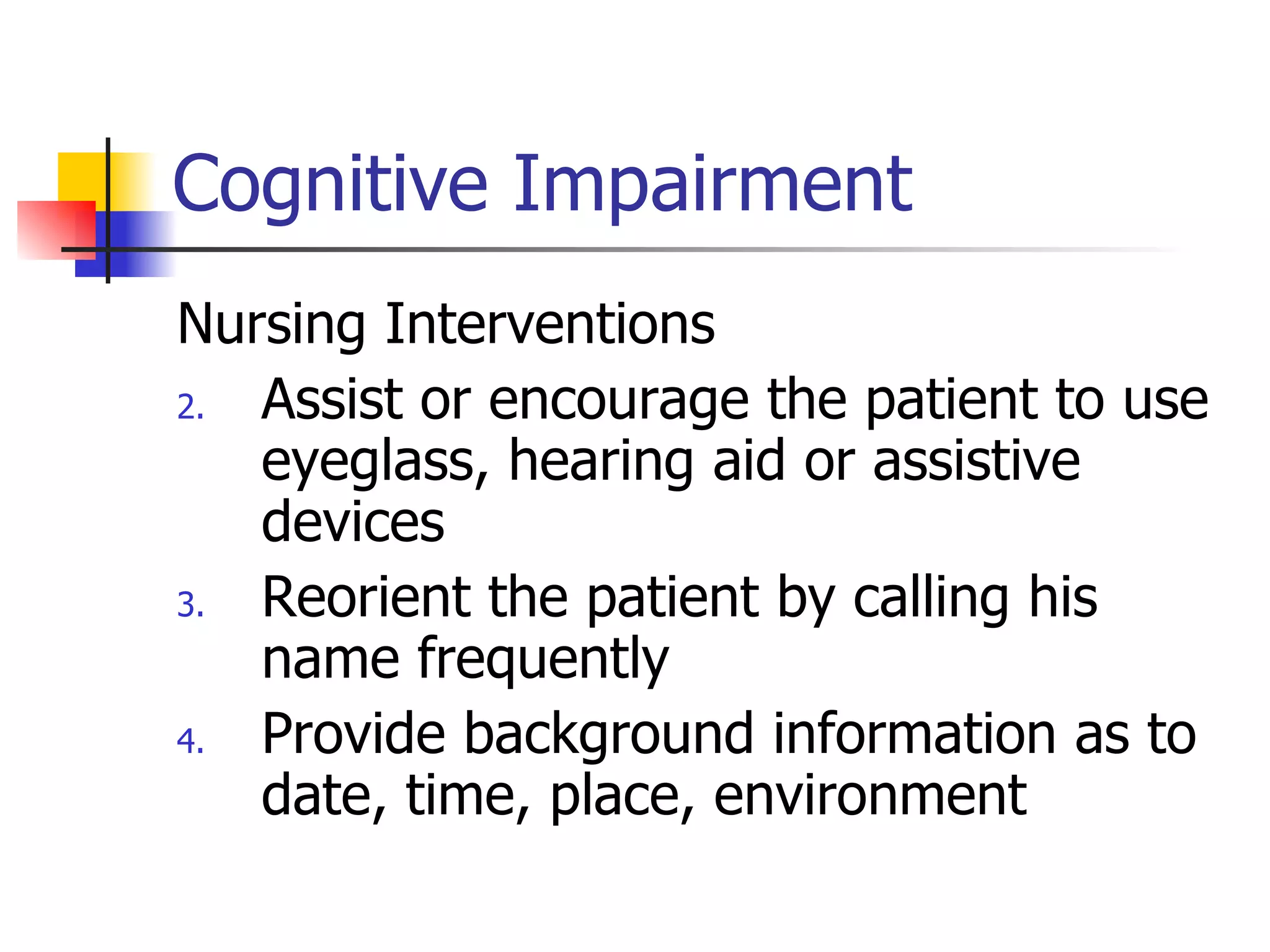
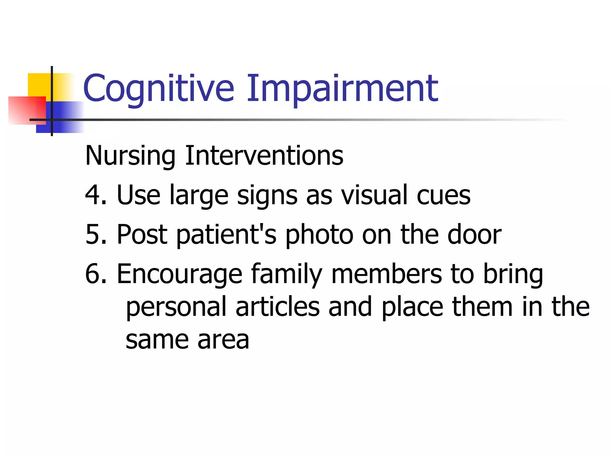
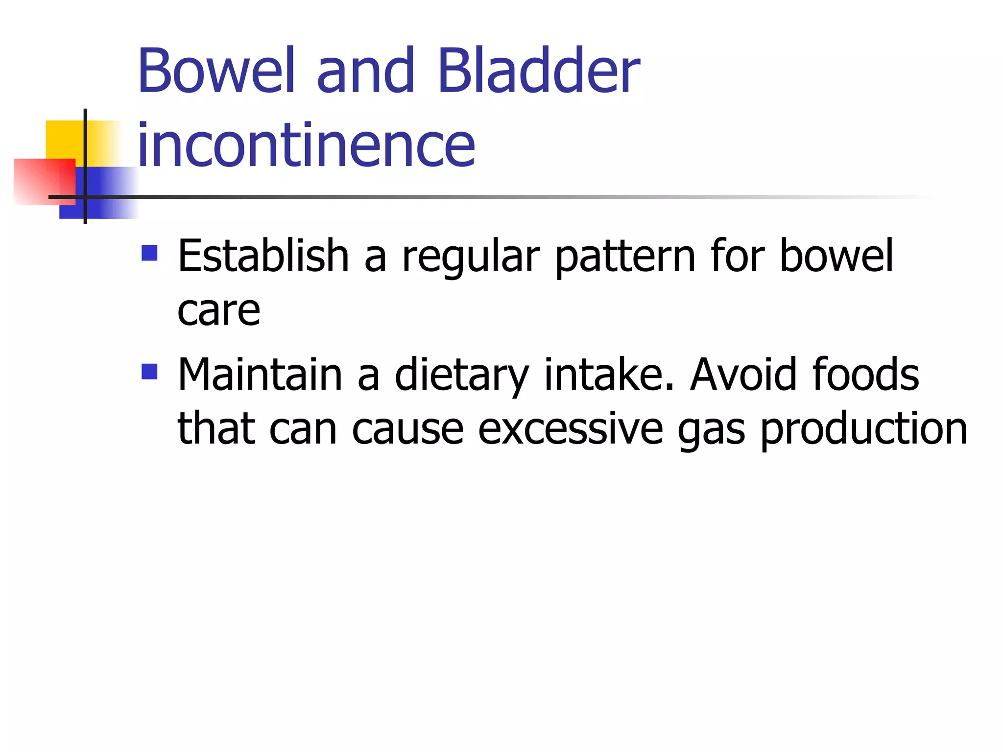
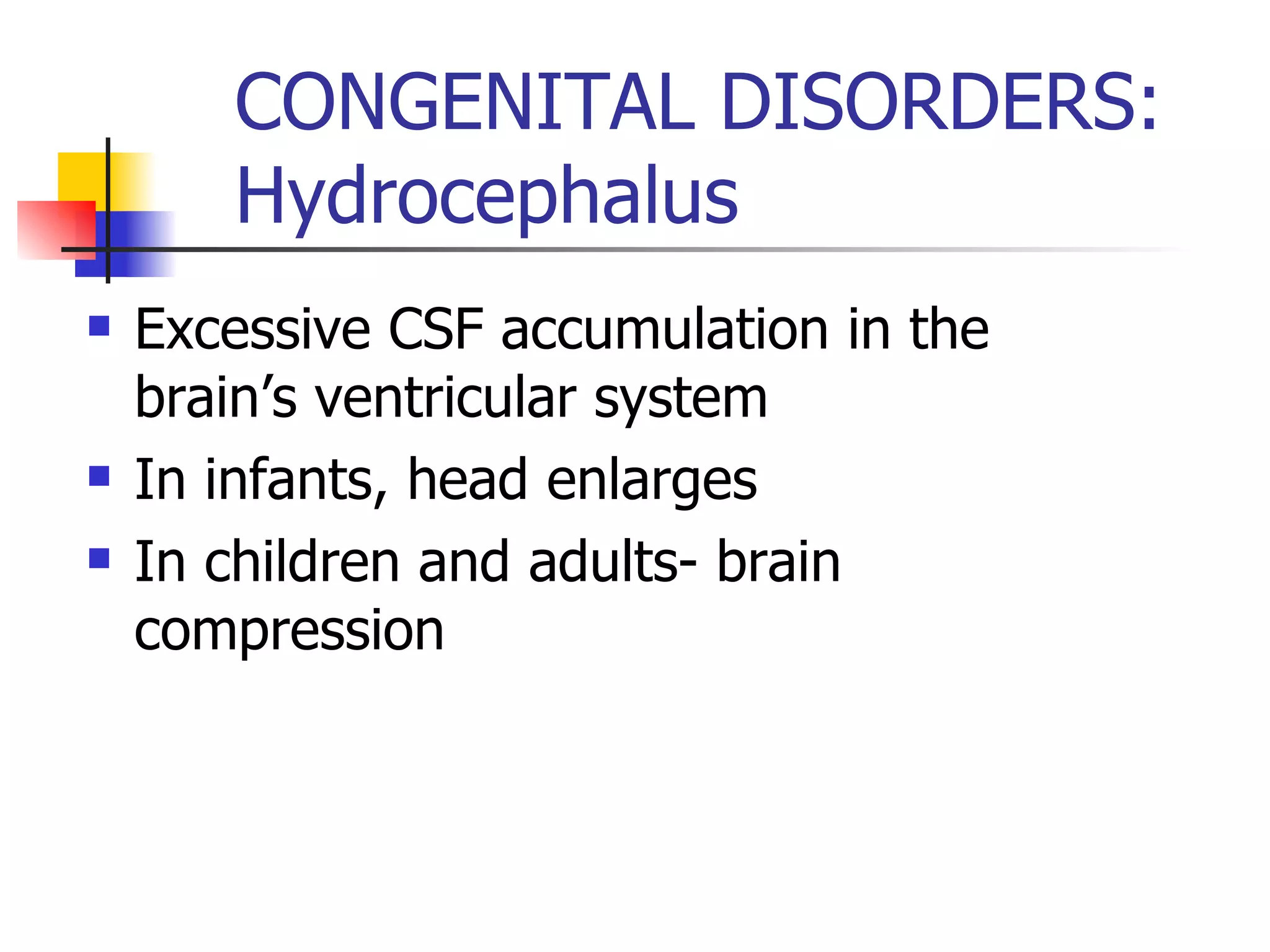
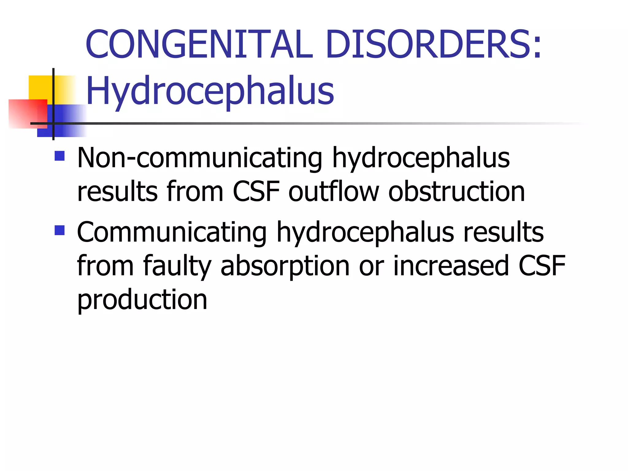
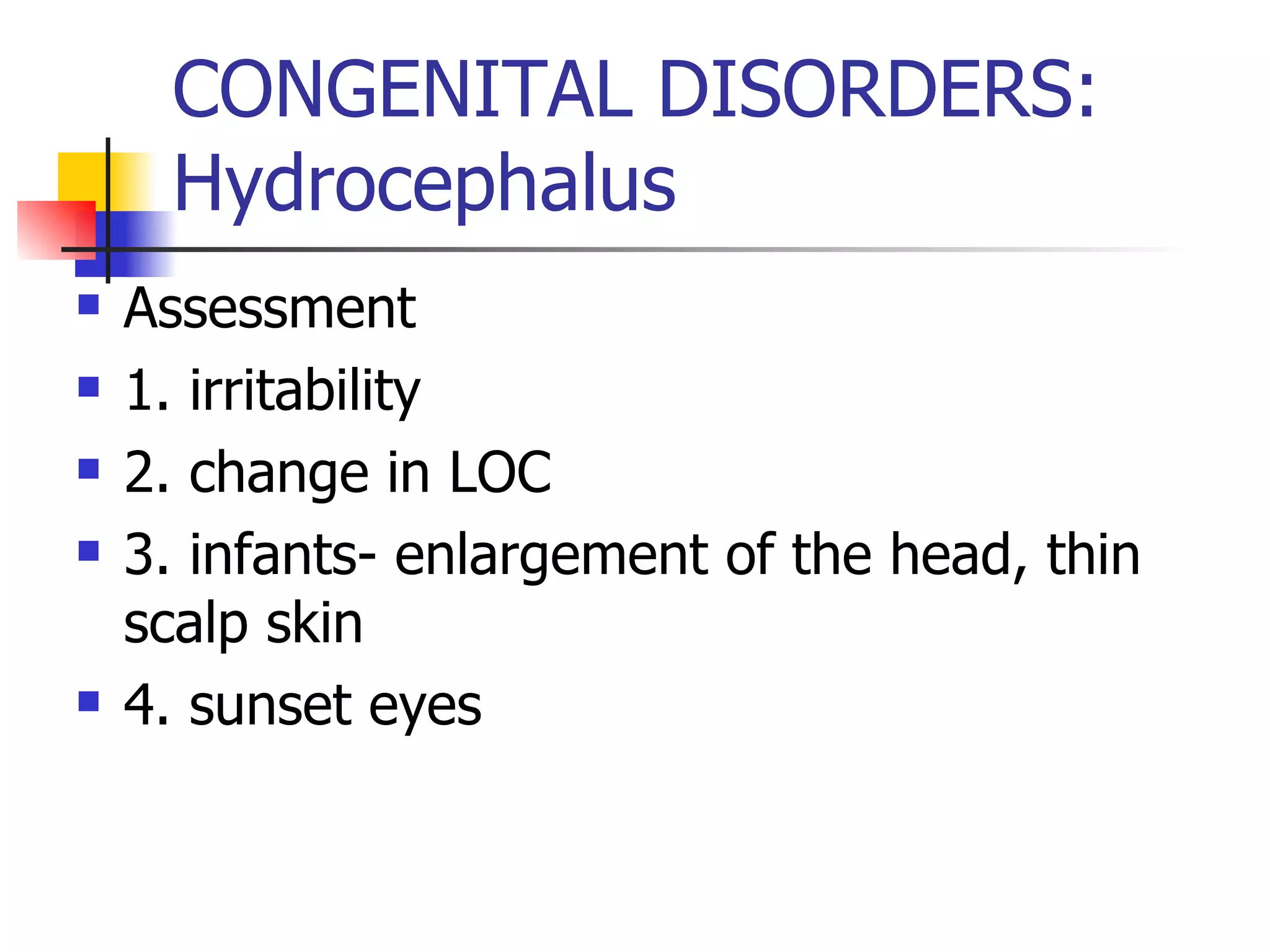
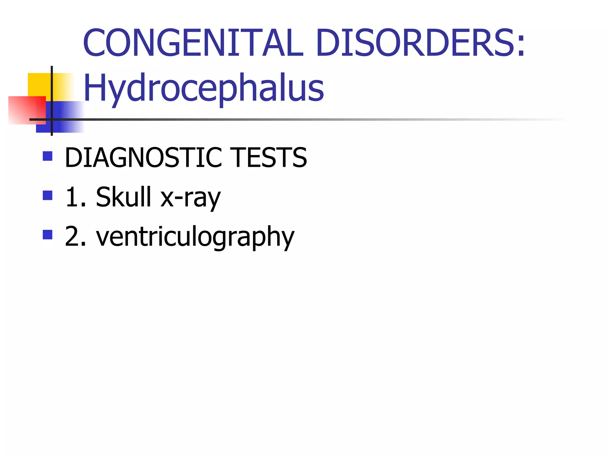
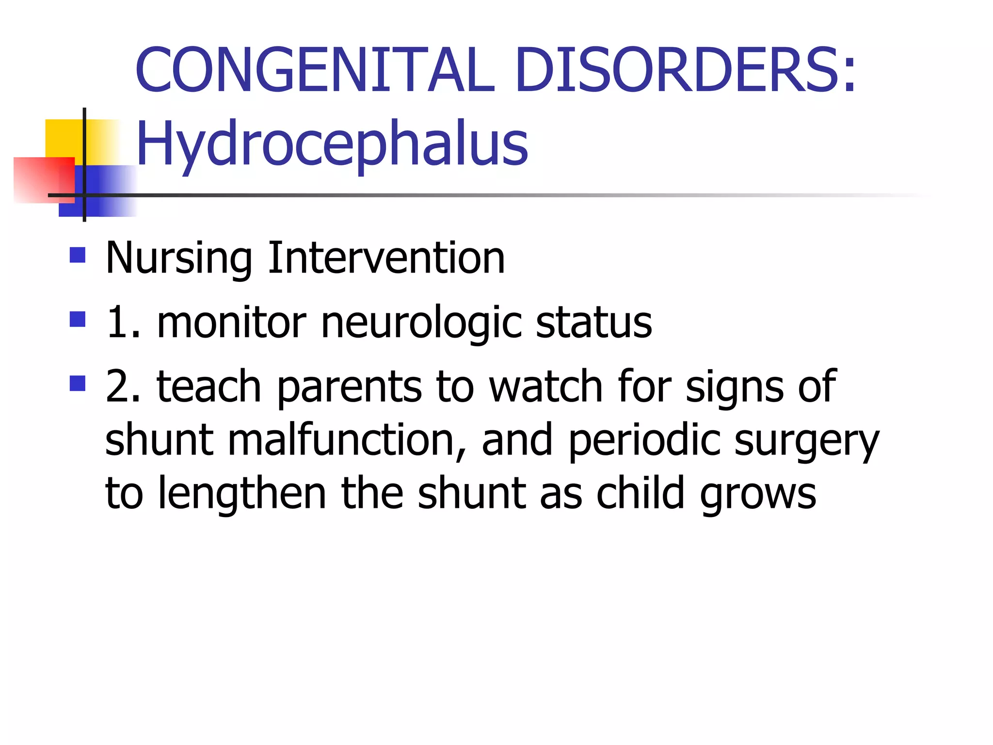
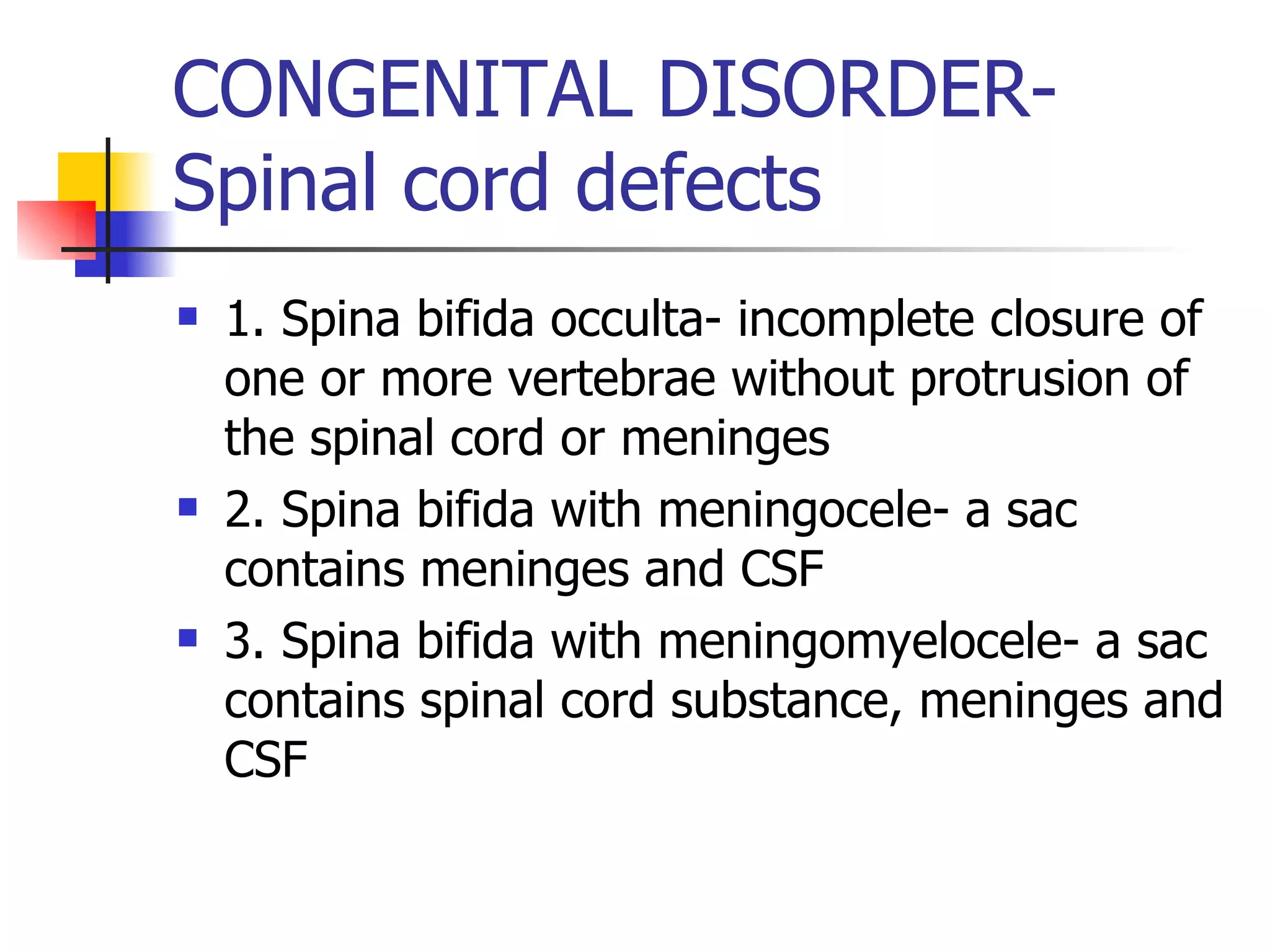
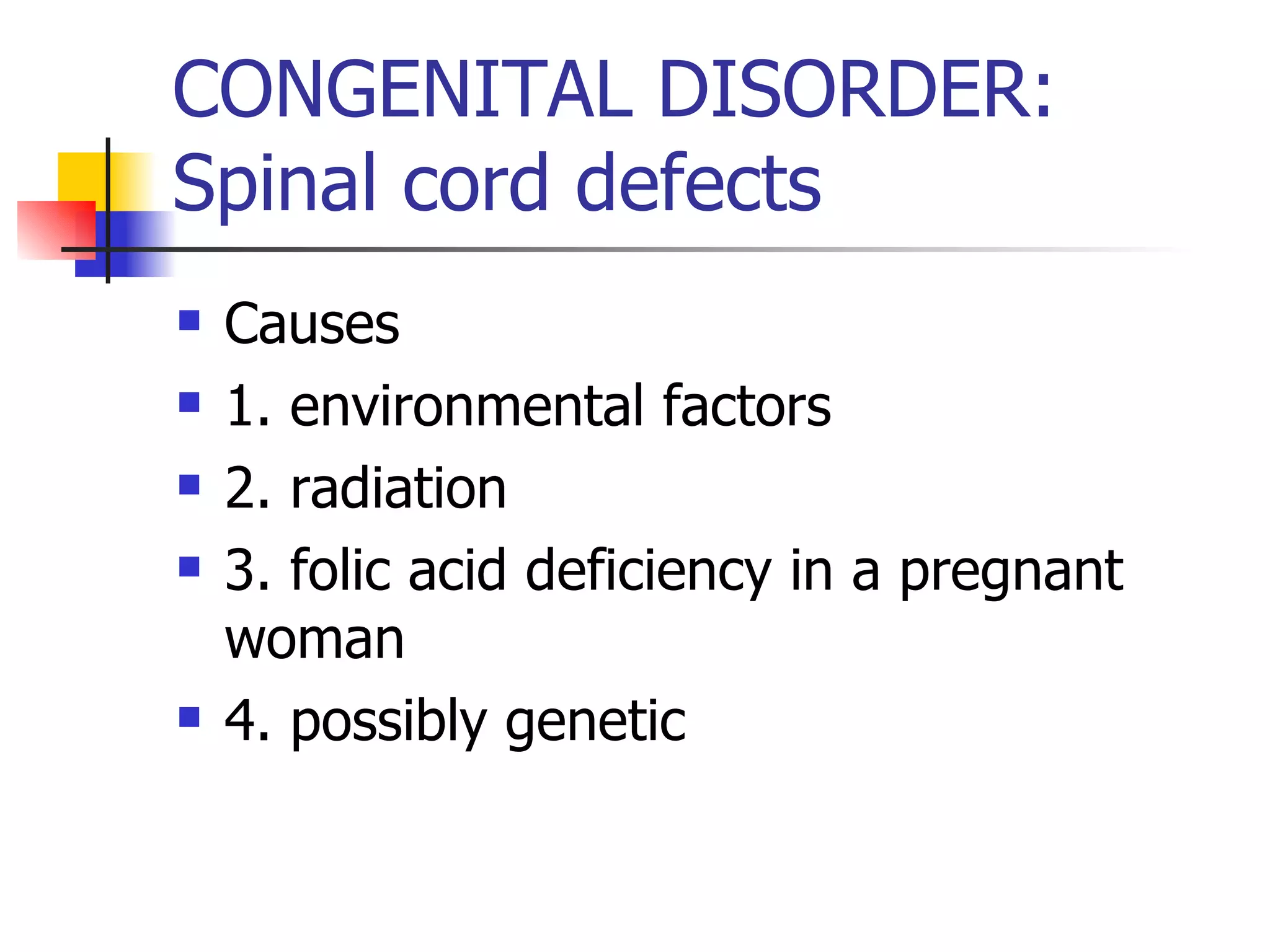
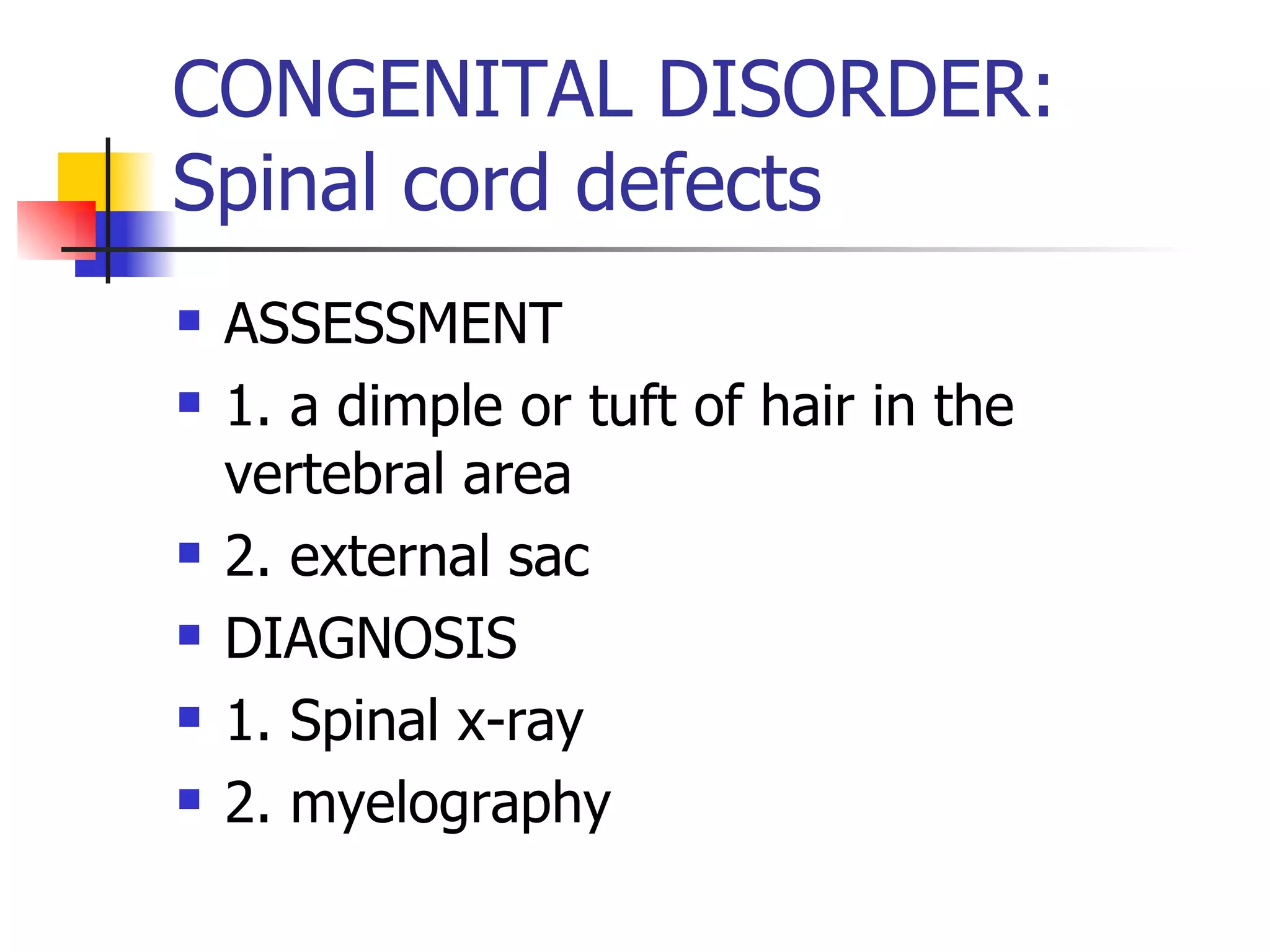
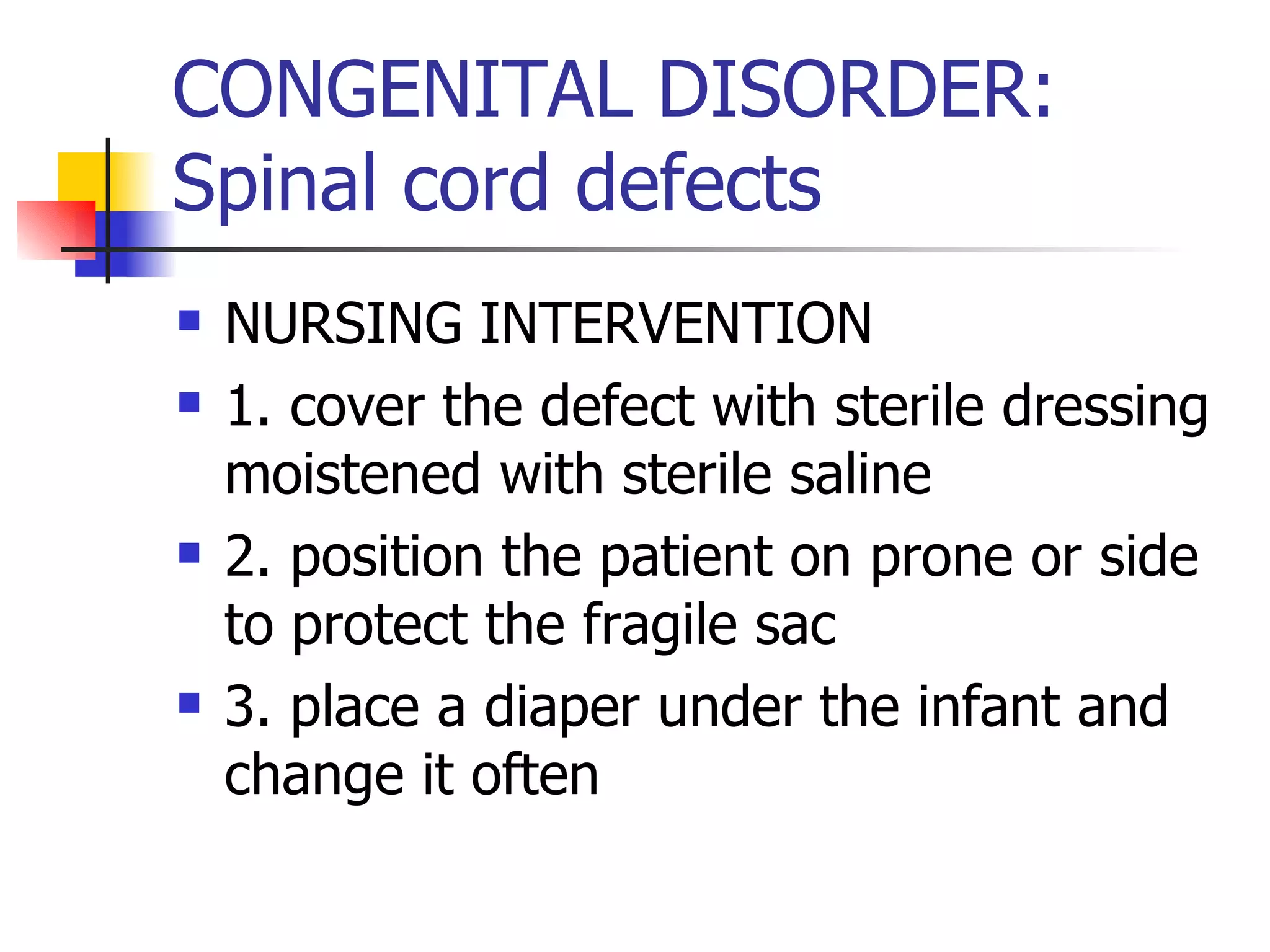
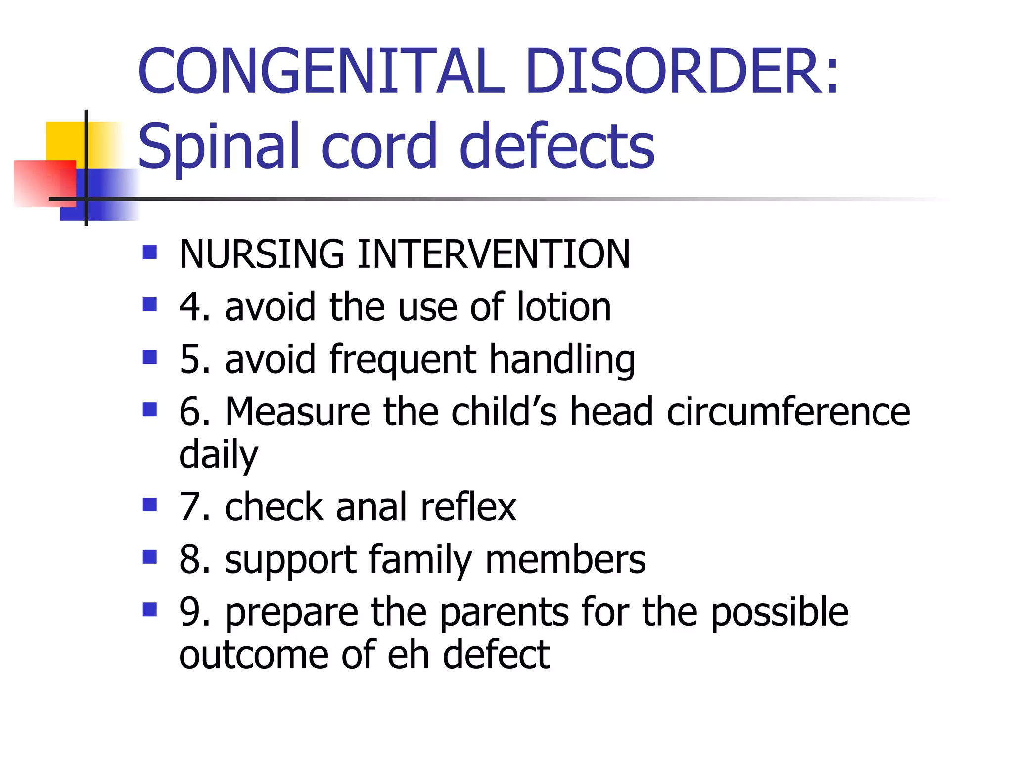
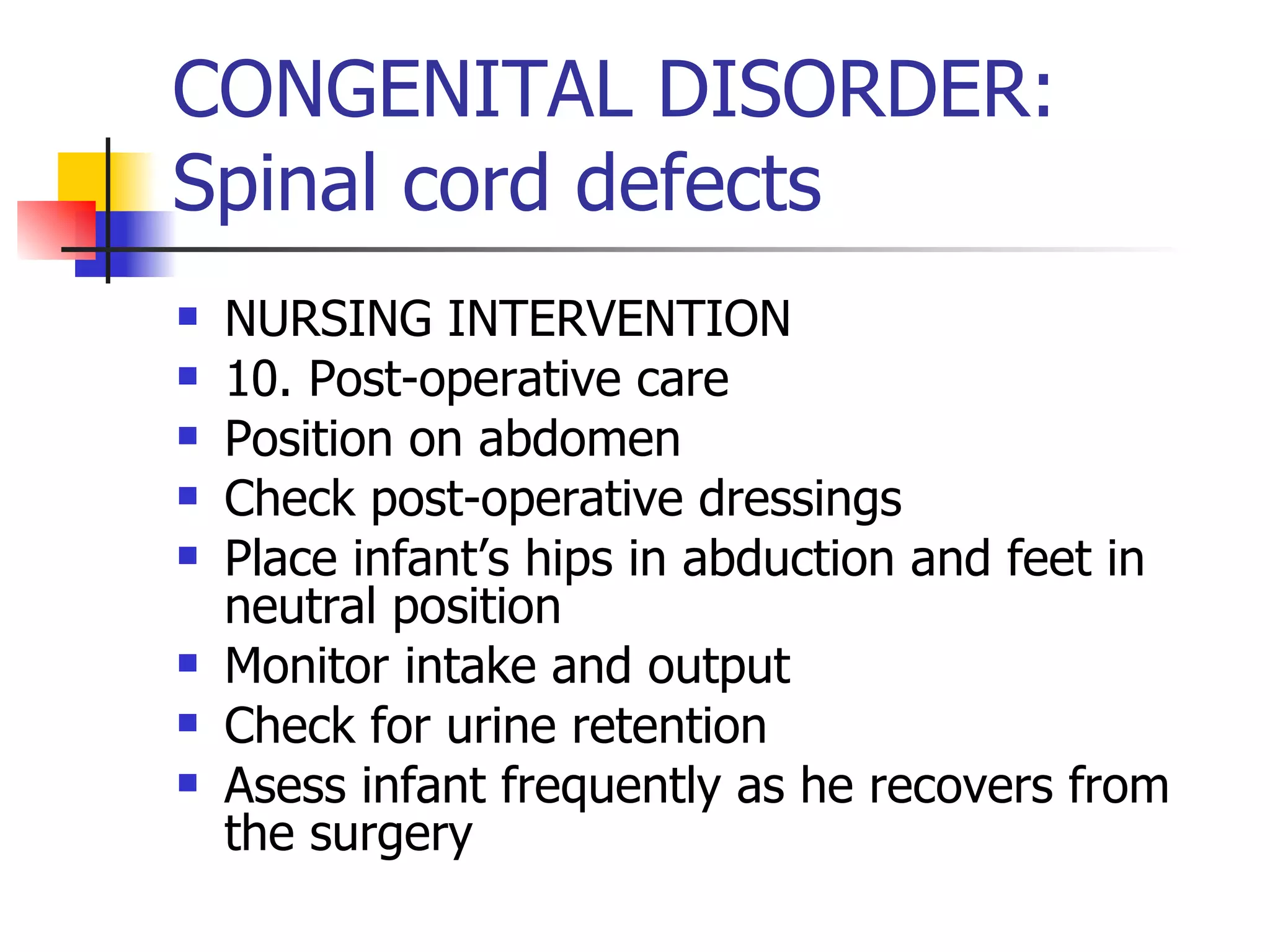
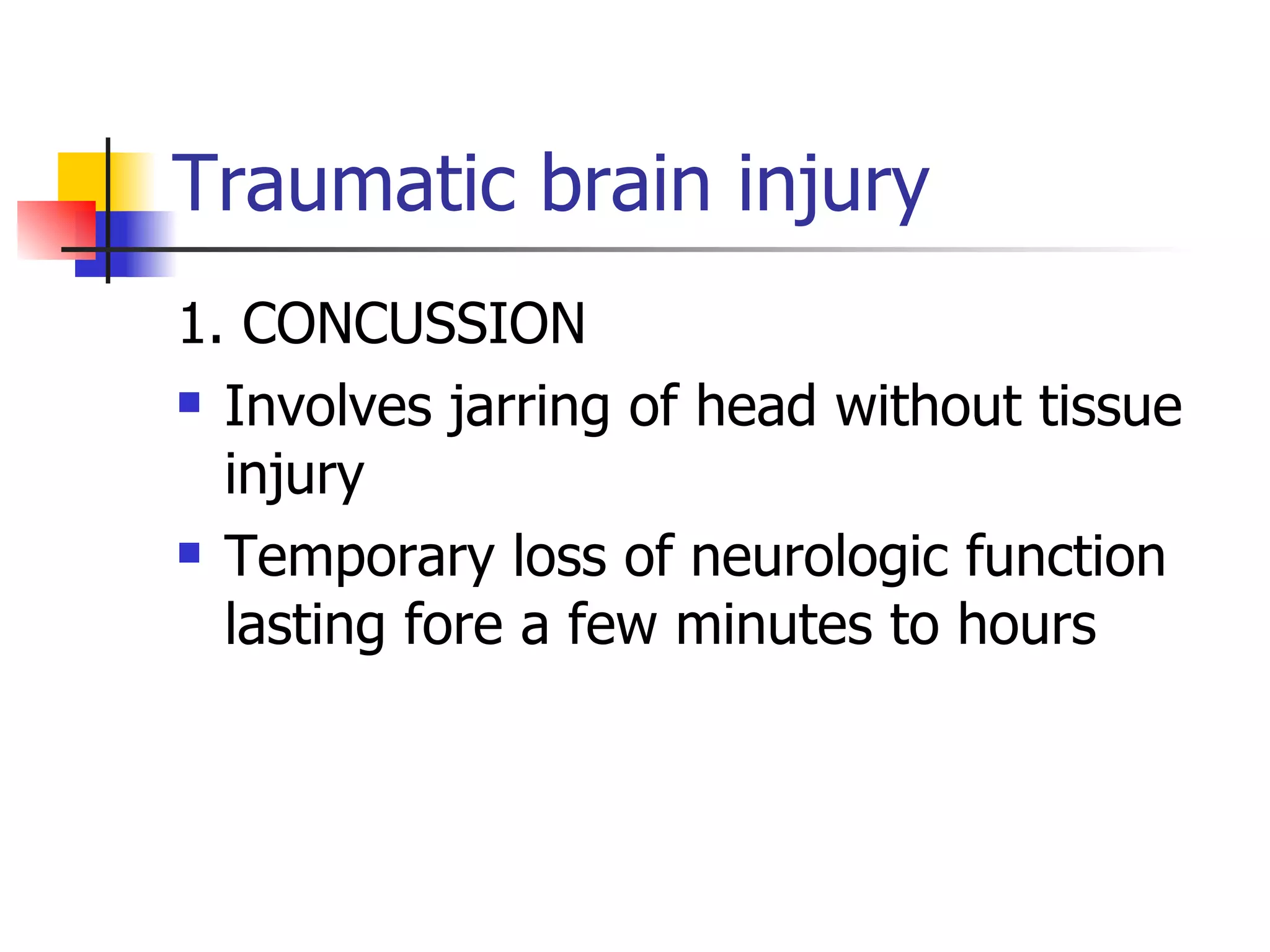
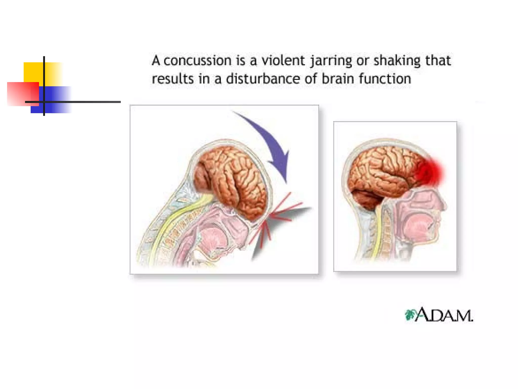
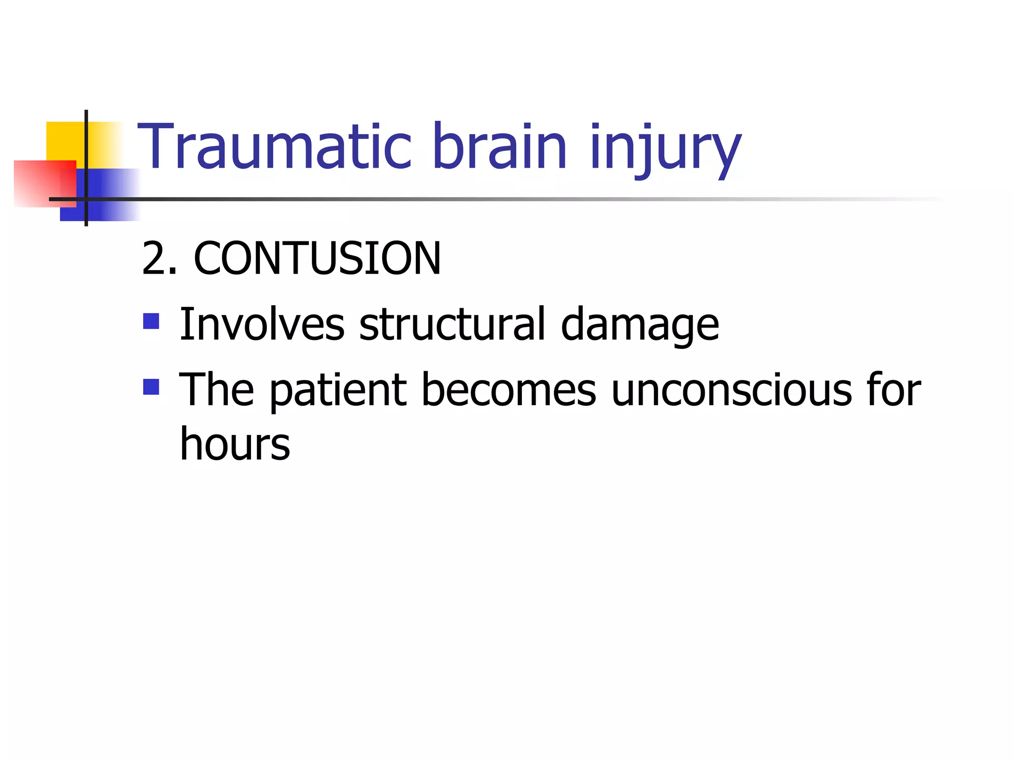
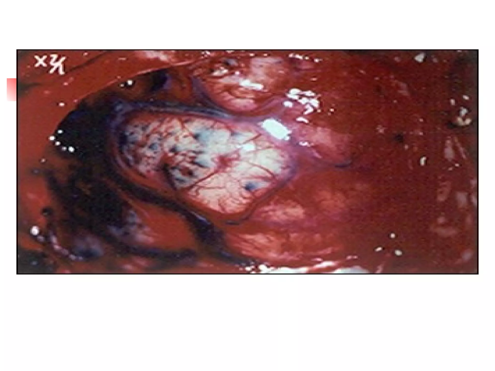
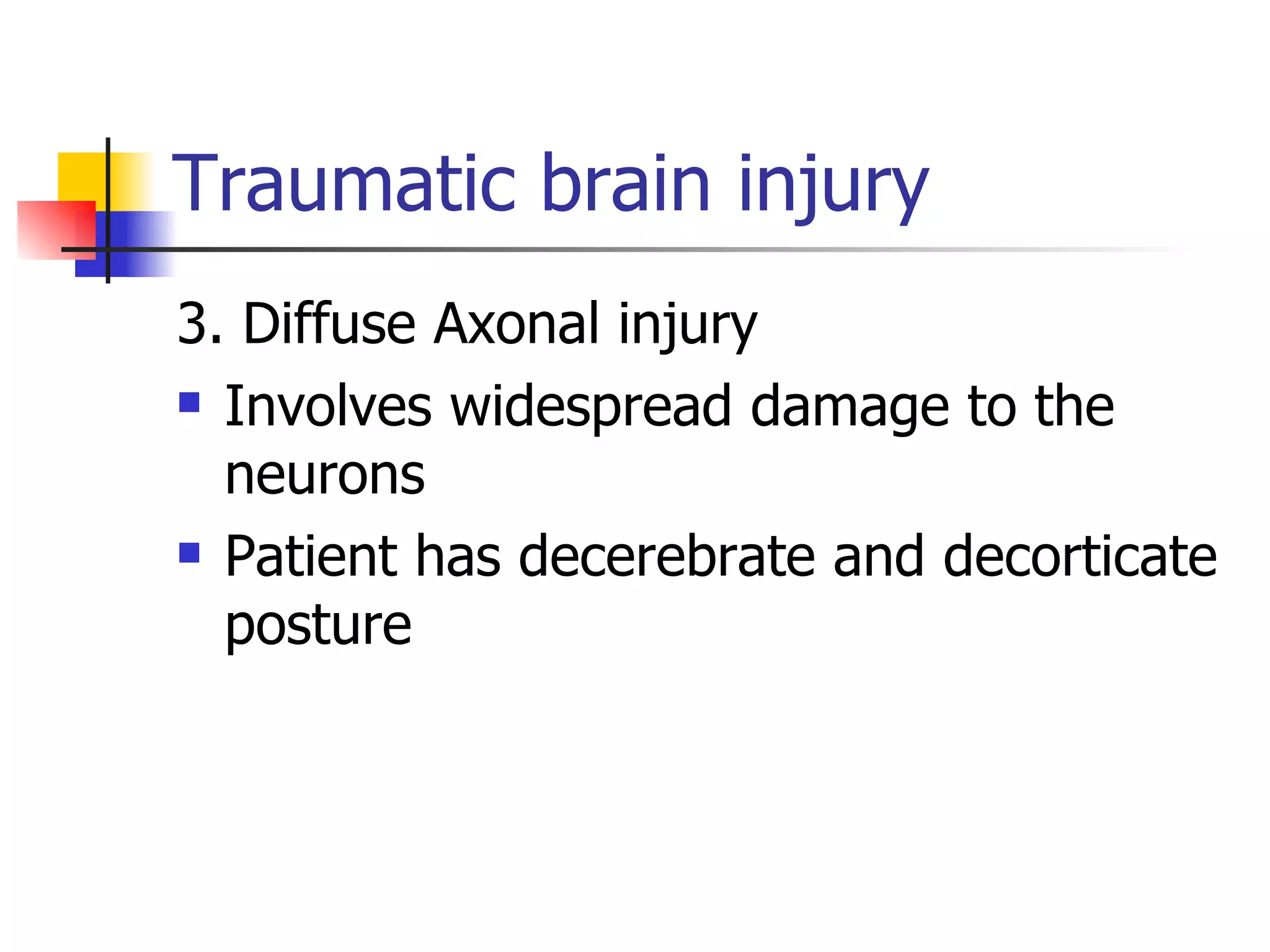
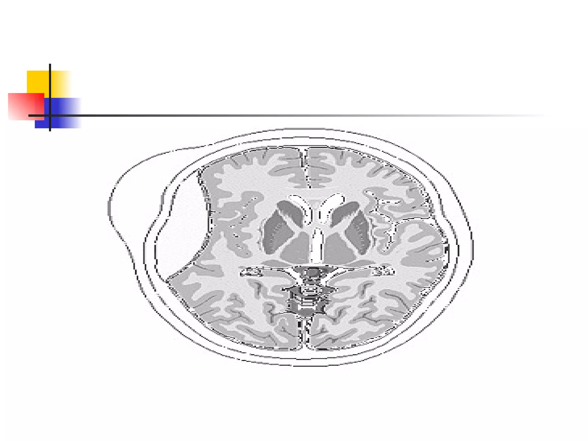
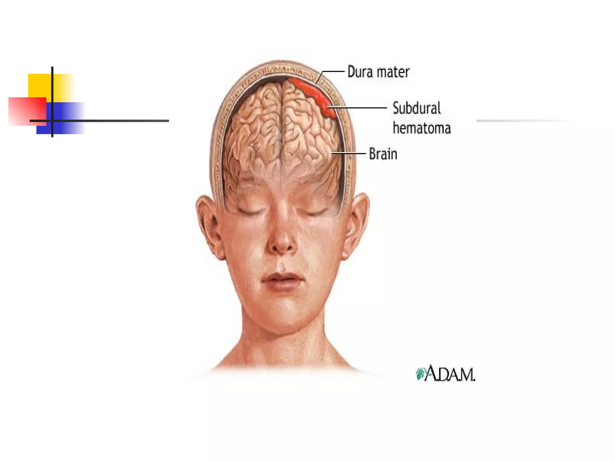
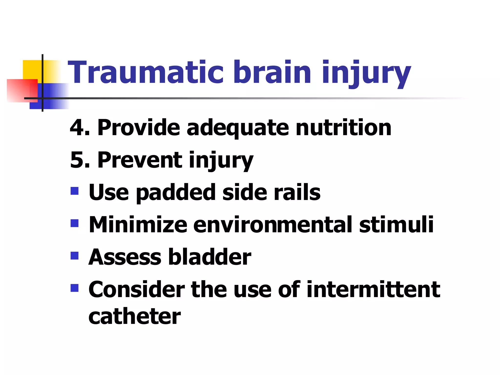
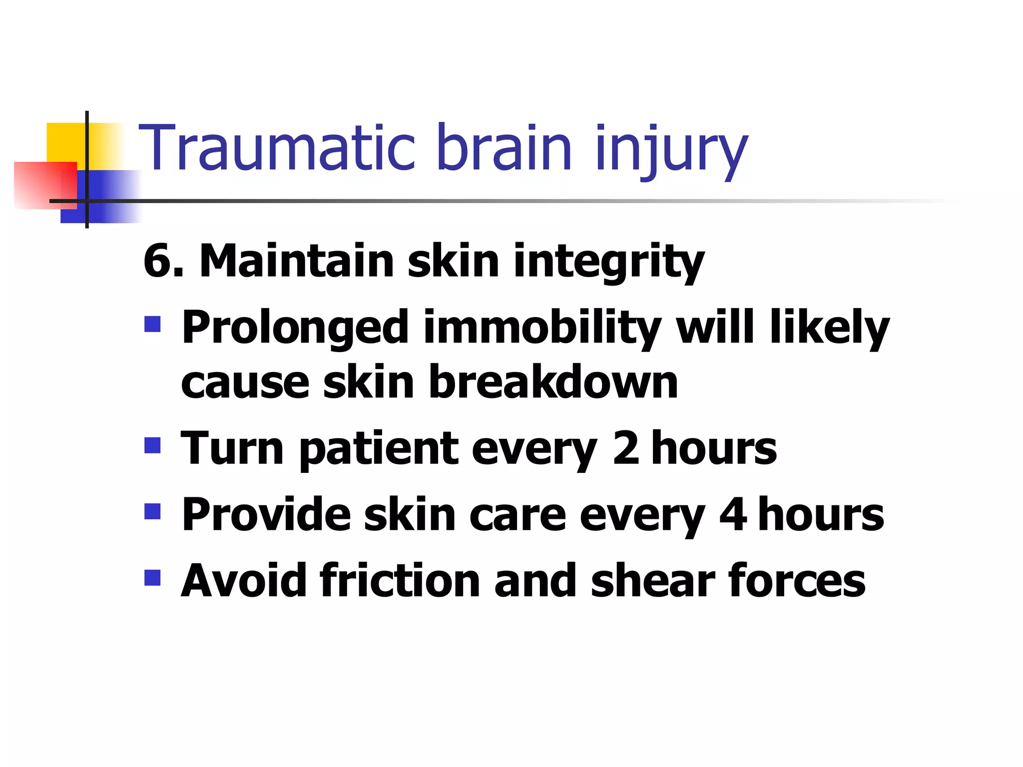
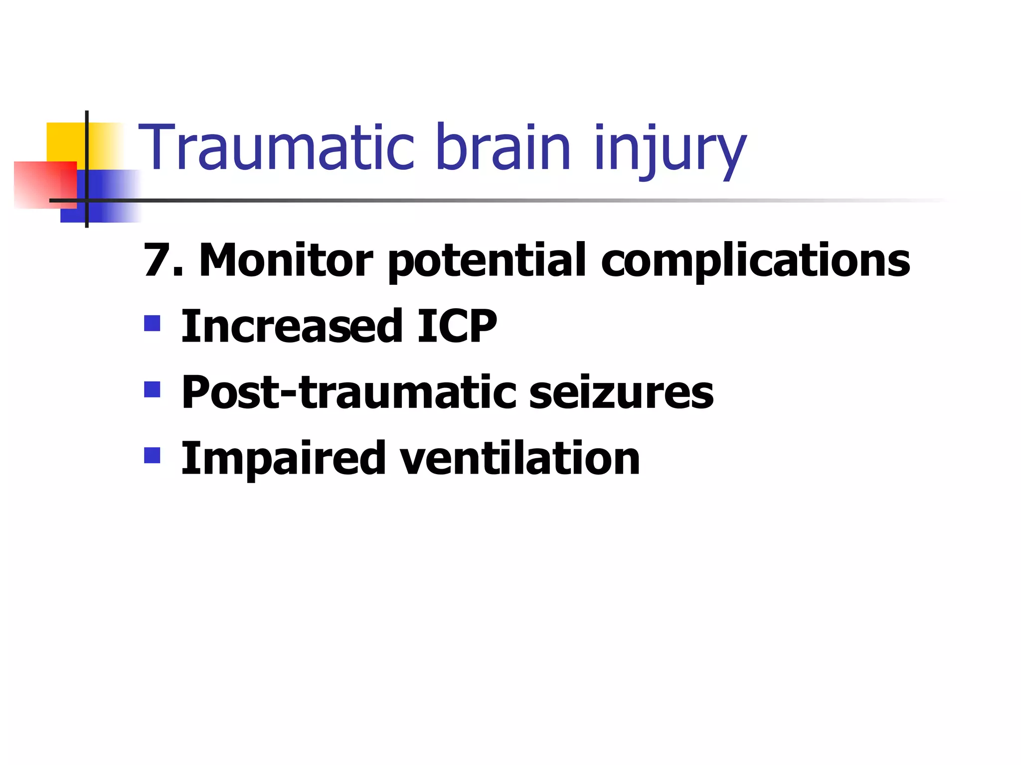
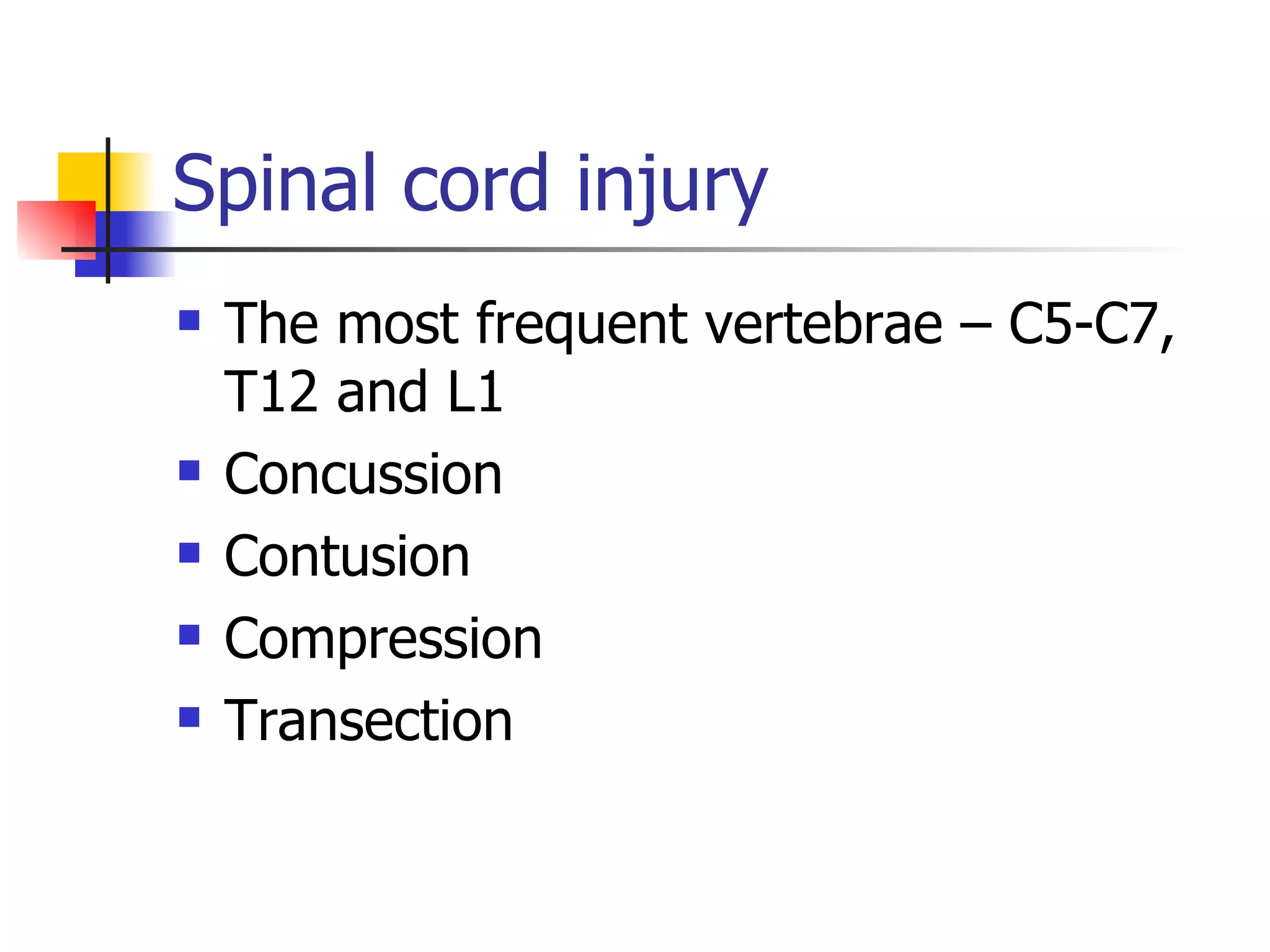
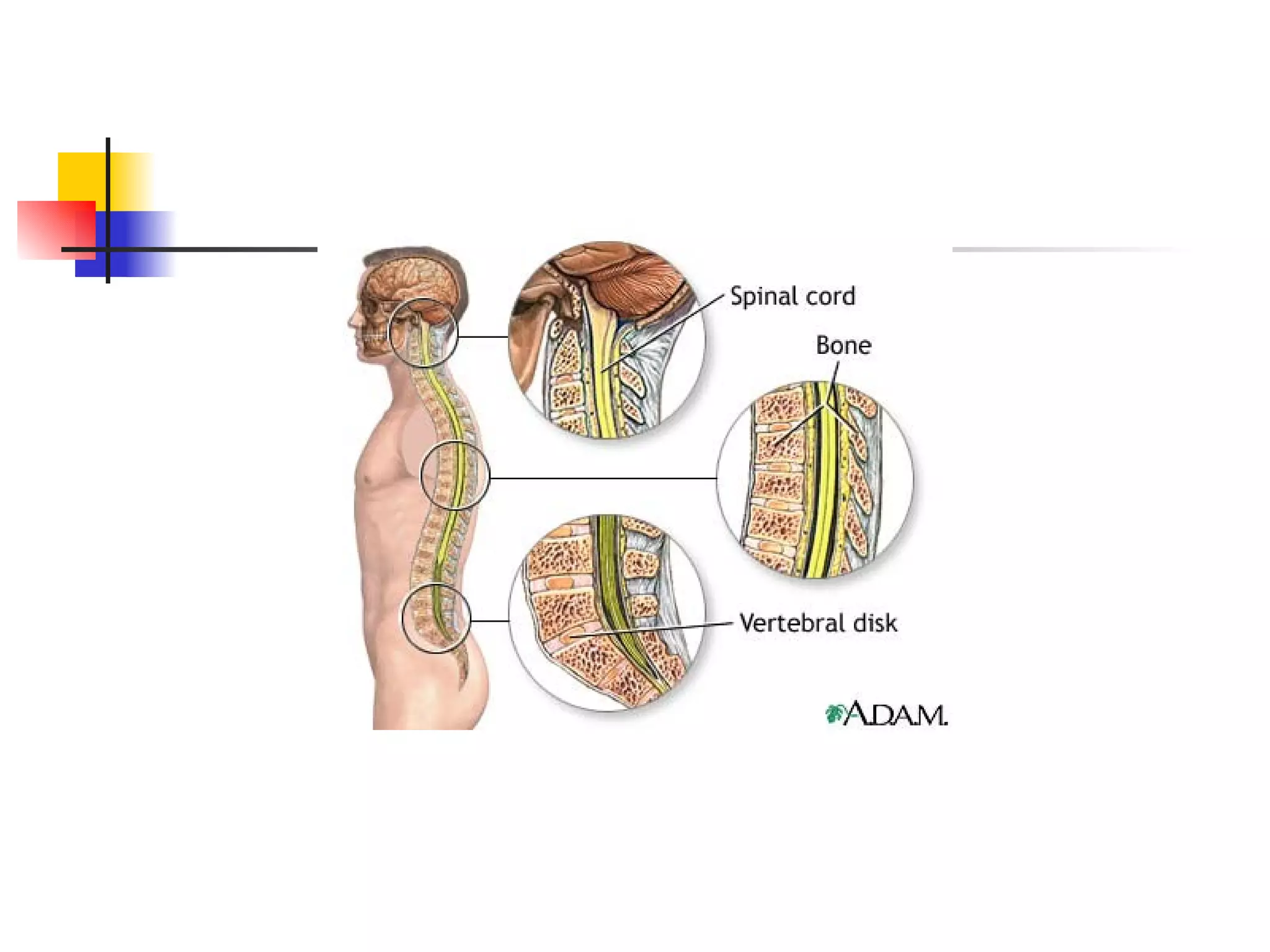
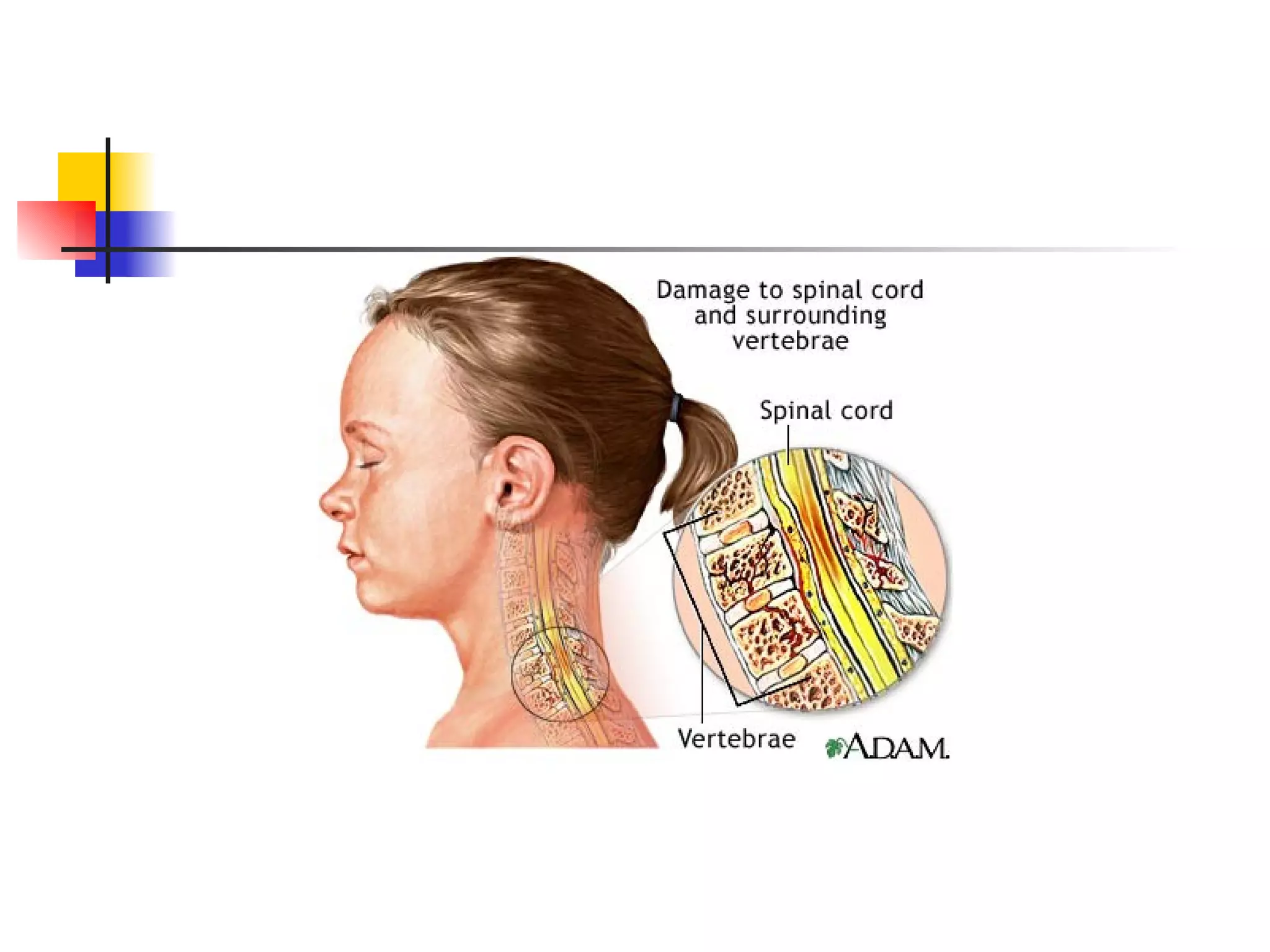
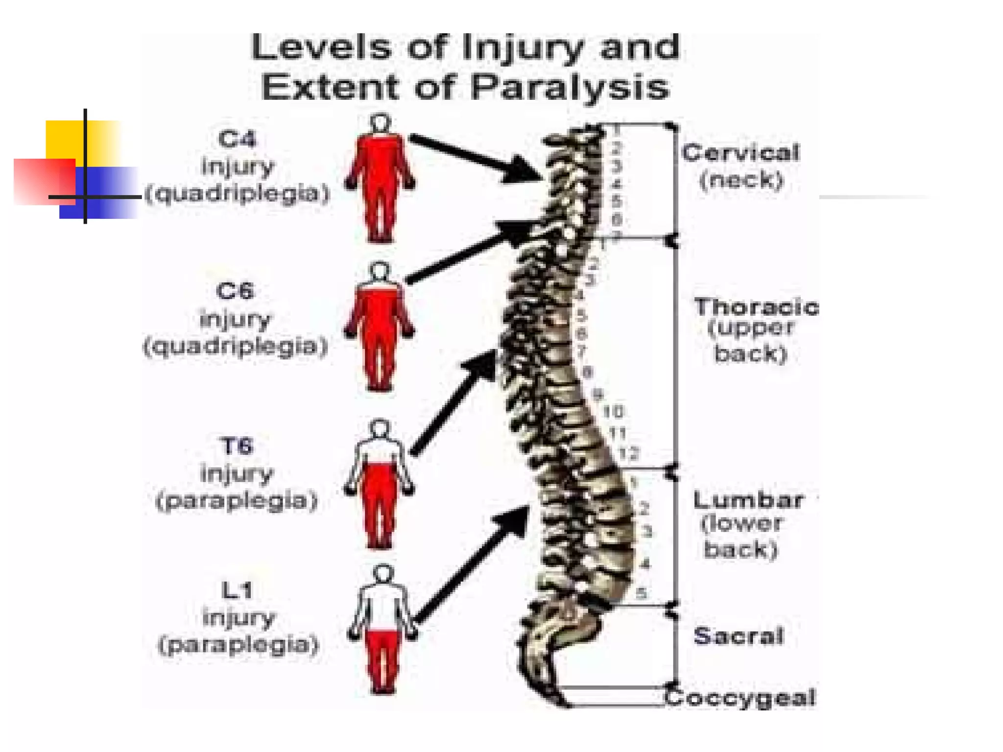
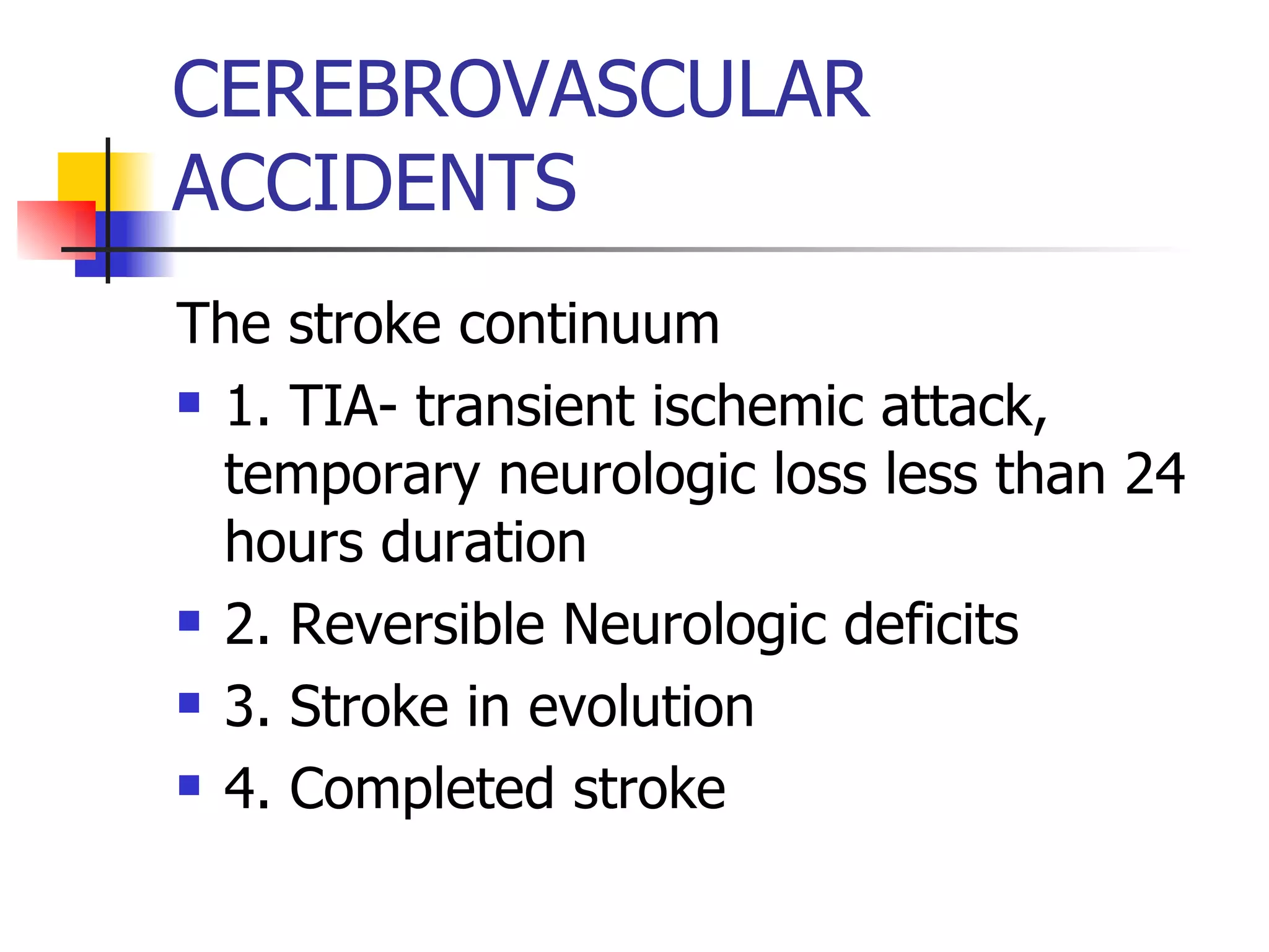
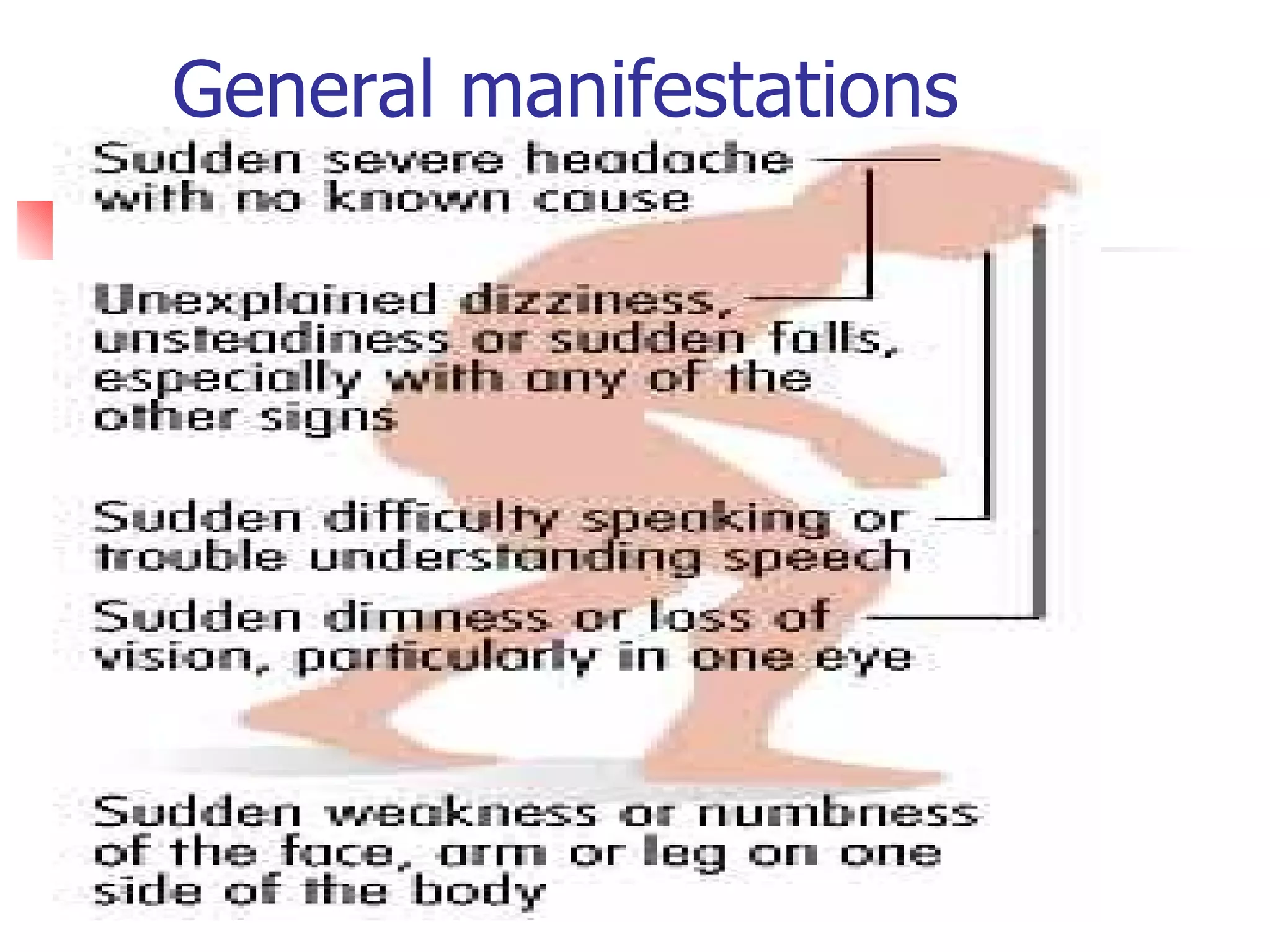
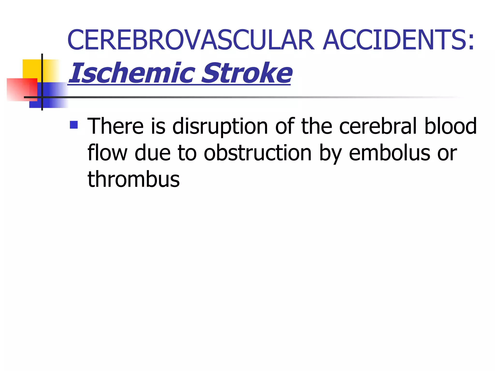
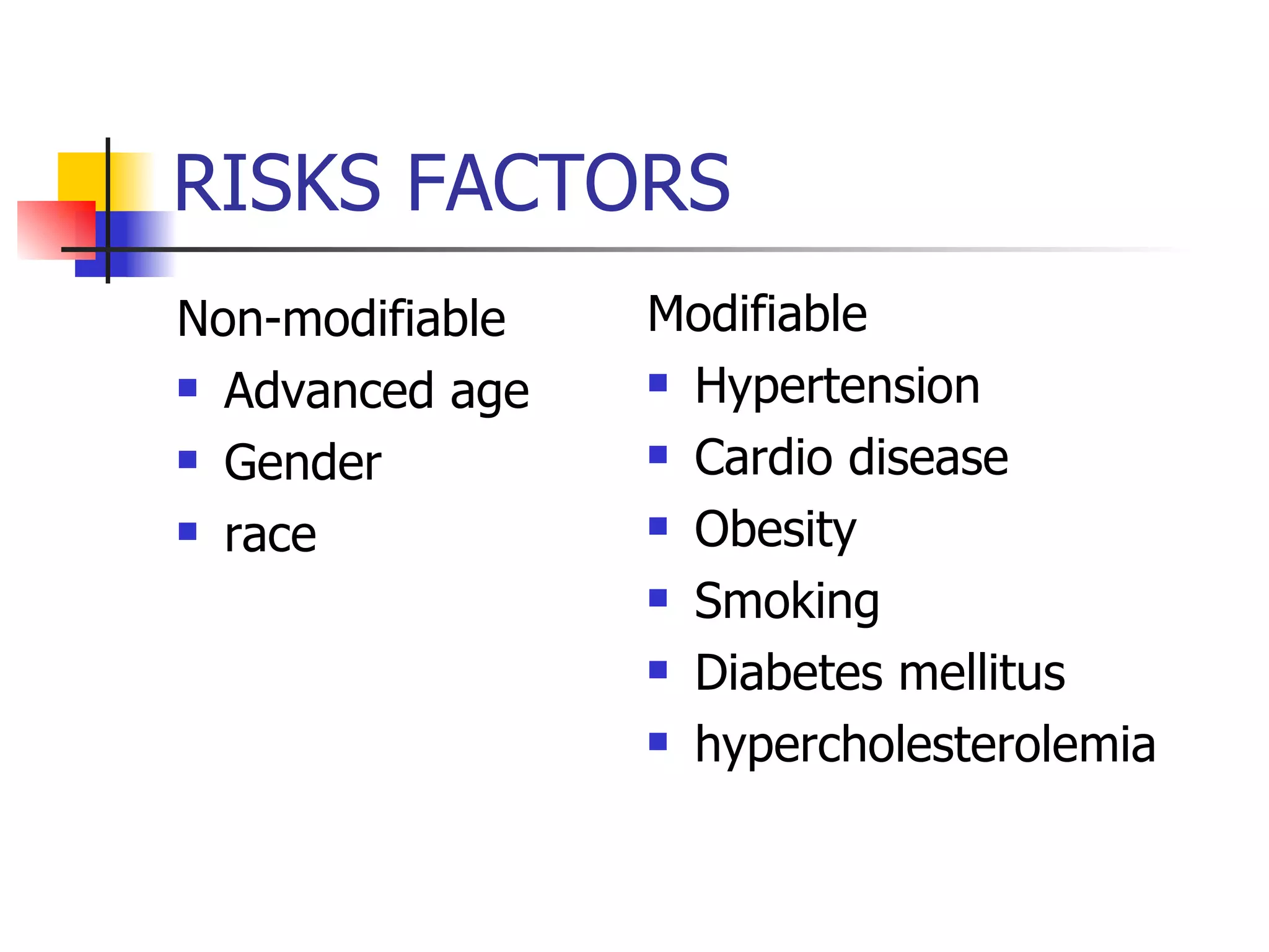
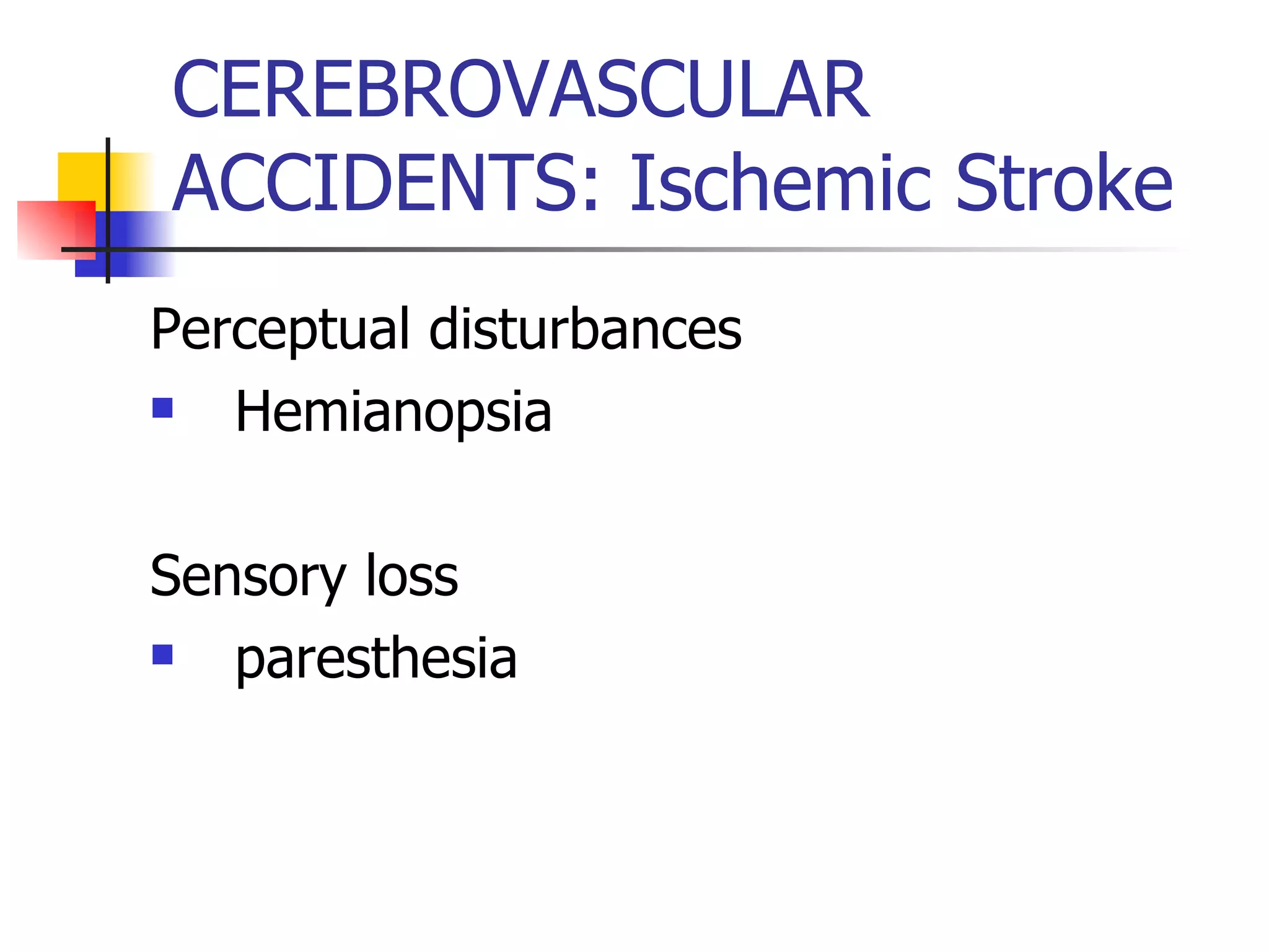
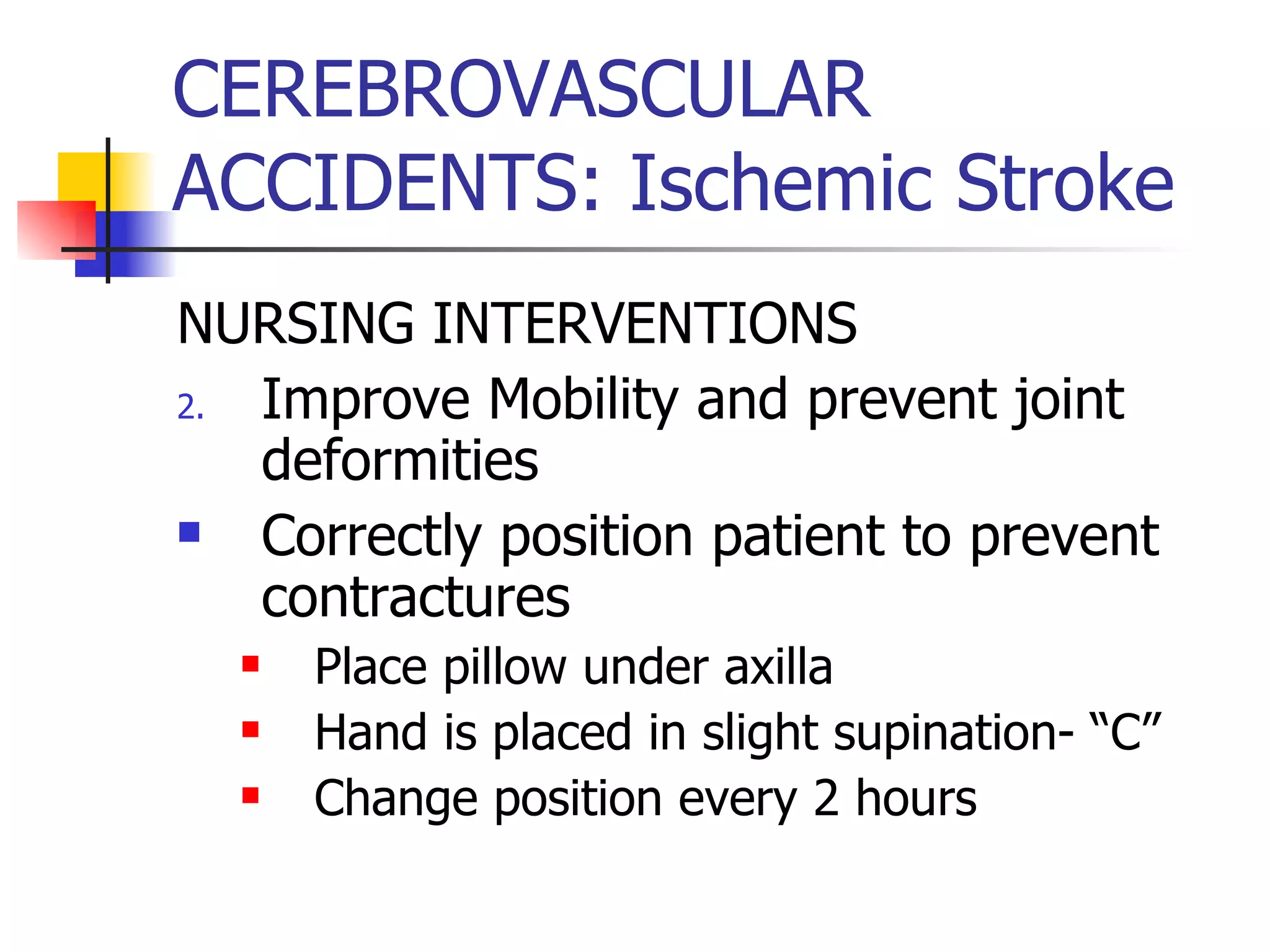
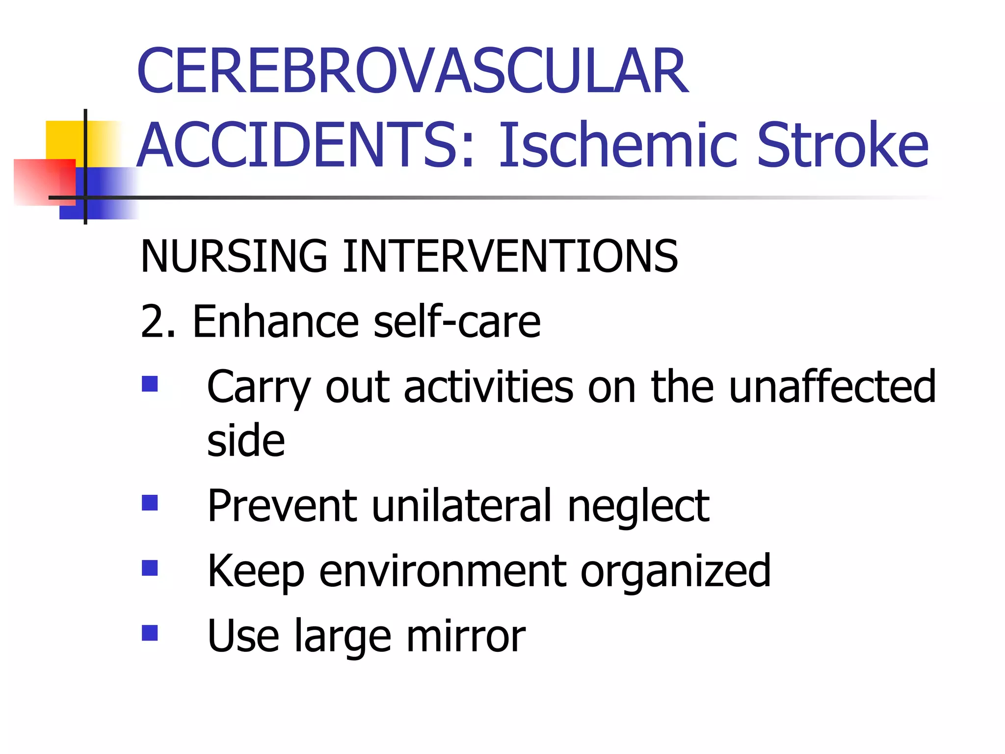
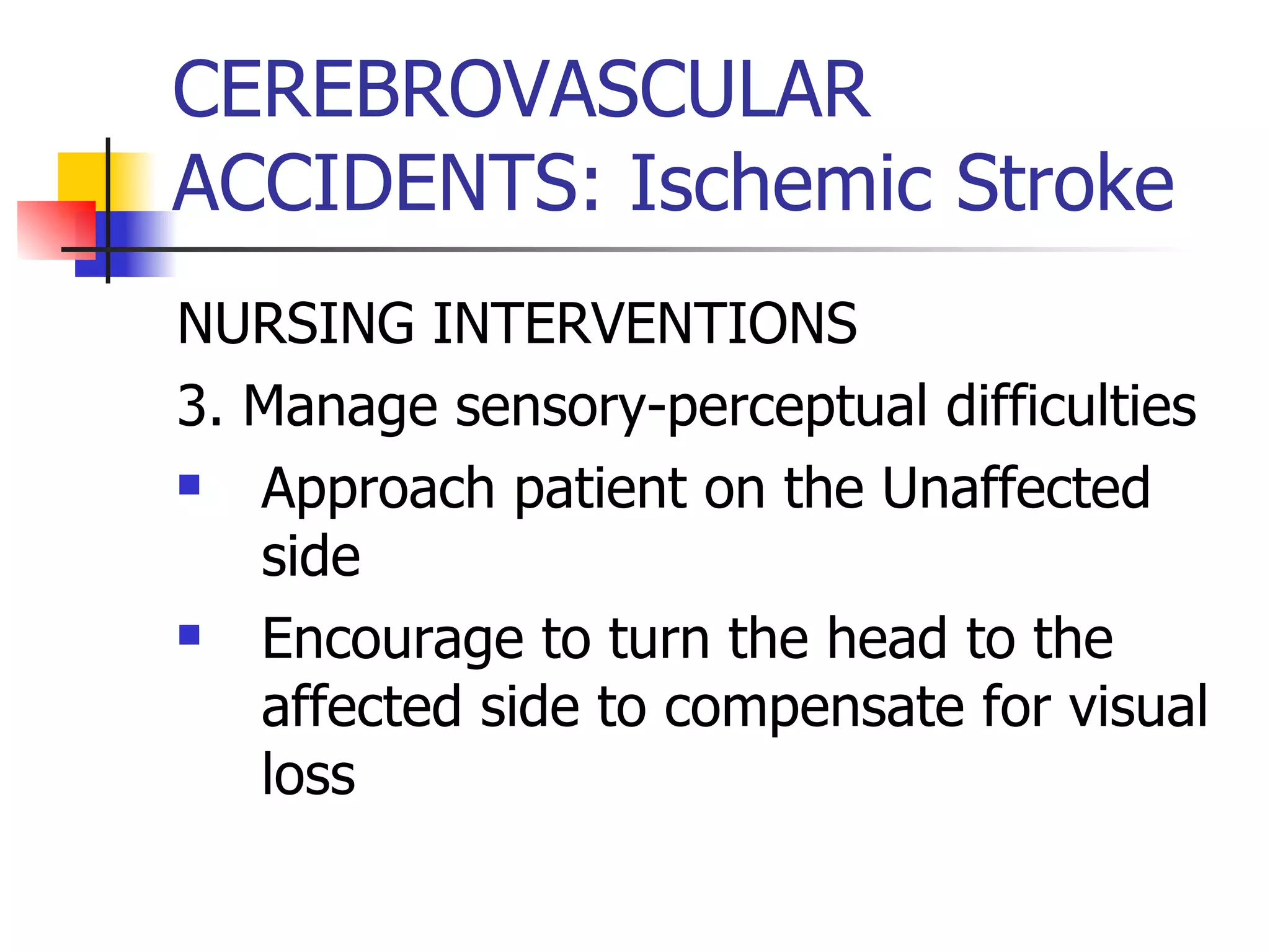
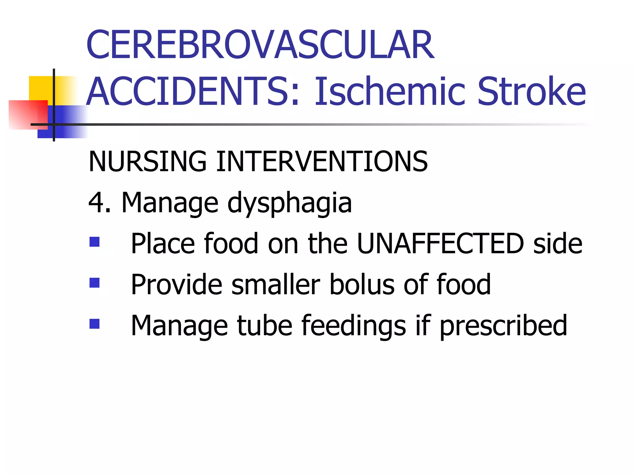
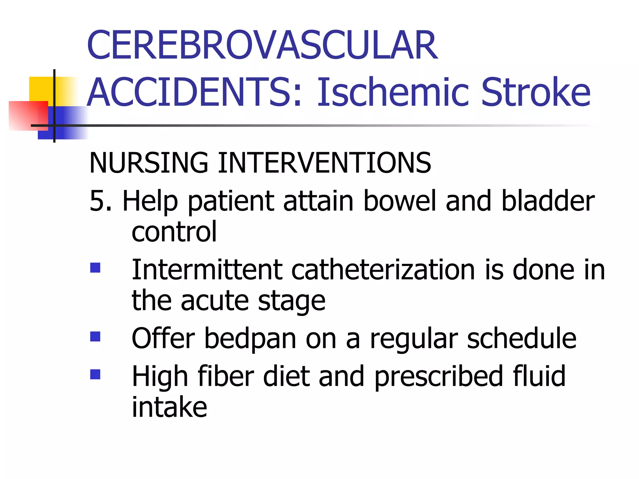
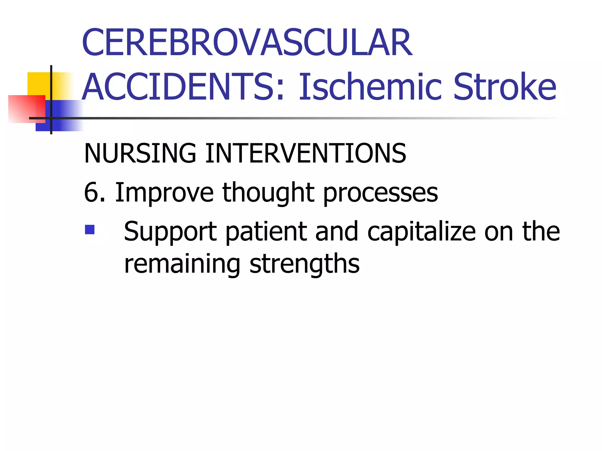
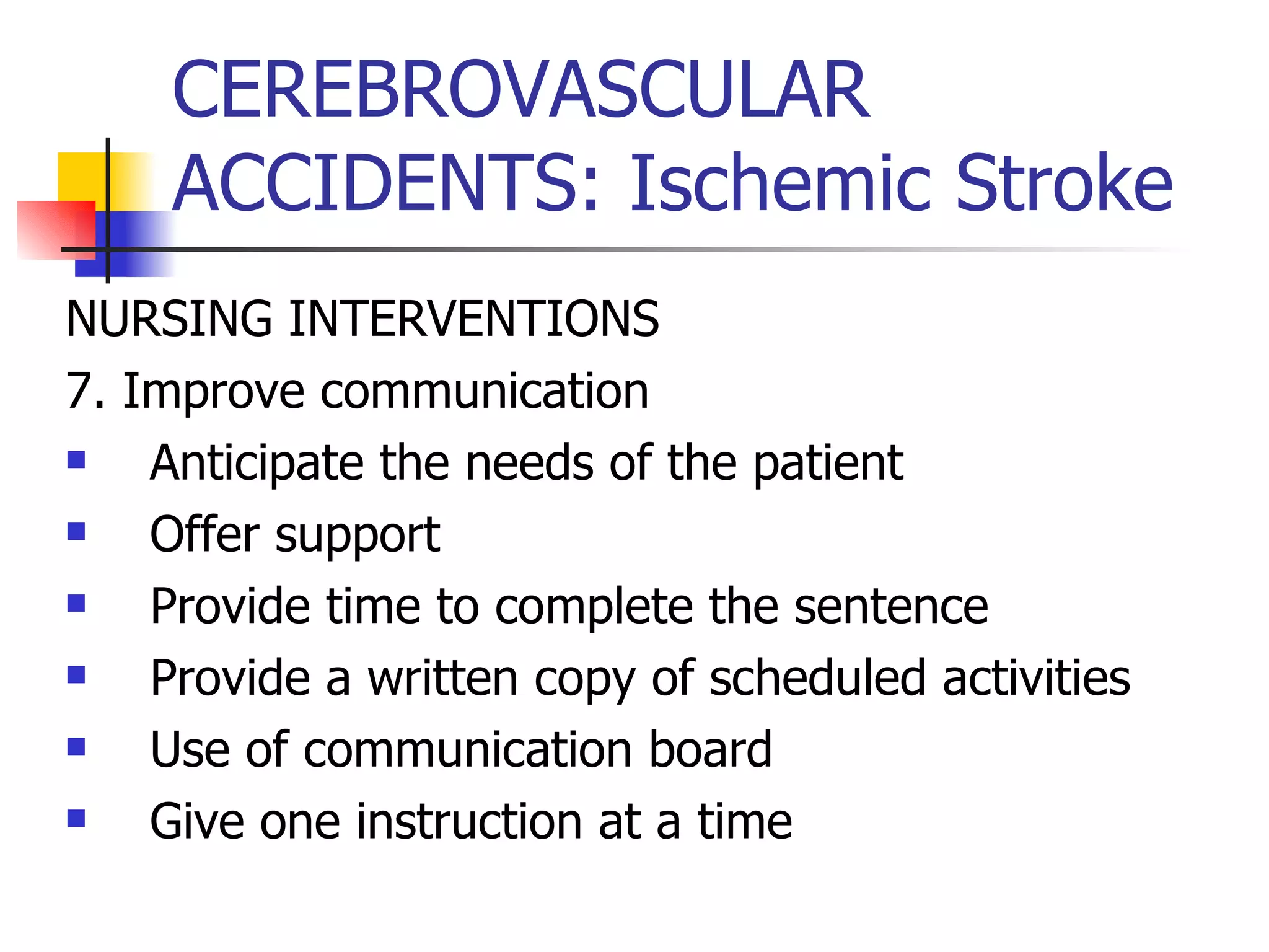
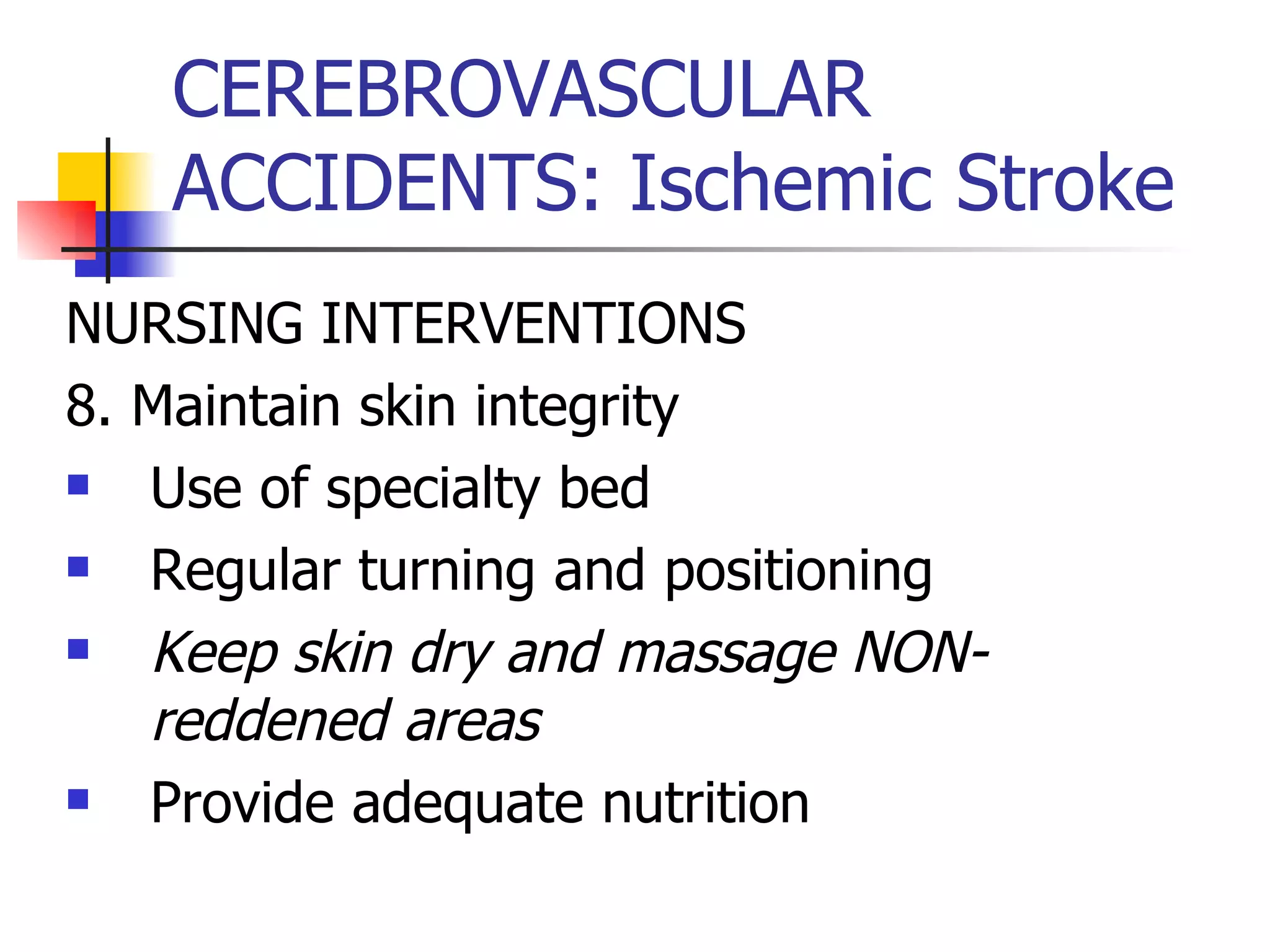
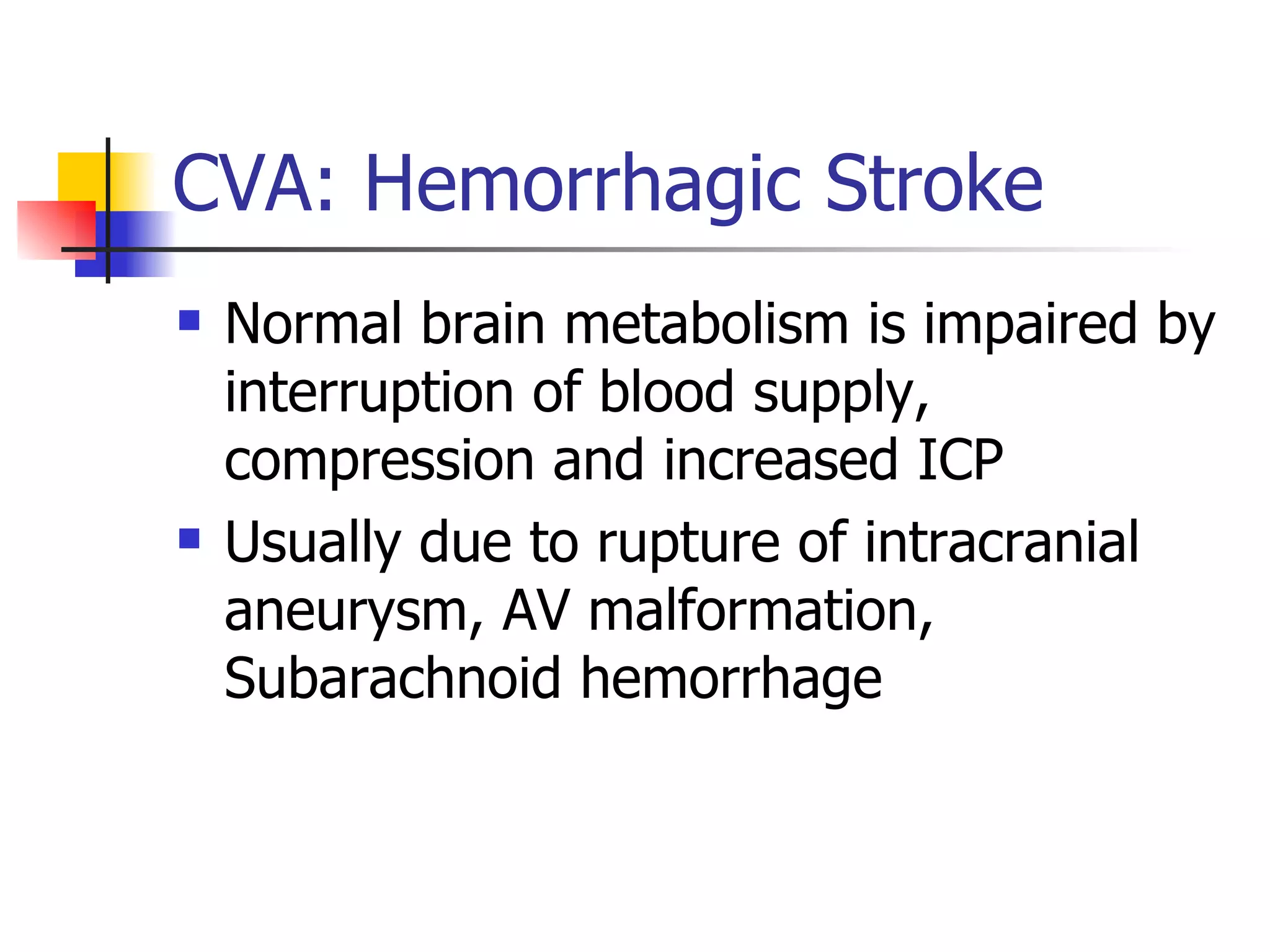
Medical-Surgical Nursing A Review of Neurologic Concepts  Nurse Licensure Examination Review pinoynursing.webkotoh.com
Key to Success! Confidence Test taking strategies Ample test preparation and study habits Review of frequent board examination topics Focus on your goals Above all- PRAYERS
Outline of Our Review Brief review of Anatomy and Physiology Application of the Nursing process in the approach of neurologic problems: ASSESSMENT – relevant techniques and lab procedures DIAGNOSIS PLANNING IMPLEMENTATION EVALUATION
Outline of the review Trauma and related accidents Traumatic brain injury Spinal cord injury Cerebrovascular Accidents
Outline of the review Degenerative disorders- demyelinating Multiple sclerosis Guillain-Barre’ syndrome Degenerative disorders-   NON- demyelinating Alzheimer’s disease Parkinson’s disease
Outline of the review Motor dysfunction- CNS Epilepsy Motor dysfunction- cranial nerve Bell’s palsy Trigeminal neuralgia Motor dysfunction- peripheral Myasthenia gravis
Outline of the review Infectious Disease Meningitis Brain abscess Encephalitis Neoplastic disease
IMPLEMENTATION PHASE Increased Intracranial pressure Altered level of consciousness Seizures Autonomic dysreflexia/hyperreflexia Spinal shock Cognitive impairment Bowel incontinence
IMPLEMENTATION PHASE Impaired physical mobility Impaired swallowing Disturbed sensory perception
Anatomy and Physiology Gross anatomy The nervous system is divided into the central and peripheral nervous system The Central nervous system consists of the BRAIN and the Spinal Cord The peripheral nervous system consists of the Spinal nerves and the cranial nerves
Anatomy and Physiology The brain is composed of lobes- Frontal lobe- personality, memory and motor function Parietal lobe- sensory function Temporal lobe- hearing and olfaction and emotion by the limbic system Occipital lobe- vision
Anatomy and Physiology The cerebellum is involved in coordination and equilibrium The diencephalon consists of the : Thalamus- the relay center of all sensory input Hypothalamus- center for endocrine regulation, sleep, temperature, thirst, sexual arousal and emotional response
Anatomy and Physiology The brainstem is composed of the: MIDBRAIN- for visual and auditory reflexes Pons- respiratory apneustic center, nucleus of cranial nerves- 5,6,7,8 Medulla oblongata- respiratory and cardiovascular centers, nucleus of cranial nerves 9,10,11,12
ASSESSMENT OF THE NEUROLOGIC SYSTEM HISTORY A confused client becomes an unreliable source of history
ASSESSMENT OF THE NEUROLOGIC SYSTEM PHYSICAL EXAMINATION 5 categories: 1. Cerebral function- LOC, mental status 2. Cranial nerves 3. Motor function 4. Sensory function 5. Reflexes
ASSESSMENT OF THE NEUROLOGIC SYSTEM Neuro Check Level of consciousness Pupillary size and response Verbal responsiveness Motor responsiveness Vital signs
CEREBRAL FUCTION Assess the degree of wakefulness/alertness Note the intensity of stimulus to cause a response Apply a painful stimulus over the nailbeds with a blunt instrument Ask questions to assess orientation to person, place and time
Cerebral function Utilize the Glasgow Coma Scale An easy method of describing mental status and abnormality detection  Tests 3 areas- eye opening, verbal response and motor response Scores are evaluated- range from 3-15 No  ZERO  score
Glasgow Coma Scale Glasgow Coma Score Eye Opening (E) Verbal Response (V) Motor Response (M)
Glasgow Coma Scale Glasgow Coma Score Eye Opening (E) 4=Spontaneous 3=To voice 2=To pain 1=None   (No response)
Glasgow Coma Scale Glasgow Coma Score Verbal Response (V) 5=Normal/oriented 4=Disoriented/ CONFUSED 3=Words, but  incoherent/ inappropriate 2=Incomprehensible/mumbled words 1=None
Glasgow Coma Scale Glasgow Coma Score Motor Response (M) 6=Normal- obeys command 5=Localizes pain 4=Withdraws to pain (Flexion) 3=Decorticate posture 2=Decerebrate posture 1=None  (flaccid)
Cranial Nerve Function: Cranial Nerve 1- Olfactory Check first for the patency of the nose Instruct to close the eyes Occlude one nostrils at a time Hold familiar substance and asks for the identification Repeat with the other nostrils PROBLEM- ANOSMIA- “loss of smell”
Cranial Nerve Function: Cranial Nerve 2- Optic Check the visual acuity with the use of the Snellen chart Check for visual field by confrontation test Check for pupillary reflex- direct and consensual Fundoscopy to check for papilledema
Snellen chart
Cranial Nerve Function: Cranial Nerve 3, 4 and 6 Assess simultaneously the movement of the extra-ocular muscles Deviations: Opthalmoplegia- inability to move the eye in a direction Diplopia- complaint of double vision
 
Cranial Nerve Function: Cranial Nerve 5 -trigeminal Sensory portion- assess for sensation of the facial skin Motor portion- assess the muscles of mastication Assess corneal reflex
Cranial Nerve Function: Cranial Nerve 7 -facial Sensory portion- prepare salt, sugar, vinegar and quinine. Place each substance in the anterior two thirds of the tongue, rinsing the mouth with water Motor portion- ask the client to make facial expressions, ask to forcefully close the eyelids
Cranial Nerve Function: Cranial Nerve 8- vestibulo-auditory Test patient’s hearing acuity Observe for nystagmus and disturbed balance
Cranial Nerve Function: Cranial Nerve 9- glossopharyngeal Together with Cranial nerve 10 –vagus Assess for gag reflex Watch the soft palate rising after instructing the client to say “AH” The posterior one-third of the tongue is supplied by the glossopharyngeal nerve
Cranial Nerve Function: Cranial Nerve 11- accessory Press down the patient’s shoulder while he attempts to shrug against resistance
Cranial Nerve Function: Cranial Nerve 12- hypoglossal Ask patient to protrude the tongue and note for symmetry
ASSESS Motor function Assess muscle tone and strength by asking patient to flex or extend the extremities while the examiner places resistance Grading of muscle strength
Assessing the motor function of the cerebellum Test for balance- heel to toe Test for coordination- rapid alternating movements and finger to nose test ROMBERG’s is actually a test for the posterior spinothalamic tract
 
Assessing the motor function of the brainstem Test for the Oculocephalic reflex- doll’s eye Normal response- eyes appear to move opposite to the movement of the head Abnormal- eyes move in the same direction
Assessing the motor function of the brainstem Test for the Oculovestibular reflex Slowly irrigate the ear with cold water and warm water Normal response- cOld- OppOsite, wArM- sAMe
Assessing the sensory function  Evaluate symmetric areas of the body Ask the patient to close the eyes while testing Use of test tubes with cold and warm water Use blunt and sharp objects Use wisp of cotton Ask to identify objects placed on the hands Test for sense of position
Assessing the reflexes Deep tendon reflexes Biceps Triceps Brachioradialis Patellar Assessing the sensory function Achilles
Assessing the reflexes Superficial reflexes Abdominal Cremasteric Anal Pathologic reflex Babinski- stroke the lateral aspect of the soles doing an inverted “J” (+)- DORSIFLEXION of the Big toe with fanning out of the little toes
Grading of reflexes Deep tendon reflex 0- absent + present but diminished ++ normal +++ increased ++++ hyperactive or clonic Superficial reflex 0 absent +present
DIAGNOSTIC TESTS EEG Withhold medications that may interfere with the results- anticonvulsants, sedatives and stimulants Wash hair thoroughly before procedure
DIAGNOSTIC TESTS CT scan With radiation risk If contrast medium will be used- ensure consent,  assess for allergies to dyes and iodine or seafood, flushing and metallic taste are expected as the dye is injected
DIAGNOSTIC TESTS MRI Uses magnetic waves Patients with pacemakers, orthopedic metal prosthesis and implanted metal devices cannot undergo this procedure
DIAGNOSTIC TESTS Cerebral arteriography Note allergies to dyes, iodine and seafood Ensure consent Keep patient at rest after procedure Maintain pressure dressing or sandbag over punctured site
DIAGNOSTIC TESTS Lumbar puncture Ensure consent, determine ability to lie still Contraindicated in patients with increased ICP Keep flat on bed after procedure Increase fluid intake after procedure
Increased Intracranial pressure Intracranial pressure  more than 15 mmHg Brunner= Normal intracranial pressure 10-20 mmHg Causes: Head injury Stroke Inflammatory lesions Brain tumor Surgical complications
Increased Intracranial pressure Pathophysiology The cranium only contains the brain substance, the CSF and the blood/blood vessels MONRO-KELLIE hypothesis- an increase in any one of the components causes a change in the volume of the other Any increase or alteration in these structures will cause increased ICP
Increased Intracranial pressure Pathophysiology Compensatory mechanisms: 1. Increased CSF absorption 2. Blood shunting 3. Decreased CSF production
Increased Intracranial pressure Pathophysiology Decompensatory mechanisms: 1. Decreased cerebral perfusion 2. Decreased PO2 leading to brain hypoxia 3. Cerebral edema 4. Brain herniation
Decreased cerebral blood flow Vasomotor reflexes are stimulated initially   slow bounding pulses Increased concentration of carbon dioxide will cause VASODILATION    increased flow   increased ICP
Cerebral Edema Abnormal accumulation of fluid in the intracellular space, extracellular space or both.
Herniation Results from an excessive increase in ICP when the pressure builds up and the brain tissue presses down on the brain stem
Cerebral response to increased ICP Steady perfusion up to 40 mmHg Cushing’s response Vasomotor center triggers rise in BP to increase ICP Sympathetic response is increased BP but the heart rate is SLOW Respiration becomes SLOW
Increased Intracranial pressure CLINICAL MANIFESTATIONS Early manifestations : Changes in the LOC- usually the earliest Pupillary changes- fixed, slowed response Headache vomiting
Increased Intracranial pressure CLINICAL MANIFESTATIONS late manifestations : Cushing reflex- systolic  hypertension ,  bradycardia  and wide pulse pressure bradypnea Hyperthermia Abnormal posturing
Increased Intracranial pressure Nursing interventions:  Maintain patent airway 1.  Elevate the head of the bed 15-30 degrees- to promote venous drainage 2. assists in  administering 100% oxygen  or controlled hyperventilation- to reduce the CO2 blood levels  constricts blood vessels  reduces edema
Increased Intracranial pressure Nursing interventions 3. Administer prescribed medications- usually  Mannitol- to produce negative fluid balance corticosteroid- to reduce edema anticonvulsants-p to prevent seizures
Increased Intracranial pressure Nursing interventions 4. Reduce environmental stimuli 5. Avoid activities that can increase ICP like valsalva, coughing, shivering, and vigorous suctioning
Increased Intracranial pressure Nursing interventions 6. Keep head on a neutral position. ACOID- extreme flexion, valsalva 7.  monitor for secondary complications Diabetes insipidus- output of >200 mL/hr SIADH
Altered level of consciousness It is a function and symptom of multiple pathophysiologic phenomena Causes: head injury, toxicity and metabolic derangement Disruption in the neuronal transmission results to improper function
Altered level of consciousness Assessment Orientation to time, place and person Motor function Decerebrate Decorticate Sensory function
Altered level of consciousness Patient is not oriented Patient does not follow command Patient needs persistent stimuli to be awake COMA= clinical state of unconsciousness where patient is NOT aware of self and environment
Altered level of consciousness Etiologic Factors Head injury  Stroke Drug overdose Alcoholic intoxication Diabetic ketoacidosis Hepatic failure
Altered level of consciousness ASSESSMENT Behavioral changes initially Pupils are slowly reactive  Then , patient becomes unresponsive and pupils become fixed dilated Glasgow Coma Scale is utilized
Altered level of consciousness Nursing Intervention 1. Maintain patent airway Elevate the head of the bed to 30 degrees Suctioning 2. Protect the patient Pad side rails Prevent injury from equipments, restraints and etc.
Altered level of consciousness Nursing Intervention 3. Maintain fluid and nutritional balance Input an output monitoring IVF therapy Feeding through NGT 4. Provide mouth care Cleansing and rinsing of mouth Petrolatum on the lips
Altered level of consciousness Nursing Intervention 5. Maintain skin integrity Regular turning every 2 hours 30 degrees bed elevation Maintain correct body alignment by using trochanter rolls, foot board 6. Preserve corneal integrity Use of artificial tears every 2 hours
Altered level of consciousness Nursing Intervention 7. Achieve thermoregulation Minimum amount of beddings Rectal or tympanic temperature Administer acetaminophen as prescribed 8. Prevent urinary retention Use of intermittent catheterization
Altered level of consciousness Nursing Intervention 9. Promote bowel function High fiber diet Stool softeners and suppository 10. Provide sensory stimulation Touch and communication Frequent reorientation
SEIZURES Episodes of abnormal motor, sensory, autonomic activity resulting from sudden excessive discharge from cerebral neurons A part or all of the brain may be involved
SEIZURES PATHOPHYSIOLOGY An electrical disturbance in the nerve cells in one brain section   EMITS ELECTRICAL IMPULSES excessively
SEIZURES ETIOLOGIC FACTORS Idiopathic Fever Head injury CNS infection Metabolic and toxic conditions
SEIZURES Nursing Interventions During seizure 1. remove harmful objects from the patient’s surrounding 2. ease the client to the floor 3. protect the head with pillows 4. Observe and note for the duration, parts of body affected, behaviors before and after the seizure
SEIZURES Nursing Interventions During seizure 5. loosen constrictive clothing 6. DO NOT restrain, or attempt to place tongue blade or insert oral airway
SEIZURES Nursing Interventions POST seizure 1. place patient to the side to drain secretions and prevent aspiration 2. help re-orient the patient if confused 3. provide care if patient became incontinent during the seizure attack 4. stress importance of medication regimen
headache Cephalgia Primary headache- no organic cause Secondary headache- with organic cause Migraine headache- periodic attacks of headache due to vascular disturbance Tension headache-the most common type- due to muscle tension
headache Migraine Prodrome stage Aura phase Headache Recovery phase
headache Nursing Interventions 1. Avoid precipitating factors 2. modify lifestyle 3. relieve pain by pharmacologic measures Beta-blockers Serotonin antagonists- “triptan"
Autonomic Dysreflexia/hyperreflexia Seen commonly in spinal cord injury  above T6 An exaggerated response by the autonomic system resulting from various stimuli most commonly distended bladder, impacted feces, pain, skin irritation
Autonomic Dysreflexia/hyperreflexia Clinical MANIFESTATIONS 1. Hypertension 2. Bradycardia 3. severe pounding headache 4. diaphoresis 5. nausea and nasal congestion
Autonomic Dysreflexia/hyperreflexia NURSING INTERVENTIONS 1.  Elevate the head of the bed immediately 2. Check for bladder distention and empty bladder with urinary catheter 3. Check for Fecal impaction and other triggering factors like skin irritation, pressure ulcer 4. Administer antihypertensive medications- usually hydralazine
Spinal Shock Pathophysiology The sudden depression of reflex activity in the spinal cord below the level of injury The muscles below the lesion are flaccid, the skin without sensation and the reflexes are absent including bowel and bladder functions
Spinal Shock Nursing Interventions 1. Assist in chest physical therapy 2. Manage potential complication- DVT
Cognitive Impairment Nursing Interventions Assist or encourage the patient to use eyeglass, hearing aid or assistive devices Reorient the patient by calling his name frequently Provide background information as to date, time, place, environment
Cognitive Impairment Nursing Interventions 4. Use large signs as visual cues 5. Post patient's photo on the door 6. Encourage family members to bring personal articles and place them in the same area
Bowel and Bladder incontinence Establish a regular pattern for bowel care Maintain a dietary intake. Avoid foods that can cause excessive gas production
CONGENITAL DISORDERS: Hydrocephalus Excessive CSF accumulation in the brain’s ventricular system In infants, head enlarges In children and adults- brain compression
CONGENITAL DISORDERS: Hydrocephalus Non-communicating hydrocephalus results from CSF outflow obstruction Communicating hydrocephalus results from faulty absorption or increased CSF production
CONGENITAL DISORDERS: Hydrocephalus Assessment 1. irritability 2. change in LOC 3. infants- enlargement of the head, thin scalp skin 4. sunset eyes
CONGENITAL DISORDERS: Hydrocephalus DIAGNOSTIC TESTS 1. Skull x-ray 2. ventriculography
CONGENITAL DISORDERS: Hydrocephalus Nursing Intervention 1. monitor neurologic status 2. teach parents to watch for signs of shunt malfunction, and periodic surgery to lengthen the shunt as child grows
CONGENITAL DISORDER- Spinal cord defects 1. Spina bifida occulta- incomplete closure of one or more vertebrae without protrusion of the spinal cord or meninges 2. Spina bifida with meningocele- a sac contains meninges and CSF 3. Spina bifida with meningomyelocele- a sac contains spinal cord substance, meninges and CSF
CONGENITAL DISORDER: Spinal cord defects Causes 1. environmental factors 2. radiation 3. folic acid deficiency in a pregnant woman 4. possibly genetic
CONGENITAL DISORDER: Spinal cord defects ASSESSMENT 1. a dimple or tuft of hair in the vertebral area 2. external sac DIAGNOSIS 1. Spinal x-ray 2. myelography
CONGENITAL DISORDER: Spinal cord defects NURSING INTERVENTION 1. cover the defect with sterile dressing moistened with sterile saline 2. position the patient on prone or side to protect the fragile sac 3. place a diaper under the infant and change it often
CONGENITAL DISORDER: Spinal cord defects NURSING INTERVENTION 4. avoid the use of lotion 5. avoid frequent handling 6. Measure the child’s head circumference daily 7. check anal reflex 8. support family members 9. prepare the parents for the possible outcome of eh defect
CONGENITAL DISORDER: Spinal cord defects NURSING INTERVENTION 10. Post-operative care Position on abdomen Check post-operative dressings Place infant’s hips in abduction and feet in neutral position Monitor intake and output Check for urine retention Asess infant frequently as he recovers from the surgery
Traumatic brain injury 1. CONCUSSION Involves jarring of head without tissue injury Temporary loss of neurologic function lasting fore a few minutes to hours
 
Traumatic brain injury 2. CONTUSION Involves structural damage The patient becomes unconscious for hours
 
Traumatic brain injury 3. Diffuse Axonal injury Involves widespread damage to the neurons Patient has decerebrate and decorticate posture
Traumatic brain injury 4. Intracranial hemorrhage Epidural Hematoma- blood collects in the epidural space between skull and dura mater. Usually due to laceration of the middle meningeal artery Symptoms develop rapidly
 
Traumatic brain injury 4. Intracranial hemorrhage Subdural hematoma- a collection of blood between the dura and the arachnoid mater caused by trauma. This is usually due to tear of dural sinuses or dural venous vessels Symptoms usually develop slowly
 
Traumatic brain injury 4. Intracranial hemorrhage Intracerebral Hemorrhage and hematoma- bleeding into the substance of the brain resulting from trauma, hypertensive rupture of aneurysm, coagulopahties, vascular abnormalities Symptoms develop insidiously, beginning with severe headache and neurologic deficits
 
Traumatic brain injury MANIFESTATIONS 1. Altered LOC 2. CSF otorrhea 3. CSF rhinorrhea 4. Racoon eyes and battle sign HALO SIGN- blood stain surrounded by a yellowish stain
Traumatic brain injury NURSING MANAGEMENT 1. Monitor for declining LOC- use of Glasgow 2. Maintain patent airway Elevate bed, suction prn, monitor ABG
Traumatic brain injury NURSING MANAGEMENT 3. Monitor F and E balance Daily weights IVF therapy Monitor possible development of DI and SIADH
Traumatic brain injury 4. Provide adequate nutrition 5. Prevent injury Use padded side rails Minimize environmental stimuli Assess bladder Consider the use of intermittent catheter
Traumatic brain injury 6. Maintain skin integrity Prolonged immobility will likely cause skin breakdown Turn patient every 2 hours Provide skin care every 4 hours Avoid friction and shear forces
Traumatic brain injury 7. Monitor potential complications Increased ICP Post-traumatic seizures Impaired ventilation
Spinal cord injury The most frequent vertebrae – C5-C7, T12 and L1 Concussion Contusion Compression Transection
 
 
Spinal cord injury Clinical manifestations 1. Paraplegia 2. quadriplegia 3. spinal shock
 
Spinal cord injury DIAGNOSTIC TEST Spinal x-ray CT scan MRI
Spinal cord injury EMERGENCY MANAGEMENT A-B-C Immobilization Immediate transfer to tertiary facility
Spinal cord injury NURSING INTERVENTION 1. Promote adequate breathing and airway clearance 2. Improve mobility and proper body alignment 3. Promote adaptation to sensory and perceptual alterations 4. Maintain skin integrity
Spinal cord injury 5. Maintain urinary elimination 6. Improve bowel function 7. Provide Comfort measures 8. Monitor and manage complications Thromboplebhitis Orthostaic hypotension Spinal shock Autonomic dysreflexia
Spinal cord injury 9. Assists with surgical reduction and stabilization of cervical vertebral column
CEREBROVASCULAR ACCIDENTS An umbrella term that refers to any functional abnormality of the CNS related to disrupted blood supply
CEREBROVASCULAR ACCIDENTS Can be divided into two major categories 1. Ischemic stroke- caused by thrombus and embolus 2. Hemorrhagic stroke- caused commonly by hypertensive bleeding
 
 
CEREBROVASCULAR ACCIDENTS The stroke continuum 1. TIA- transient ischemic attack, temporary neurologic loss less than 24 hours duration 2. Reversible Neurologic deficits 3. Stroke in evolution 4. Completed stroke
General manifestations
CEREBROVASCULAR ACCIDENTS:  Ischemic Stroke There is disruption of the cerebral blood flow due to obstruction by embolus or thrombus
RISKS FACTORS Non-modifiable Advanced age Gender race Modifiable Hypertension Cardio disease Obesity Smoking Diabetes mellitus hypercholesterolemia
Pathophysiology of ischemic stroke Disruption of blood supply Anaerobic metabolism ensues Decreased ATP production leads to impaired membrane function Cellular injury and death can occur
CEREBROVASCULAR ACCIDENTS: Ischemic Stroke DIAGNOSTIC test 1. CT scan 2. MRI 3. Angiography
CEREBROVASCULAR ACCIDENTS: Ischemic Stroke CLINICAL MANIFESTATIONS 1. Numbness or weakness 2. confusion or change of LOC 3. motor and speech difficulties 4. Visual disturbance 5. Severe headache
CEREBROVASCULAR ACCIDENTS: Ischemic Stroke Motor Loss Hemiplegia Hemiparesis
CEREBROVASCULAR ACCIDENTS: Ischemic Stroke Communication loss Dysarthria= difficulty in speaking Aphasia= Loss of speech Apraxia= inability to perform a previously learned action
CEREBROVASCULAR ACCIDENTS: Ischemic Stroke Perceptual disturbances Hemianopsia Sensory loss paresthesia
CEREBROVASCULAR ACCIDENTS: Ischemic Stroke NURSING INTERVENTIONS Improve Mobility and prevent joint deformities Correctly position patient to prevent contractures Place pillow under axilla Hand is placed in slight supination- “C” Change position every 2 hours
CEREBROVASCULAR ACCIDENTS: Ischemic Stroke NURSING INTERVENTIONS 2. Enhance self-care Carry out activities on the unaffected side Prevent unilateral neglect Keep environment organized Use large mirror
CEREBROVASCULAR ACCIDENTS: Ischemic Stroke NURSING INTERVENTIONS 3. Manage sensory-perceptual difficulties Approach patient on the Unaffected side Encourage to turn the head to the affected side to compensate for visual loss
CEREBROVASCULAR ACCIDENTS: Ischemic Stroke NURSING INTERVENTIONS 4. Manage dysphagia Place food on the UNAFFECTED side Provide smaller bolus of food Manage tube feedings if prescribed
CEREBROVASCULAR ACCIDENTS: Ischemic Stroke NURSING INTERVENTIONS 5. Help patient attain bowel and bladder control Intermittent catheterization is done in the acute stage Offer bedpan on a regular schedule High fiber diet and prescribed fluid intake
CEREBROVASCULAR ACCIDENTS: Ischemic Stroke NURSING INTERVENTIONS 6. Improve thought processes Support patient and capitalize on the remaining strengths
CEREBROVASCULAR ACCIDENTS: Ischemic Stroke NURSING INTERVENTIONS 7. Improve communication Anticipate the needs of the patient Offer support Provide time to complete the sentence Provide a written copy of scheduled activities Use of communication board Give one instruction at a time
CEREBROVASCULAR ACCIDENTS: Ischemic Stroke NURSING INTERVENTIONS 8. Maintain skin integrity Use of specialty bed Regular turning and positioning Keep skin dry and massage NON-reddened areas Provide adequate nutrition
CEREBROVASCULAR ACCIDENTS: Ischemic Stroke NURSING INTERVENTIONS 9. Promote continuing care Referral to other health care providers
CEREBROVASCULAR ACCIDENTS: Ischemic Stroke NURSING INTERVENTIONS 10. Improve family coping 11. Help patient cope with sexual dysfunction
CVA: Hemorrhagic Stroke Normal brain metabolism is impaired by interruption of blood supply, compression and increased ICP Usually due to rupture of intracranial aneurysm, AV malformation, Subarachnoid hemorrhage
CVA: Hemorrhagic Stroke Sudden and severe headache Same neurologic deficits as ischemic stroke Loss of consciousness Meningeal irritation Visual disturbances
CVA: Hemorrhagic Stroke DIAGNOSTIC TESTS 1. CT scan 2. MRI 3. Lumbar puncture (only if with no increased ICP)
CVA: Hemorrhagic Stroke NURSING INTERVENTIONS 1. Optimize cerebral tissue perfusion 2. relieve Sensory deprivation and anxiety 3. Monitor and manage potential complications
 

Neurology Part 1

  • 1.
    Medical-Surgical Nursing AReview of Neurologic Concepts Nurse Licensure Examination Review pinoynursing.webkotoh.com
  • 2.
    Key to Success!Confidence Test taking strategies Ample test preparation and study habits Review of frequent board examination topics Focus on your goals Above all- PRAYERS
  • 3.
    Outline of OurReview Brief review of Anatomy and Physiology Application of the Nursing process in the approach of neurologic problems: ASSESSMENT – relevant techniques and lab procedures DIAGNOSIS PLANNING IMPLEMENTATION EVALUATION
  • 4.
    Outline of thereview Trauma and related accidents Traumatic brain injury Spinal cord injury Cerebrovascular Accidents
  • 5.
    Outline of thereview Degenerative disorders- demyelinating Multiple sclerosis Guillain-Barre’ syndrome Degenerative disorders- NON- demyelinating Alzheimer’s disease Parkinson’s disease
  • 6.
    Outline of thereview Motor dysfunction- CNS Epilepsy Motor dysfunction- cranial nerve Bell’s palsy Trigeminal neuralgia Motor dysfunction- peripheral Myasthenia gravis
  • 7.
    Outline of thereview Infectious Disease Meningitis Brain abscess Encephalitis Neoplastic disease
  • 8.
    IMPLEMENTATION PHASE IncreasedIntracranial pressure Altered level of consciousness Seizures Autonomic dysreflexia/hyperreflexia Spinal shock Cognitive impairment Bowel incontinence
  • 9.
    IMPLEMENTATION PHASE Impairedphysical mobility Impaired swallowing Disturbed sensory perception
  • 10.
    Anatomy and PhysiologyGross anatomy The nervous system is divided into the central and peripheral nervous system The Central nervous system consists of the BRAIN and the Spinal Cord The peripheral nervous system consists of the Spinal nerves and the cranial nerves
  • 11.
    Anatomy and PhysiologyThe brain is composed of lobes- Frontal lobe- personality, memory and motor function Parietal lobe- sensory function Temporal lobe- hearing and olfaction and emotion by the limbic system Occipital lobe- vision
  • 12.
    Anatomy and PhysiologyThe cerebellum is involved in coordination and equilibrium The diencephalon consists of the : Thalamus- the relay center of all sensory input Hypothalamus- center for endocrine regulation, sleep, temperature, thirst, sexual arousal and emotional response
  • 13.
    Anatomy and PhysiologyThe brainstem is composed of the: MIDBRAIN- for visual and auditory reflexes Pons- respiratory apneustic center, nucleus of cranial nerves- 5,6,7,8 Medulla oblongata- respiratory and cardiovascular centers, nucleus of cranial nerves 9,10,11,12
  • 14.
    ASSESSMENT OF THENEUROLOGIC SYSTEM HISTORY A confused client becomes an unreliable source of history
  • 15.
    ASSESSMENT OF THENEUROLOGIC SYSTEM PHYSICAL EXAMINATION 5 categories: 1. Cerebral function- LOC, mental status 2. Cranial nerves 3. Motor function 4. Sensory function 5. Reflexes
  • 16.
    ASSESSMENT OF THENEUROLOGIC SYSTEM Neuro Check Level of consciousness Pupillary size and response Verbal responsiveness Motor responsiveness Vital signs
  • 17.
    CEREBRAL FUCTION Assessthe degree of wakefulness/alertness Note the intensity of stimulus to cause a response Apply a painful stimulus over the nailbeds with a blunt instrument Ask questions to assess orientation to person, place and time
  • 18.
    Cerebral function Utilizethe Glasgow Coma Scale An easy method of describing mental status and abnormality detection Tests 3 areas- eye opening, verbal response and motor response Scores are evaluated- range from 3-15 No ZERO score
  • 19.
    Glasgow Coma ScaleGlasgow Coma Score Eye Opening (E) Verbal Response (V) Motor Response (M)
  • 20.
    Glasgow Coma ScaleGlasgow Coma Score Eye Opening (E) 4=Spontaneous 3=To voice 2=To pain 1=None (No response)
  • 21.
    Glasgow Coma ScaleGlasgow Coma Score Verbal Response (V) 5=Normal/oriented 4=Disoriented/ CONFUSED 3=Words, but incoherent/ inappropriate 2=Incomprehensible/mumbled words 1=None
  • 22.
    Glasgow Coma ScaleGlasgow Coma Score Motor Response (M) 6=Normal- obeys command 5=Localizes pain 4=Withdraws to pain (Flexion) 3=Decorticate posture 2=Decerebrate posture 1=None (flaccid)
  • 23.
    Cranial Nerve Function:Cranial Nerve 1- Olfactory Check first for the patency of the nose Instruct to close the eyes Occlude one nostrils at a time Hold familiar substance and asks for the identification Repeat with the other nostrils PROBLEM- ANOSMIA- “loss of smell”
  • 24.
    Cranial Nerve Function:Cranial Nerve 2- Optic Check the visual acuity with the use of the Snellen chart Check for visual field by confrontation test Check for pupillary reflex- direct and consensual Fundoscopy to check for papilledema
  • 25.
  • 26.
    Cranial Nerve Function:Cranial Nerve 3, 4 and 6 Assess simultaneously the movement of the extra-ocular muscles Deviations: Opthalmoplegia- inability to move the eye in a direction Diplopia- complaint of double vision
  • 27.
  • 28.
    Cranial Nerve Function:Cranial Nerve 5 -trigeminal Sensory portion- assess for sensation of the facial skin Motor portion- assess the muscles of mastication Assess corneal reflex
  • 29.
    Cranial Nerve Function:Cranial Nerve 7 -facial Sensory portion- prepare salt, sugar, vinegar and quinine. Place each substance in the anterior two thirds of the tongue, rinsing the mouth with water Motor portion- ask the client to make facial expressions, ask to forcefully close the eyelids
  • 30.
    Cranial Nerve Function:Cranial Nerve 8- vestibulo-auditory Test patient’s hearing acuity Observe for nystagmus and disturbed balance
  • 31.
    Cranial Nerve Function:Cranial Nerve 9- glossopharyngeal Together with Cranial nerve 10 –vagus Assess for gag reflex Watch the soft palate rising after instructing the client to say “AH” The posterior one-third of the tongue is supplied by the glossopharyngeal nerve
  • 32.
    Cranial Nerve Function:Cranial Nerve 11- accessory Press down the patient’s shoulder while he attempts to shrug against resistance
  • 33.
    Cranial Nerve Function:Cranial Nerve 12- hypoglossal Ask patient to protrude the tongue and note for symmetry
  • 34.
    ASSESS Motor functionAssess muscle tone and strength by asking patient to flex or extend the extremities while the examiner places resistance Grading of muscle strength
  • 35.
    Assessing the motorfunction of the cerebellum Test for balance- heel to toe Test for coordination- rapid alternating movements and finger to nose test ROMBERG’s is actually a test for the posterior spinothalamic tract
  • 36.
  • 37.
    Assessing the motorfunction of the brainstem Test for the Oculocephalic reflex- doll’s eye Normal response- eyes appear to move opposite to the movement of the head Abnormal- eyes move in the same direction
  • 38.
    Assessing the motorfunction of the brainstem Test for the Oculovestibular reflex Slowly irrigate the ear with cold water and warm water Normal response- cOld- OppOsite, wArM- sAMe
  • 39.
    Assessing the sensoryfunction Evaluate symmetric areas of the body Ask the patient to close the eyes while testing Use of test tubes with cold and warm water Use blunt and sharp objects Use wisp of cotton Ask to identify objects placed on the hands Test for sense of position
  • 40.
    Assessing the reflexesDeep tendon reflexes Biceps Triceps Brachioradialis Patellar Assessing the sensory function Achilles
  • 41.
    Assessing the reflexesSuperficial reflexes Abdominal Cremasteric Anal Pathologic reflex Babinski- stroke the lateral aspect of the soles doing an inverted “J” (+)- DORSIFLEXION of the Big toe with fanning out of the little toes
  • 42.
    Grading of reflexesDeep tendon reflex 0- absent + present but diminished ++ normal +++ increased ++++ hyperactive or clonic Superficial reflex 0 absent +present
  • 43.
    DIAGNOSTIC TESTS EEGWithhold medications that may interfere with the results- anticonvulsants, sedatives and stimulants Wash hair thoroughly before procedure
  • 44.
    DIAGNOSTIC TESTS CTscan With radiation risk If contrast medium will be used- ensure consent, assess for allergies to dyes and iodine or seafood, flushing and metallic taste are expected as the dye is injected
  • 45.
    DIAGNOSTIC TESTS MRIUses magnetic waves Patients with pacemakers, orthopedic metal prosthesis and implanted metal devices cannot undergo this procedure
  • 46.
    DIAGNOSTIC TESTS Cerebralarteriography Note allergies to dyes, iodine and seafood Ensure consent Keep patient at rest after procedure Maintain pressure dressing or sandbag over punctured site
  • 47.
    DIAGNOSTIC TESTS Lumbarpuncture Ensure consent, determine ability to lie still Contraindicated in patients with increased ICP Keep flat on bed after procedure Increase fluid intake after procedure
  • 48.
    Increased Intracranial pressureIntracranial pressure more than 15 mmHg Brunner= Normal intracranial pressure 10-20 mmHg Causes: Head injury Stroke Inflammatory lesions Brain tumor Surgical complications
  • 49.
    Increased Intracranial pressurePathophysiology The cranium only contains the brain substance, the CSF and the blood/blood vessels MONRO-KELLIE hypothesis- an increase in any one of the components causes a change in the volume of the other Any increase or alteration in these structures will cause increased ICP
  • 50.
    Increased Intracranial pressurePathophysiology Compensatory mechanisms: 1. Increased CSF absorption 2. Blood shunting 3. Decreased CSF production
  • 51.
    Increased Intracranial pressurePathophysiology Decompensatory mechanisms: 1. Decreased cerebral perfusion 2. Decreased PO2 leading to brain hypoxia 3. Cerebral edema 4. Brain herniation
  • 52.
    Decreased cerebral bloodflow Vasomotor reflexes are stimulated initially  slow bounding pulses Increased concentration of carbon dioxide will cause VASODILATION  increased flow  increased ICP
  • 53.
    Cerebral Edema Abnormalaccumulation of fluid in the intracellular space, extracellular space or both.
  • 54.
    Herniation Results froman excessive increase in ICP when the pressure builds up and the brain tissue presses down on the brain stem
  • 55.
    Cerebral response toincreased ICP Steady perfusion up to 40 mmHg Cushing’s response Vasomotor center triggers rise in BP to increase ICP Sympathetic response is increased BP but the heart rate is SLOW Respiration becomes SLOW
  • 56.
    Increased Intracranial pressureCLINICAL MANIFESTATIONS Early manifestations : Changes in the LOC- usually the earliest Pupillary changes- fixed, slowed response Headache vomiting
  • 57.
    Increased Intracranial pressureCLINICAL MANIFESTATIONS late manifestations : Cushing reflex- systolic hypertension , bradycardia and wide pulse pressure bradypnea Hyperthermia Abnormal posturing
  • 58.
    Increased Intracranial pressureNursing interventions: Maintain patent airway 1. Elevate the head of the bed 15-30 degrees- to promote venous drainage 2. assists in administering 100% oxygen or controlled hyperventilation- to reduce the CO2 blood levels  constricts blood vessels  reduces edema
  • 59.
    Increased Intracranial pressureNursing interventions 3. Administer prescribed medications- usually Mannitol- to produce negative fluid balance corticosteroid- to reduce edema anticonvulsants-p to prevent seizures
  • 60.
    Increased Intracranial pressureNursing interventions 4. Reduce environmental stimuli 5. Avoid activities that can increase ICP like valsalva, coughing, shivering, and vigorous suctioning
  • 61.
    Increased Intracranial pressureNursing interventions 6. Keep head on a neutral position. ACOID- extreme flexion, valsalva 7. monitor for secondary complications Diabetes insipidus- output of >200 mL/hr SIADH
  • 62.
    Altered level ofconsciousness It is a function and symptom of multiple pathophysiologic phenomena Causes: head injury, toxicity and metabolic derangement Disruption in the neuronal transmission results to improper function
  • 63.
    Altered level ofconsciousness Assessment Orientation to time, place and person Motor function Decerebrate Decorticate Sensory function
  • 64.
    Altered level ofconsciousness Patient is not oriented Patient does not follow command Patient needs persistent stimuli to be awake COMA= clinical state of unconsciousness where patient is NOT aware of self and environment
  • 65.
    Altered level ofconsciousness Etiologic Factors Head injury Stroke Drug overdose Alcoholic intoxication Diabetic ketoacidosis Hepatic failure
  • 66.
    Altered level ofconsciousness ASSESSMENT Behavioral changes initially Pupils are slowly reactive Then , patient becomes unresponsive and pupils become fixed dilated Glasgow Coma Scale is utilized
  • 67.
    Altered level ofconsciousness Nursing Intervention 1. Maintain patent airway Elevate the head of the bed to 30 degrees Suctioning 2. Protect the patient Pad side rails Prevent injury from equipments, restraints and etc.
  • 68.
    Altered level ofconsciousness Nursing Intervention 3. Maintain fluid and nutritional balance Input an output monitoring IVF therapy Feeding through NGT 4. Provide mouth care Cleansing and rinsing of mouth Petrolatum on the lips
  • 69.
    Altered level ofconsciousness Nursing Intervention 5. Maintain skin integrity Regular turning every 2 hours 30 degrees bed elevation Maintain correct body alignment by using trochanter rolls, foot board 6. Preserve corneal integrity Use of artificial tears every 2 hours
  • 70.
    Altered level ofconsciousness Nursing Intervention 7. Achieve thermoregulation Minimum amount of beddings Rectal or tympanic temperature Administer acetaminophen as prescribed 8. Prevent urinary retention Use of intermittent catheterization
  • 71.
    Altered level ofconsciousness Nursing Intervention 9. Promote bowel function High fiber diet Stool softeners and suppository 10. Provide sensory stimulation Touch and communication Frequent reorientation
  • 72.
    SEIZURES Episodes ofabnormal motor, sensory, autonomic activity resulting from sudden excessive discharge from cerebral neurons A part or all of the brain may be involved
  • 73.
    SEIZURES PATHOPHYSIOLOGY Anelectrical disturbance in the nerve cells in one brain section  EMITS ELECTRICAL IMPULSES excessively
  • 74.
    SEIZURES ETIOLOGIC FACTORSIdiopathic Fever Head injury CNS infection Metabolic and toxic conditions
  • 75.
    SEIZURES Nursing InterventionsDuring seizure 1. remove harmful objects from the patient’s surrounding 2. ease the client to the floor 3. protect the head with pillows 4. Observe and note for the duration, parts of body affected, behaviors before and after the seizure
  • 76.
    SEIZURES Nursing InterventionsDuring seizure 5. loosen constrictive clothing 6. DO NOT restrain, or attempt to place tongue blade or insert oral airway
  • 77.
    SEIZURES Nursing InterventionsPOST seizure 1. place patient to the side to drain secretions and prevent aspiration 2. help re-orient the patient if confused 3. provide care if patient became incontinent during the seizure attack 4. stress importance of medication regimen
  • 78.
    headache Cephalgia Primaryheadache- no organic cause Secondary headache- with organic cause Migraine headache- periodic attacks of headache due to vascular disturbance Tension headache-the most common type- due to muscle tension
  • 79.
    headache Migraine Prodromestage Aura phase Headache Recovery phase
  • 80.
    headache Nursing Interventions1. Avoid precipitating factors 2. modify lifestyle 3. relieve pain by pharmacologic measures Beta-blockers Serotonin antagonists- “triptan"
  • 81.
    Autonomic Dysreflexia/hyperreflexia Seencommonly in spinal cord injury above T6 An exaggerated response by the autonomic system resulting from various stimuli most commonly distended bladder, impacted feces, pain, skin irritation
  • 82.
    Autonomic Dysreflexia/hyperreflexia ClinicalMANIFESTATIONS 1. Hypertension 2. Bradycardia 3. severe pounding headache 4. diaphoresis 5. nausea and nasal congestion
  • 83.
    Autonomic Dysreflexia/hyperreflexia NURSINGINTERVENTIONS 1. Elevate the head of the bed immediately 2. Check for bladder distention and empty bladder with urinary catheter 3. Check for Fecal impaction and other triggering factors like skin irritation, pressure ulcer 4. Administer antihypertensive medications- usually hydralazine
  • 84.
    Spinal Shock PathophysiologyThe sudden depression of reflex activity in the spinal cord below the level of injury The muscles below the lesion are flaccid, the skin without sensation and the reflexes are absent including bowel and bladder functions
  • 85.
    Spinal Shock NursingInterventions 1. Assist in chest physical therapy 2. Manage potential complication- DVT
  • 86.
    Cognitive Impairment NursingInterventions Assist or encourage the patient to use eyeglass, hearing aid or assistive devices Reorient the patient by calling his name frequently Provide background information as to date, time, place, environment
  • 87.
    Cognitive Impairment NursingInterventions 4. Use large signs as visual cues 5. Post patient's photo on the door 6. Encourage family members to bring personal articles and place them in the same area
  • 88.
    Bowel and Bladderincontinence Establish a regular pattern for bowel care Maintain a dietary intake. Avoid foods that can cause excessive gas production
  • 89.
    CONGENITAL DISORDERS: HydrocephalusExcessive CSF accumulation in the brain’s ventricular system In infants, head enlarges In children and adults- brain compression
  • 90.
    CONGENITAL DISORDERS: HydrocephalusNon-communicating hydrocephalus results from CSF outflow obstruction Communicating hydrocephalus results from faulty absorption or increased CSF production
  • 91.
    CONGENITAL DISORDERS: HydrocephalusAssessment 1. irritability 2. change in LOC 3. infants- enlargement of the head, thin scalp skin 4. sunset eyes
  • 92.
    CONGENITAL DISORDERS: HydrocephalusDIAGNOSTIC TESTS 1. Skull x-ray 2. ventriculography
  • 93.
    CONGENITAL DISORDERS: HydrocephalusNursing Intervention 1. monitor neurologic status 2. teach parents to watch for signs of shunt malfunction, and periodic surgery to lengthen the shunt as child grows
  • 94.
    CONGENITAL DISORDER- Spinalcord defects 1. Spina bifida occulta- incomplete closure of one or more vertebrae without protrusion of the spinal cord or meninges 2. Spina bifida with meningocele- a sac contains meninges and CSF 3. Spina bifida with meningomyelocele- a sac contains spinal cord substance, meninges and CSF
  • 95.
    CONGENITAL DISORDER: Spinalcord defects Causes 1. environmental factors 2. radiation 3. folic acid deficiency in a pregnant woman 4. possibly genetic
  • 96.
    CONGENITAL DISORDER: Spinalcord defects ASSESSMENT 1. a dimple or tuft of hair in the vertebral area 2. external sac DIAGNOSIS 1. Spinal x-ray 2. myelography
  • 97.
    CONGENITAL DISORDER: Spinalcord defects NURSING INTERVENTION 1. cover the defect with sterile dressing moistened with sterile saline 2. position the patient on prone or side to protect the fragile sac 3. place a diaper under the infant and change it often
  • 98.
    CONGENITAL DISORDER: Spinalcord defects NURSING INTERVENTION 4. avoid the use of lotion 5. avoid frequent handling 6. Measure the child’s head circumference daily 7. check anal reflex 8. support family members 9. prepare the parents for the possible outcome of eh defect
  • 99.
    CONGENITAL DISORDER: Spinalcord defects NURSING INTERVENTION 10. Post-operative care Position on abdomen Check post-operative dressings Place infant’s hips in abduction and feet in neutral position Monitor intake and output Check for urine retention Asess infant frequently as he recovers from the surgery
  • 100.
    Traumatic brain injury1. CONCUSSION Involves jarring of head without tissue injury Temporary loss of neurologic function lasting fore a few minutes to hours
  • 101.
  • 102.
    Traumatic brain injury2. CONTUSION Involves structural damage The patient becomes unconscious for hours
  • 103.
  • 104.
    Traumatic brain injury3. Diffuse Axonal injury Involves widespread damage to the neurons Patient has decerebrate and decorticate posture
  • 105.
    Traumatic brain injury4. Intracranial hemorrhage Epidural Hematoma- blood collects in the epidural space between skull and dura mater. Usually due to laceration of the middle meningeal artery Symptoms develop rapidly
  • 106.
  • 107.
    Traumatic brain injury4. Intracranial hemorrhage Subdural hematoma- a collection of blood between the dura and the arachnoid mater caused by trauma. This is usually due to tear of dural sinuses or dural venous vessels Symptoms usually develop slowly
  • 108.
  • 109.
    Traumatic brain injury4. Intracranial hemorrhage Intracerebral Hemorrhage and hematoma- bleeding into the substance of the brain resulting from trauma, hypertensive rupture of aneurysm, coagulopahties, vascular abnormalities Symptoms develop insidiously, beginning with severe headache and neurologic deficits
  • 110.
  • 111.
    Traumatic brain injuryMANIFESTATIONS 1. Altered LOC 2. CSF otorrhea 3. CSF rhinorrhea 4. Racoon eyes and battle sign HALO SIGN- blood stain surrounded by a yellowish stain
  • 112.
    Traumatic brain injuryNURSING MANAGEMENT 1. Monitor for declining LOC- use of Glasgow 2. Maintain patent airway Elevate bed, suction prn, monitor ABG
  • 113.
    Traumatic brain injuryNURSING MANAGEMENT 3. Monitor F and E balance Daily weights IVF therapy Monitor possible development of DI and SIADH
  • 114.
    Traumatic brain injury4. Provide adequate nutrition 5. Prevent injury Use padded side rails Minimize environmental stimuli Assess bladder Consider the use of intermittent catheter
  • 115.
    Traumatic brain injury6. Maintain skin integrity Prolonged immobility will likely cause skin breakdown Turn patient every 2 hours Provide skin care every 4 hours Avoid friction and shear forces
  • 116.
    Traumatic brain injury7. Monitor potential complications Increased ICP Post-traumatic seizures Impaired ventilation
  • 117.
    Spinal cord injuryThe most frequent vertebrae – C5-C7, T12 and L1 Concussion Contusion Compression Transection
  • 118.
  • 119.
  • 120.
    Spinal cord injuryClinical manifestations 1. Paraplegia 2. quadriplegia 3. spinal shock
  • 121.
  • 122.
    Spinal cord injuryDIAGNOSTIC TEST Spinal x-ray CT scan MRI
  • 123.
    Spinal cord injuryEMERGENCY MANAGEMENT A-B-C Immobilization Immediate transfer to tertiary facility
  • 124.
    Spinal cord injuryNURSING INTERVENTION 1. Promote adequate breathing and airway clearance 2. Improve mobility and proper body alignment 3. Promote adaptation to sensory and perceptual alterations 4. Maintain skin integrity
  • 125.
    Spinal cord injury5. Maintain urinary elimination 6. Improve bowel function 7. Provide Comfort measures 8. Monitor and manage complications Thromboplebhitis Orthostaic hypotension Spinal shock Autonomic dysreflexia
  • 126.
    Spinal cord injury9. Assists with surgical reduction and stabilization of cervical vertebral column
  • 127.
    CEREBROVASCULAR ACCIDENTS Anumbrella term that refers to any functional abnormality of the CNS related to disrupted blood supply
  • 128.
    CEREBROVASCULAR ACCIDENTS Canbe divided into two major categories 1. Ischemic stroke- caused by thrombus and embolus 2. Hemorrhagic stroke- caused commonly by hypertensive bleeding
  • 129.
  • 130.
  • 131.
    CEREBROVASCULAR ACCIDENTS Thestroke continuum 1. TIA- transient ischemic attack, temporary neurologic loss less than 24 hours duration 2. Reversible Neurologic deficits 3. Stroke in evolution 4. Completed stroke
  • 132.
  • 133.
    CEREBROVASCULAR ACCIDENTS: Ischemic Stroke There is disruption of the cerebral blood flow due to obstruction by embolus or thrombus
  • 134.
    RISKS FACTORS Non-modifiableAdvanced age Gender race Modifiable Hypertension Cardio disease Obesity Smoking Diabetes mellitus hypercholesterolemia
  • 135.
    Pathophysiology of ischemicstroke Disruption of blood supply Anaerobic metabolism ensues Decreased ATP production leads to impaired membrane function Cellular injury and death can occur
  • 136.
    CEREBROVASCULAR ACCIDENTS: IschemicStroke DIAGNOSTIC test 1. CT scan 2. MRI 3. Angiography
  • 137.
    CEREBROVASCULAR ACCIDENTS: IschemicStroke CLINICAL MANIFESTATIONS 1. Numbness or weakness 2. confusion or change of LOC 3. motor and speech difficulties 4. Visual disturbance 5. Severe headache
  • 138.
    CEREBROVASCULAR ACCIDENTS: IschemicStroke Motor Loss Hemiplegia Hemiparesis
  • 139.
    CEREBROVASCULAR ACCIDENTS: IschemicStroke Communication loss Dysarthria= difficulty in speaking Aphasia= Loss of speech Apraxia= inability to perform a previously learned action
  • 140.
    CEREBROVASCULAR ACCIDENTS: IschemicStroke Perceptual disturbances Hemianopsia Sensory loss paresthesia
  • 141.
    CEREBROVASCULAR ACCIDENTS: IschemicStroke NURSING INTERVENTIONS Improve Mobility and prevent joint deformities Correctly position patient to prevent contractures Place pillow under axilla Hand is placed in slight supination- “C” Change position every 2 hours
  • 142.
    CEREBROVASCULAR ACCIDENTS: IschemicStroke NURSING INTERVENTIONS 2. Enhance self-care Carry out activities on the unaffected side Prevent unilateral neglect Keep environment organized Use large mirror
  • 143.
    CEREBROVASCULAR ACCIDENTS: IschemicStroke NURSING INTERVENTIONS 3. Manage sensory-perceptual difficulties Approach patient on the Unaffected side Encourage to turn the head to the affected side to compensate for visual loss
  • 144.
    CEREBROVASCULAR ACCIDENTS: IschemicStroke NURSING INTERVENTIONS 4. Manage dysphagia Place food on the UNAFFECTED side Provide smaller bolus of food Manage tube feedings if prescribed
  • 145.
    CEREBROVASCULAR ACCIDENTS: IschemicStroke NURSING INTERVENTIONS 5. Help patient attain bowel and bladder control Intermittent catheterization is done in the acute stage Offer bedpan on a regular schedule High fiber diet and prescribed fluid intake
  • 146.
    CEREBROVASCULAR ACCIDENTS: IschemicStroke NURSING INTERVENTIONS 6. Improve thought processes Support patient and capitalize on the remaining strengths
  • 147.
    CEREBROVASCULAR ACCIDENTS: IschemicStroke NURSING INTERVENTIONS 7. Improve communication Anticipate the needs of the patient Offer support Provide time to complete the sentence Provide a written copy of scheduled activities Use of communication board Give one instruction at a time
  • 148.
    CEREBROVASCULAR ACCIDENTS: IschemicStroke NURSING INTERVENTIONS 8. Maintain skin integrity Use of specialty bed Regular turning and positioning Keep skin dry and massage NON-reddened areas Provide adequate nutrition
  • 149.
    CEREBROVASCULAR ACCIDENTS: IschemicStroke NURSING INTERVENTIONS 9. Promote continuing care Referral to other health care providers
  • 150.
    CEREBROVASCULAR ACCIDENTS: IschemicStroke NURSING INTERVENTIONS 10. Improve family coping 11. Help patient cope with sexual dysfunction
  • 151.
    CVA: Hemorrhagic StrokeNormal brain metabolism is impaired by interruption of blood supply, compression and increased ICP Usually due to rupture of intracranial aneurysm, AV malformation, Subarachnoid hemorrhage
  • 152.
    CVA: Hemorrhagic StrokeSudden and severe headache Same neurologic deficits as ischemic stroke Loss of consciousness Meningeal irritation Visual disturbances
  • 153.
    CVA: Hemorrhagic StrokeDIAGNOSTIC TESTS 1. CT scan 2. MRI 3. Lumbar puncture (only if with no increased ICP)
  • 154.
    CVA: Hemorrhagic StrokeNURSING INTERVENTIONS 1. Optimize cerebral tissue perfusion 2. relieve Sensory deprivation and anxiety 3. Monitor and manage potential complications
  • 155.