MUSCULAR DYSTROPHY
• Causes
• Inheritance
• Dominant genes
• Recessive gene
Depends on the age when symptoms appear, and the
types of symptoms that develop.
• Risk
• Because these are inherited disorders, risk include a
family history of muscular dystrophy
How Many People Are Affected
It is estimated that between 50,000 -250,000 are
affected annually. 1 per 3500 live male births
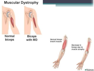
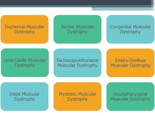
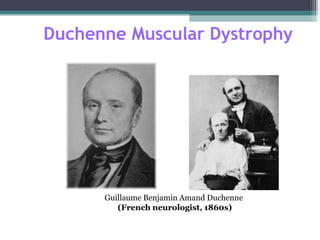
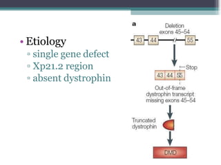
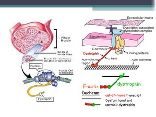
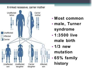
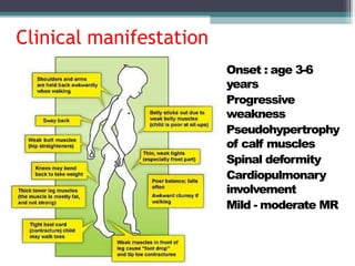
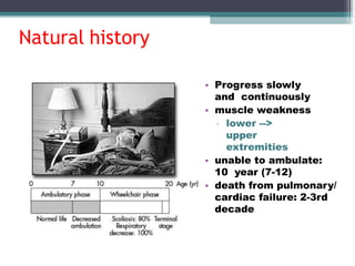
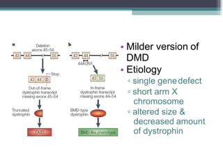
• Muscular dystrophy is a heterogeneous
group of inherited disorders recognized
weakness and loss of muscle
by progressive degenerative muscle
tissue
(started in childhood).
• Affect muscles strength and action.
• Generalized or localized.
• Skeletal muscle and other organs may
involve
• Limitation: Difficulties with walking or Maintaining posture,
Muscle spasms. Neurological, Behavioral, Cardiac, or other
Functional limitations.
Classification
DMD, BMD,
• Sex-linked:
EDMD
• Autosomal recessive: LGMD,
infantile FSHD
• Autosomal dominant: FSHD,
distalMD, ocular MD,
oculopharyngeal MD.
Duchenne Muscular Dystrophy
Guillaume Benjamin Amand Duchenne
(French neurologist, 1860s)
• Etiology
▫ single gene defect
▫ Xp21.2 region
▫ absent dystrophin
• Most common
• male, Turner
syndrome
• 1:3500 live
male birth
• 1/3 new
mutation
• 65% family
history
Clinical manifestation
• Onset : age 3-6
years
• Progressive
weakness
• Pseudohypertrophy
of calf muscles
• Spinal deformity
• Cardiopulmonary
involvement
• Mild - moderate MR
Natural history
• Progress slowly
and continuously
• muscle weakness
▫ lower -->
upper
extremities
• unable to ambulate:
10 year (7-12)
• death from pulmonary/
cardiac failure: 2-3rd
decade
Pseudohypertrhophy of calf muscle, Tip toe gait
forward tilt of pelvis, compensatory lordosis
Disappearance of
lordosis while sitting
DMD: Diagnosis
• Gait
• Absent DTR
• Ober test
• Thomas test
• Meyeron sign - child
slips through truncal
grasp
• Macroglossia
• IQ ~ 80
• IncreaseCPK
(200x)
• Myopathicchangein
EMG
Bx:m. degeneration
• Immunoblotting:
Absencedystrophin
• Myocardial deterioration • DNA mutation
analysis
Becker Muscular Dystrophy
Peter Emil Becker
(German doctor, 1950s)
• Milder version of
DMD
• Etiology
▫ single genedefect
▫ short arm X
chromosome
▫ altered size &
decreased amount
of dystrophin
• Less common
▫ 1: 30000 live male birth
• Less severe
• Family history: atypical MD
• Similar & less severe than DMD
• Onset: age > 7 years
• Pseudohypertrophy of calf
• Equinous and varus foot
• High rate of scoliosis
• Less frequent cardiac involvement
Clinical features
Diagnosis
• The same as DMD
• Increase CPK (<200x)
• Decrease dystrophin and/or altered size
Natural history
▫ Slower progression
▫ ambulate until adolescence
▫ longer life expectancy
Treatment
▫ the same as in DMD
▫ forefoot equinous: plantar release, midfoot dorsal-
wedge osteotomy
muscular dystrophy.pptx

muscular dystrophy.pptx

  • 1.
  • 2.
    • Causes • Inheritance •Dominant genes • Recessive gene Depends on the age when symptoms appear, and the types of symptoms that develop. • Risk • Because these are inherited disorders, risk include a family history of muscular dystrophy How Many People Are Affected It is estimated that between 50,000 -250,000 are affected annually. 1 per 3500 live male births
  • 4.
    • Muscular dystrophyis a heterogeneous group of inherited disorders recognized weakness and loss of muscle by progressive degenerative muscle tissue (started in childhood). • Affect muscles strength and action. • Generalized or localized. • Skeletal muscle and other organs may involve • Limitation: Difficulties with walking or Maintaining posture, Muscle spasms. Neurological, Behavioral, Cardiac, or other Functional limitations.
  • 8.
    Classification DMD, BMD, • Sex-linked: EDMD •Autosomal recessive: LGMD, infantile FSHD • Autosomal dominant: FSHD, distalMD, ocular MD, oculopharyngeal MD.
  • 11.
    Duchenne Muscular Dystrophy GuillaumeBenjamin Amand Duchenne (French neurologist, 1860s)
  • 12.
    • Etiology ▫ singlegene defect ▫ Xp21.2 region ▫ absent dystrophin
  • 14.
    • Most common •male, Turner syndrome • 1:3500 live male birth • 1/3 new mutation • 65% family history
  • 15.
    Clinical manifestation • Onset: age 3-6 years • Progressive weakness • Pseudohypertrophy of calf muscles • Spinal deformity • Cardiopulmonary involvement • Mild - moderate MR
  • 16.
    Natural history • Progressslowly and continuously • muscle weakness ▫ lower --> upper extremities • unable to ambulate: 10 year (7-12) • death from pulmonary/ cardiac failure: 2-3rd decade
  • 19.
    Pseudohypertrhophy of calfmuscle, Tip toe gait forward tilt of pelvis, compensatory lordosis
  • 20.
  • 21.
    DMD: Diagnosis • Gait •Absent DTR • Ober test • Thomas test • Meyeron sign - child slips through truncal grasp • Macroglossia • IQ ~ 80 • IncreaseCPK (200x) • Myopathicchangein EMG Bx:m. degeneration • Immunoblotting: Absencedystrophin • Myocardial deterioration • DNA mutation analysis
  • 23.
    Becker Muscular Dystrophy PeterEmil Becker (German doctor, 1950s)
  • 24.
    • Milder versionof DMD • Etiology ▫ single genedefect ▫ short arm X chromosome ▫ altered size & decreased amount of dystrophin
  • 25.
    • Less common ▫1: 30000 live male birth • Less severe • Family history: atypical MD • Similar & less severe than DMD • Onset: age > 7 years • Pseudohypertrophy of calf • Equinous and varus foot • High rate of scoliosis • Less frequent cardiac involvement Clinical features
  • 26.
    Diagnosis • The sameas DMD • Increase CPK (<200x) • Decrease dystrophin and/or altered size Natural history ▫ Slower progression ▫ ambulate until adolescence ▫ longer life expectancy Treatment ▫ the same as in DMD ▫ forefoot equinous: plantar release, midfoot dorsal- wedge osteotomy