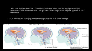
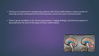
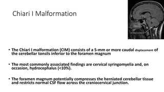
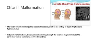
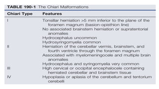
CHIARI
MALFORMATIONS
Journal Title
Clinical Presentation of Chiari I Malformation
and Syringomyelia in Children
Jonathan Pindrik, James M. Johnston Jr
Department of Neurosurgery, Children’s of
Alabama, University of Alabama, Birmingham (UAB)
Neurosurgery Clinics of North America- 2015
• The Chiari malformations are a collection of hindbrain abnormalities ranging from simple
herniation of the cerebellar tonsils through the foramen magnum to complete agenesis of the
cerebellum.
• It is unlikely that a unifying pathophysiology underlies all of these findings
• The focus of treatment for symptomatic patients with Chiari malformations mainly consists of
restoring normal cerebrospinal fluid (CSF) dynamics across the cranio cervical junction.
• There is great variability in the clinical presentation, imaging findings, and technical aspects of
decompression for each of the types of Chiari malformation.
Chiari I Malformation
• The Chiari I malformation (CIM) consists of a 5-mm or more caudal displacement of
the cerebellar tonsils inferior to the foramen magnum
• The most commonly associated findings are cervical syringomyelia and, on
occasion, hydrocephalus (<10%).
• The foramen magnum potentially compresses the herniated cerebellar tissue
and restricts normal CSF flow across the craniocervical junction.
• This form of hindbrain herniation may present with commonly recognized or rarely reported
clinical symptoms, and also may be found incidentally
• Incidental finding in radiography is 3% to 4%.
• Approximately 15% to 37% of pediatric patients with radiographic evidence of inferior
cerebellar tonsillar ectopia may be asymptomatic, based on large retrospective reviews.
Chiari II Malformation
• The Chiari II malformation (CIIM) is seen almost exclusively in the setting of myelodysplasia and
hydrocephalus.
• In type II malformations, the structures herniating through the foramen magnum include the
cerebellar vermis, brainstem, and fourth ventricle
Chiari II Malformation
• In addition to these neural structures, the accompanying choroid
plexus and the associated basilar artery and posterior inferior
cerebellar arteries may also be caudally displaced.
• The posterior fossa is often small and the foramen magnum larger
than normal, and syringomyelia is seen in many of these patients.
Chiari III Malformation
• The Chiari III malformation is the rarest of the Chiari malformations,
with herniation of cerebellum and brainstem into a posterior
encephalocele and other intracranial anomalies that are seen in CIIM
• This is the most severe form of hindbrain herniation, and its
management is often problematic from both a technical and an ethical
point of view.
• Patients with a Chiari III malformation generally have a poor prognosis
Chiari III Malformation
• Severe neurological, developmental, and cranial nerve defects, in
conjunction with seizures and respiratory insufficiency, are
common
• Surgical planning follows the same basic principles of any
encephalocele closure.
Chiari IV Malformation
• Chiari expanded his initial classification in a follow-up study published in
1896, in which he added a fourth subgroup consisting of cerebellar
hypoplasia or aplasia (i.e., the Chiari IV malformation).
• In these patients, the posterior fossa is relatively normal in size, and there
is an absence of any hindbrain herniation.
• Although this was included in Chiari’s classification of rhombencephalic
malformations, it is more appropriate to include this in the category of
posterior fossa cysts
Chiari 0 Malformation
• The Chiari 0 malformation is defined as syringomyelia without tonsillar
herniation that responds to posterior fossa decompression.
• MRI of the entire neuraxis rules out other causes of a syrinx.
• Ultimately, abnormal CSF flow at the posterior fossa or foramen magnum is the
suspected cause.
• Patients undergo a posterior fossa decompression and duraplasty without direct
fenestration or management of the syrinx, which is followed by symptom
resolution
Chiari 1.5 Malformation
• Implying that only cerebellar tonsillar tissue is herniated through the
foramen magnum, the CIM does not fully encompass all varieties of
hindbrain herniation that also include descent of the brainstem.
• Severe form of Chiari 1. Entire cervicomedullary junction (and obex) is
situated below the foramen magnum.
• Many of these cases have platybasia. Clinical manifestations and response
to suboccipital decompression are similar to Chiari I with the exception
that syringomyelia persisted in almost twice as many Chiari 1.5 cases
(13.6%) as Chiari I (6.9%).
SIGNS AND SYMPTOMS Chiari I Malformation
• The most common presenting symptom is pain (60% to 70%), usually
occipital and upper cervical in location, and often induced by Valsalva
maneuvers such as laughing, sneezing, and coughing.
• In infants and children who are unable to communicate verbally,
headaches may manifest simply as crying and irritability.
• Careful investigation reveals that many patients have some type of
ophthalmologic or otologic disturbance at diagnosis.
SIGNS AND SYMPTOMS Chiari I Malformation
• Ophthalmologic symptoms include blurry vision, nystagmus, extraocular
muscle palsies, diplopia, and visual field deficits
• Otologic complaints consist of tinnitus, fluctuating hearing loss, vertigo,
and nausea.
• Signs at presentation have included weakness, atrophy, hyperreflexia,
cape-like sensory loss, ataxia, and lower cranial nerve dysfunction.
SIGNS AND SYMPTOMS Chiari I Malformation
• By stretching the centrally located ventral white commissure through
a syrinx, pain and temperature signals cannot cross to the
contralateral spinothalamic tract. The loss of pain and temperature
sensation occurs only at the levels that are served by stretched
spinothalamic fibers.
• Down-beat nystagmus is reported to be specific for lesions involving
the cervicomedullary junction.
SIGNS AND SYMPTOMS Chiari I Malformation
• Spinal cord dysfunction is the result of direct cord compression or syringomyelia.
• Characterized objectively by Cobb angles, scoliosis demonstrates strong association with
syringomyelia in CM.
• In general, the following rules apply.
Most (but not all) pediatric CM1 patients with scoliosis have underlying syringomyelia; and Not
all CM1 patients with syringomyelia have scoliosis.
• Syringomyelia in CM1 demonstrates predilection for the following regions:
Cervical spinal cord (15%–21%),
Cervicothoracic spinal cord (12%–25%);
Thoracic spinal cord (15%–16%);
Lumbar spinal cord (3%–4%); and
Holocord (39%–44%).
CLINICAL PRESENTATION DURING THE NEONATAL PERIOD AND INFANCY
(AGE 0 TO 3 YEARS)
• Owing to limitations of communication, neonates or infants with
symptomatic CM1 often present with generalized irritability.
• Clinical signs
1} Opisthotonus, frequent neck extension/arching, and/or apneic
episodes
2} Medullary compression with lower cranial nerve dysfunction
may manifest as : Dysphagia, choking, or aspiration; Poor feeding,
failure to thrive; Gastroesophageal reflux; Persistent cough; Snoring
or episodic sleep apnea; Stridor; and Recurrent respiratory
infections
CLINICAL PRESENTATION IN TODDLERS (AGE 3 TO 5 YEARS)
• Toddlers with adequate verbal skills may report headache pain or
discomfort in the upper neck. Occipital headaches represent a
component of the clinical presentation of CM1 in up to 40% to 57%
of patients during the toddler stages and early childhood.
• They may present frequently with syringomyelia and/or scoliosis -
back or shoulder pain, paresthesia, gait disturbance, and/or
clumsiness
• Symptoms or signs of medullary and lower cranial nerve dysfunction
(sleep apnea, oropharyngeal dysphagia, dysarthria, absent gag
reflex) may be present
CLINICAL PRESENTATION DURING CHILDHOOD AND ADOLESCENCE (AGE
5 YEARS AND OLDER)
• These patients report classic symptoms of CM1
• occipital headaches and/or neck pain, often induced by Valsalva-type maneuvers (straining for
a bowel movement, laughing, coughing, sneezing, etc)
• Scoliosis, typically associated with syringomyelia (present in 19%–76% of CM1 patients),
represents another important component of the clinical presentation in this group of patients
• Classic symptoms and physical examination
signs suggesting syringomyelia include:
A) Upper extremity weakness ( intrinsic
muscles of the hand;)
B) Pain and temperature sensory loss in a
‘cape- like’ distribution;
C) Preservation of light touch sensation and
proprioception;
D) Absence of superficial abdominal reflexes
ipsilateral to the convexity of scoliosis
CLINICAL CONDITIONS ASSOCIATED WITH CHIARI I
MALFORMATION
1) craniocervical osseous abnormalities
33% - 38%
2) Hydrocephalus (8%–10%)
3) Neurofibromatosis type I (up to 5%)
4) Growth hormone deficiency
(idiopathic; around 4%)
5) Sprengel deformity (abnormal
elevation of the scapula)
6) Pierre Robin syndrome.
Imaging Chiari I Malformations
• The diagnosis of CIM should include the absence of an intracranial mass
lesion, Dandy-Walker malformation, or hydrocephalus— all of which may
cause tonsillar displacement secondary to raised intracranial pressure.
• The imaging method of choice is MRI
• The tonsillar herniation was noted to be clearly patho- logic when it
exceeded 5 mm and borderline between 3 and 5 mm.
Cerebrospinal Fluid Flow Studies and Cine-
ModeMagnetic Resonance Imaging
• One technique to assist in the surgical decision making of
patients with Chiari malformations is motion-sensitive MRI, or
cine- mode MRI.
• As opposed to static MRI, cine MRI may demonstrate lack of CSF
flow patterns that can occur in the setting of CIM.
PATHOPHYSIOLOGY
• The theories of pathogenesis of the Chiari malformations may be
grouped into
1.The hindbrain dysgenesis and developmental arrest theory
2.The caudal traction theory,
3.The hydrocephalus and hydrodynamic theory of Gardner,
4.The small posterior fossa/ hindbrain overgrowth theory, and
5.The lack of embryologic ventricular distention theory.
PATHOPHYSIOLOGY
• It is unlikely that Chiari types I and II malformations share a
common pathoembryologic origin.
• Nonetheless, the result of cerebellar descent, crowded foramen
magnum, and impaired CSF flow across the craniocervical
junction may share a common etiology in the development of
syringomyelia.
PATHOPHYSIOLOGY Chiari I Malformation
• Chiari’s belief that hydrocephalus causes CIM has been abandoned
because hydrocephalus is present in only a minority of cases.
• Idiopathic CIM may be the result of mesodermal defects that create a
congenitally small posterior fossa, leading to compression of the neural
elements and herniation through the foramen magnum.
• Neural dysfunction and deleterious alterations of CSF flow may ensue,
resulting in the symptoms associated with CIM.
PATHOPHYSIOLOGY Chiari I Malformation
• Negative CSF pressure in the spinal compartment relative to the
intracranial compartment creates a “sump effect” that forces the
tonsils down through the foramen magnum.
• Once CSF flow is blocked at the foramen magnum, low intraspinal
pressures can be accentuated and perpetuated by continuous
absorption of CSF through spinal
pathways, further worsening the clinical situation.
PATHOPHYSIOLOGY Chiari I Malformation
• Lumboperitoneal shunting, repetitive lumbar punctures, lumbar
drainage, and chronic spinal CSF leaks of an iatrogenic nature are
all familiar causes of an acquired CIM.
• Although CIM is thought to occur sporadically, inheritable genetic
factors may be responsible for a minority of cases. This conclusion
is based on studies in which disease incidence is abnormally higher
in some families than in the general population.
TREATMENT Chiari I Malformation
• There is no effective nonsurgical alternative to operative decompression
for patients with symptomatic CIM.
• A 2004 opinion survey of neurosurgeons concluded there was
widespread agreement to treat patients with a syrinx and progressive
scoliosis or symptoms.
• the farther away (e.g., lower thoracic, lumbar) from the craniocervical
junction a syrinx is, the less likely it is to respond to posterior fossa
decompression.
TREATMENT Chiari I Malformation
• Patients suspected of having CIM should undergo MRI of the posterior
fossa and entire cervical spine to assess tonsillar ectopia and screen for a
syrinx.
• Furthermore, patients with long-tract signs and no evidence of a cervical
syrinx should also have their thoracic and lumbar spine screened.
• Also necessary is imaging to rule out hydrocephalus.
TREATMENT Chiari I Malformation
• Flexion-extension radiographs of the cervical spine are suggested for
individuals in whom there is a concern for instability. This is especially
relevant in this group because cervical spine anomalies are very common.
• All patients with a syrinx (not a patent central canal), regardless of the
size, location, or other associated symptoms, are offered surgical
intervention. This strategy is based on the belief that a syrinx is indicative
of pathologic forces acting on the spinal cord that should be corrected to
prevent permanent cord damage.
TREATMENT Chiari I Malformation
• The decision about whether to operate is a bit more complicated in patients
without a syrinx.
• Individuals with minimal tonsillar ectopia, no objective neurological findings,
and mild headaches that are not lifestyle limiting are observed.
• Surgery is deferred until symptoms worsen and become unacceptable to the
patient and headaches are refractory to multidisciplinary management.
• Patients with lifestyle-limiting headaches or objective neurological
abnormalities, especially respiratory or cranial nerve dysfunction, receive earlier
surgical intervention.
TREATMENT Chiari I Malformation
• If hydrocephalus coexists or any question of raised intracranial pressure is
present, this situation should be resolved before consideration is given to a
decompression.
• The purpose of the Chiari Decompression operation is to enlarge the bony
area of the craniocervical junction and expand the dura surrounding the
brainstem.
• By doing this, the surgeon wishes to relieve any direct compression and
facilitate adequate outflow of CSF from the fourth ventricle.
COMPLICATIONS
• Posterior fossa decompression is relatively safe but not without
complications.
• Direct vascular or neural injury, pseudomeningocele, CSF leaks, and
meningitis are well recognized.
• Bleeding from dural venous lakes can be profuse at times.
• Less common complications include occipital-cervical instability, acute
postoperative hydrocephalus secondary to infratentorial hygromas, and
anterior brainstem compression from a retroflexed odontoid.
COMPLICATIONS
• A complication unique to posterior fossa craniectomies is cerebellar
slump or ptosis, which results from extending a craniectomy so far
laterally that the cerebellum herniates through the craniectomy defect.
• This can cause headaches (different from typical Chiari I headaches),
obstruction of CSF flow with syrinx formation, and a variety of motor,
sensory, and cranial nerve deficits.
• Cranioplasty to buttress the cerebellum into place is the most definitive
treatment.
CONCLUSION
• Chiari presented his series of hindbrain herniations more than 100 years
ago and attempted to elucidate a unifying theory as to their formation.
• Today, we understand the Chiari malformations to involve an abnormality
at the craniocervical junction resulting in impaired neural function and CSF
hydrodynamics
• However, the pathophysiology of each malformation is likely very
different, and the management is tailored to each individual.
CONCLUSION
• There are some consistent simple tenets in the management of
these patients:
• (1) patient selection is important in determining who will benefit
from surgical intervention
• (2) assurance of normal intracranial physiology before suboccipital
decompression is paramount
CONCLUSION
• (3) restoration of normal CSF dynamic flow from the fourth ventricle
to the subarachnoid space and relief of direct brainstem
compression are the goals of surgery.
• Adequate decompression can be accomplished in a variety of ways
but must allow free flow of CSF between the intracranial
compartment and spinal subarachnoid space to successfully treat
this patient population.

Arnold chiari malformation and types.pptx

  • 1.
  • 2.
    Journal Title Clinical Presentationof Chiari I Malformation and Syringomyelia in Children Jonathan Pindrik, James M. Johnston Jr Department of Neurosurgery, Children’s of Alabama, University of Alabama, Birmingham (UAB) Neurosurgery Clinics of North America- 2015
  • 3.
    • The Chiarimalformations are a collection of hindbrain abnormalities ranging from simple herniation of the cerebellar tonsils through the foramen magnum to complete agenesis of the cerebellum. • It is unlikely that a unifying pathophysiology underlies all of these findings
  • 4.
    • The focusof treatment for symptomatic patients with Chiari malformations mainly consists of restoring normal cerebrospinal fluid (CSF) dynamics across the cranio cervical junction. • There is great variability in the clinical presentation, imaging findings, and technical aspects of decompression for each of the types of Chiari malformation.
  • 5.
    Chiari I Malformation •The Chiari I malformation (CIM) consists of a 5-mm or more caudal displacement of the cerebellar tonsils inferior to the foramen magnum • The most commonly associated findings are cervical syringomyelia and, on occasion, hydrocephalus (<10%). • The foramen magnum potentially compresses the herniated cerebellar tissue and restricts normal CSF flow across the craniocervical junction.
  • 6.
    • This formof hindbrain herniation may present with commonly recognized or rarely reported clinical symptoms, and also may be found incidentally • Incidental finding in radiography is 3% to 4%. • Approximately 15% to 37% of pediatric patients with radiographic evidence of inferior cerebellar tonsillar ectopia may be asymptomatic, based on large retrospective reviews.
  • 7.
    Chiari II Malformation •The Chiari II malformation (CIIM) is seen almost exclusively in the setting of myelodysplasia and hydrocephalus. • In type II malformations, the structures herniating through the foramen magnum include the cerebellar vermis, brainstem, and fourth ventricle
  • 8.
    Chiari II Malformation •In addition to these neural structures, the accompanying choroid plexus and the associated basilar artery and posterior inferior cerebellar arteries may also be caudally displaced. • The posterior fossa is often small and the foramen magnum larger than normal, and syringomyelia is seen in many of these patients.
  • 9.
    Chiari III Malformation •The Chiari III malformation is the rarest of the Chiari malformations, with herniation of cerebellum and brainstem into a posterior encephalocele and other intracranial anomalies that are seen in CIIM • This is the most severe form of hindbrain herniation, and its management is often problematic from both a technical and an ethical point of view. • Patients with a Chiari III malformation generally have a poor prognosis
  • 10.
    Chiari III Malformation •Severe neurological, developmental, and cranial nerve defects, in conjunction with seizures and respiratory insufficiency, are common • Surgical planning follows the same basic principles of any encephalocele closure.
  • 11.
    Chiari IV Malformation •Chiari expanded his initial classification in a follow-up study published in 1896, in which he added a fourth subgroup consisting of cerebellar hypoplasia or aplasia (i.e., the Chiari IV malformation). • In these patients, the posterior fossa is relatively normal in size, and there is an absence of any hindbrain herniation. • Although this was included in Chiari’s classification of rhombencephalic malformations, it is more appropriate to include this in the category of posterior fossa cysts
  • 12.
    Chiari 0 Malformation •The Chiari 0 malformation is defined as syringomyelia without tonsillar herniation that responds to posterior fossa decompression. • MRI of the entire neuraxis rules out other causes of a syrinx. • Ultimately, abnormal CSF flow at the posterior fossa or foramen magnum is the suspected cause. • Patients undergo a posterior fossa decompression and duraplasty without direct fenestration or management of the syrinx, which is followed by symptom resolution
  • 16.
    Chiari 1.5 Malformation •Implying that only cerebellar tonsillar tissue is herniated through the foramen magnum, the CIM does not fully encompass all varieties of hindbrain herniation that also include descent of the brainstem. • Severe form of Chiari 1. Entire cervicomedullary junction (and obex) is situated below the foramen magnum. • Many of these cases have platybasia. Clinical manifestations and response to suboccipital decompression are similar to Chiari I with the exception that syringomyelia persisted in almost twice as many Chiari 1.5 cases (13.6%) as Chiari I (6.9%).
  • 17.
    SIGNS AND SYMPTOMSChiari I Malformation • The most common presenting symptom is pain (60% to 70%), usually occipital and upper cervical in location, and often induced by Valsalva maneuvers such as laughing, sneezing, and coughing. • In infants and children who are unable to communicate verbally, headaches may manifest simply as crying and irritability. • Careful investigation reveals that many patients have some type of ophthalmologic or otologic disturbance at diagnosis.
  • 18.
    SIGNS AND SYMPTOMSChiari I Malformation • Ophthalmologic symptoms include blurry vision, nystagmus, extraocular muscle palsies, diplopia, and visual field deficits • Otologic complaints consist of tinnitus, fluctuating hearing loss, vertigo, and nausea. • Signs at presentation have included weakness, atrophy, hyperreflexia, cape-like sensory loss, ataxia, and lower cranial nerve dysfunction.
  • 19.
    SIGNS AND SYMPTOMSChiari I Malformation • By stretching the centrally located ventral white commissure through a syrinx, pain and temperature signals cannot cross to the contralateral spinothalamic tract. The loss of pain and temperature sensation occurs only at the levels that are served by stretched spinothalamic fibers. • Down-beat nystagmus is reported to be specific for lesions involving the cervicomedullary junction.
  • 20.
    SIGNS AND SYMPTOMSChiari I Malformation • Spinal cord dysfunction is the result of direct cord compression or syringomyelia. • Characterized objectively by Cobb angles, scoliosis demonstrates strong association with syringomyelia in CM. • In general, the following rules apply. Most (but not all) pediatric CM1 patients with scoliosis have underlying syringomyelia; and Not all CM1 patients with syringomyelia have scoliosis.
  • 21.
    • Syringomyelia inCM1 demonstrates predilection for the following regions: Cervical spinal cord (15%–21%), Cervicothoracic spinal cord (12%–25%); Thoracic spinal cord (15%–16%); Lumbar spinal cord (3%–4%); and Holocord (39%–44%).
  • 22.
    CLINICAL PRESENTATION DURINGTHE NEONATAL PERIOD AND INFANCY (AGE 0 TO 3 YEARS) • Owing to limitations of communication, neonates or infants with symptomatic CM1 often present with generalized irritability. • Clinical signs 1} Opisthotonus, frequent neck extension/arching, and/or apneic episodes 2} Medullary compression with lower cranial nerve dysfunction may manifest as : Dysphagia, choking, or aspiration; Poor feeding, failure to thrive; Gastroesophageal reflux; Persistent cough; Snoring or episodic sleep apnea; Stridor; and Recurrent respiratory infections
  • 23.
    CLINICAL PRESENTATION INTODDLERS (AGE 3 TO 5 YEARS) • Toddlers with adequate verbal skills may report headache pain or discomfort in the upper neck. Occipital headaches represent a component of the clinical presentation of CM1 in up to 40% to 57% of patients during the toddler stages and early childhood. • They may present frequently with syringomyelia and/or scoliosis - back or shoulder pain, paresthesia, gait disturbance, and/or clumsiness • Symptoms or signs of medullary and lower cranial nerve dysfunction (sleep apnea, oropharyngeal dysphagia, dysarthria, absent gag reflex) may be present
  • 24.
    CLINICAL PRESENTATION DURINGCHILDHOOD AND ADOLESCENCE (AGE 5 YEARS AND OLDER) • These patients report classic symptoms of CM1 • occipital headaches and/or neck pain, often induced by Valsalva-type maneuvers (straining for a bowel movement, laughing, coughing, sneezing, etc) • Scoliosis, typically associated with syringomyelia (present in 19%–76% of CM1 patients), represents another important component of the clinical presentation in this group of patients
  • 25.
    • Classic symptomsand physical examination signs suggesting syringomyelia include: A) Upper extremity weakness ( intrinsic muscles of the hand;) B) Pain and temperature sensory loss in a ‘cape- like’ distribution; C) Preservation of light touch sensation and proprioception; D) Absence of superficial abdominal reflexes ipsilateral to the convexity of scoliosis
  • 26.
    CLINICAL CONDITIONS ASSOCIATEDWITH CHIARI I MALFORMATION 1) craniocervical osseous abnormalities 33% - 38% 2) Hydrocephalus (8%–10%) 3) Neurofibromatosis type I (up to 5%) 4) Growth hormone deficiency (idiopathic; around 4%) 5) Sprengel deformity (abnormal elevation of the scapula) 6) Pierre Robin syndrome.
  • 27.
    Imaging Chiari IMalformations • The diagnosis of CIM should include the absence of an intracranial mass lesion, Dandy-Walker malformation, or hydrocephalus— all of which may cause tonsillar displacement secondary to raised intracranial pressure. • The imaging method of choice is MRI • The tonsillar herniation was noted to be clearly patho- logic when it exceeded 5 mm and borderline between 3 and 5 mm.
  • 31.
    Cerebrospinal Fluid FlowStudies and Cine- ModeMagnetic Resonance Imaging • One technique to assist in the surgical decision making of patients with Chiari malformations is motion-sensitive MRI, or cine- mode MRI. • As opposed to static MRI, cine MRI may demonstrate lack of CSF flow patterns that can occur in the setting of CIM.
  • 32.
    PATHOPHYSIOLOGY • The theoriesof pathogenesis of the Chiari malformations may be grouped into 1.The hindbrain dysgenesis and developmental arrest theory 2.The caudal traction theory, 3.The hydrocephalus and hydrodynamic theory of Gardner, 4.The small posterior fossa/ hindbrain overgrowth theory, and 5.The lack of embryologic ventricular distention theory.
  • 33.
    PATHOPHYSIOLOGY • It isunlikely that Chiari types I and II malformations share a common pathoembryologic origin. • Nonetheless, the result of cerebellar descent, crowded foramen magnum, and impaired CSF flow across the craniocervical junction may share a common etiology in the development of syringomyelia.
  • 34.
    PATHOPHYSIOLOGY Chiari IMalformation • Chiari’s belief that hydrocephalus causes CIM has been abandoned because hydrocephalus is present in only a minority of cases. • Idiopathic CIM may be the result of mesodermal defects that create a congenitally small posterior fossa, leading to compression of the neural elements and herniation through the foramen magnum. • Neural dysfunction and deleterious alterations of CSF flow may ensue, resulting in the symptoms associated with CIM.
  • 35.
    PATHOPHYSIOLOGY Chiari IMalformation • Negative CSF pressure in the spinal compartment relative to the intracranial compartment creates a “sump effect” that forces the tonsils down through the foramen magnum. • Once CSF flow is blocked at the foramen magnum, low intraspinal pressures can be accentuated and perpetuated by continuous absorption of CSF through spinal pathways, further worsening the clinical situation.
  • 36.
    PATHOPHYSIOLOGY Chiari IMalformation • Lumboperitoneal shunting, repetitive lumbar punctures, lumbar drainage, and chronic spinal CSF leaks of an iatrogenic nature are all familiar causes of an acquired CIM. • Although CIM is thought to occur sporadically, inheritable genetic factors may be responsible for a minority of cases. This conclusion is based on studies in which disease incidence is abnormally higher in some families than in the general population.
  • 37.
    TREATMENT Chiari IMalformation • There is no effective nonsurgical alternative to operative decompression for patients with symptomatic CIM. • A 2004 opinion survey of neurosurgeons concluded there was widespread agreement to treat patients with a syrinx and progressive scoliosis or symptoms. • the farther away (e.g., lower thoracic, lumbar) from the craniocervical junction a syrinx is, the less likely it is to respond to posterior fossa decompression.
  • 38.
    TREATMENT Chiari IMalformation • Patients suspected of having CIM should undergo MRI of the posterior fossa and entire cervical spine to assess tonsillar ectopia and screen for a syrinx. • Furthermore, patients with long-tract signs and no evidence of a cervical syrinx should also have their thoracic and lumbar spine screened. • Also necessary is imaging to rule out hydrocephalus.
  • 39.
    TREATMENT Chiari IMalformation • Flexion-extension radiographs of the cervical spine are suggested for individuals in whom there is a concern for instability. This is especially relevant in this group because cervical spine anomalies are very common. • All patients with a syrinx (not a patent central canal), regardless of the size, location, or other associated symptoms, are offered surgical intervention. This strategy is based on the belief that a syrinx is indicative of pathologic forces acting on the spinal cord that should be corrected to prevent permanent cord damage.
  • 40.
    TREATMENT Chiari IMalformation • The decision about whether to operate is a bit more complicated in patients without a syrinx. • Individuals with minimal tonsillar ectopia, no objective neurological findings, and mild headaches that are not lifestyle limiting are observed. • Surgery is deferred until symptoms worsen and become unacceptable to the patient and headaches are refractory to multidisciplinary management. • Patients with lifestyle-limiting headaches or objective neurological abnormalities, especially respiratory or cranial nerve dysfunction, receive earlier surgical intervention.
  • 41.
    TREATMENT Chiari IMalformation • If hydrocephalus coexists or any question of raised intracranial pressure is present, this situation should be resolved before consideration is given to a decompression. • The purpose of the Chiari Decompression operation is to enlarge the bony area of the craniocervical junction and expand the dura surrounding the brainstem. • By doing this, the surgeon wishes to relieve any direct compression and facilitate adequate outflow of CSF from the fourth ventricle.
  • 42.
    COMPLICATIONS • Posterior fossadecompression is relatively safe but not without complications. • Direct vascular or neural injury, pseudomeningocele, CSF leaks, and meningitis are well recognized. • Bleeding from dural venous lakes can be profuse at times. • Less common complications include occipital-cervical instability, acute postoperative hydrocephalus secondary to infratentorial hygromas, and anterior brainstem compression from a retroflexed odontoid.
  • 43.
    COMPLICATIONS • A complicationunique to posterior fossa craniectomies is cerebellar slump or ptosis, which results from extending a craniectomy so far laterally that the cerebellum herniates through the craniectomy defect. • This can cause headaches (different from typical Chiari I headaches), obstruction of CSF flow with syrinx formation, and a variety of motor, sensory, and cranial nerve deficits. • Cranioplasty to buttress the cerebellum into place is the most definitive treatment.
  • 44.
    CONCLUSION • Chiari presentedhis series of hindbrain herniations more than 100 years ago and attempted to elucidate a unifying theory as to their formation. • Today, we understand the Chiari malformations to involve an abnormality at the craniocervical junction resulting in impaired neural function and CSF hydrodynamics • However, the pathophysiology of each malformation is likely very different, and the management is tailored to each individual.
  • 45.
    CONCLUSION • There aresome consistent simple tenets in the management of these patients: • (1) patient selection is important in determining who will benefit from surgical intervention • (2) assurance of normal intracranial physiology before suboccipital decompression is paramount
  • 46.
    CONCLUSION • (3) restorationof normal CSF dynamic flow from the fourth ventricle to the subarachnoid space and relief of direct brainstem compression are the goals of surgery. • Adequate decompression can be accomplished in a variety of ways but must allow free flow of CSF between the intracranial compartment and spinal subarachnoid space to successfully treat this patient population.

Editor's Notes

  • #14 Macrae line , chamberlain line, mcgregor line
  • #27 KLIPPEL FIEL – abnormal fusion of 2 or more vertebra in cervical region, short neck , low hairline
  • #28 Dandy walker – agenesis or hypoplasia of vermis and cystic enlargement of 4th ventricle