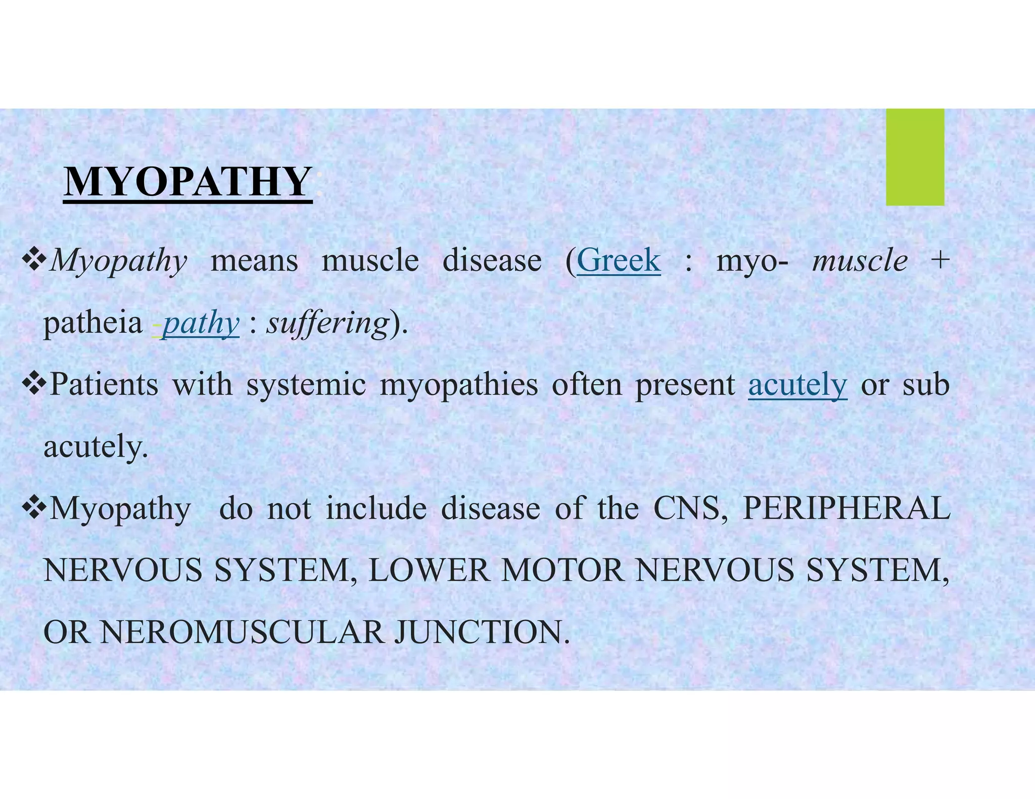
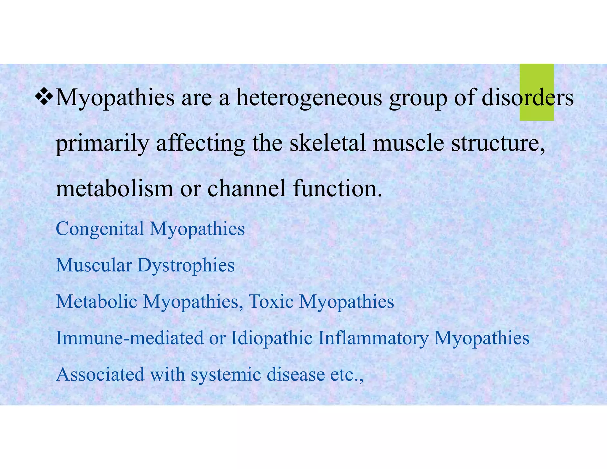
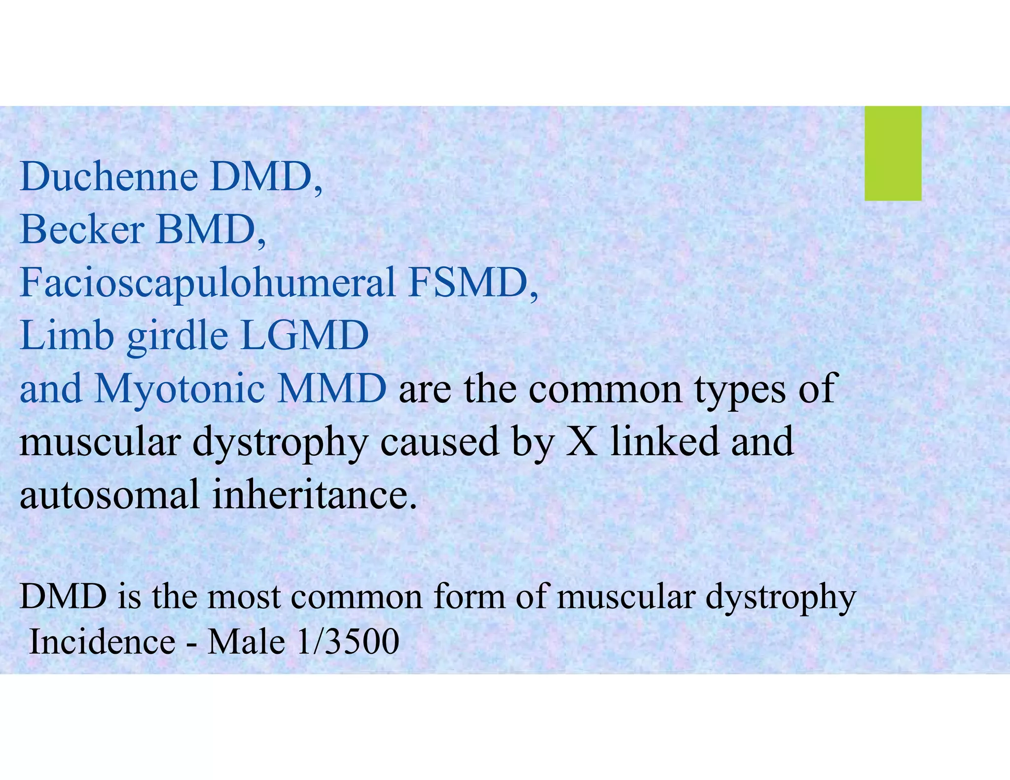
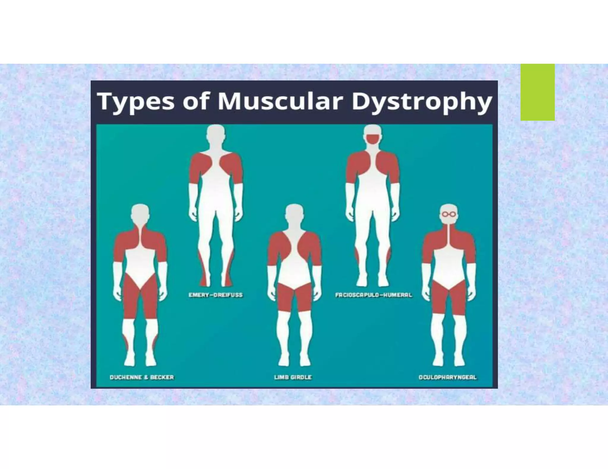
MUSCULAR DYSTROPHY
DR. K. MALATHI
M.D. SCHOLAR
DEPARTMENT OF KAYACHIKITSA
MYOPATHY:
Myopathy means muscle disease (Greek : myo- muscle +
patheia -pathy : suffering).
Patients with systemic myopathies often present acutely or sub
acutely.
Myopathy do not include disease of the CNS, PERIPHERAL
NERVOUS SYSTEM, LOWER MOTOR NERVOUS SYSTEM,
OR NEROMUSCULAR JUNCTION.
Myopathies are a heterogeneous group of disorders
primarily affecting the skeletal muscle structure,
metabolism or channel function.
Congenital Myopathies
Muscular Dystrophies
Metabolic Myopathies, Toxic Myopathies
Immune-mediated or Idiopathic Inflammatory Myopathies
Associated with systemic disease etc.,
MUSCULAR DYSTROPHY
Muscular dystrophies are inherited myopathies
characterized by progressive muscle weakness and
degeneration with subsequent replacement by
connective tissue and fat.
Also known as
myodystrophy and myodystrophia.
Duchenne DMD,
Becker BMD,
Facioscapulohumeral FSMD,
Limb girdle LGMD
and Myotonic MMD are the common types of
muscular dystrophy caused by X linked and
autosomal inheritance.
DMD is the most common form of muscular dystrophy
Incidence - Male 1/3500
Duchenne muscular dystrophy DMD
X linked recessive disorder – the gene
Dystrophin, which is located in the short arm
of the X-chromosome 21, is absent or
grossly deficient
Mainly in boys before the age of 5years
Lifespan rarely exceeds 30years
Duchenne is worse than the BeckerMD
 Calf muscle pseudohypertrophy
Gower’s sign – boy dependent
on his arms in order to stand up
 Waddling gait
 Khyphoscoliosis
Mental retardation
Gastro intestinal –Pseudo
Obstruction occurs
Cause of death – Dialated
Cardiomyopathy
 Muscle weakness over abdominal muscles
Weakness of pelvic girdle and proximal muscles
Thin thighs - thick calf muscles (pseudo
hypertrophied),
Poor balance
 Shoulders arms bent backwards, sways back
Pathophysiology
Dystrophin
deficiency
Abnormal cell membrane
with increased transient
local membrane disruption
and inflows of calcium
Altered calcium
channel activity,
increases inflow
of calcium, out
flow of CK
Impaired
homeostasis of
calcium and cell
death leads to
Cell necrosis
Muscular
dystrophy
Becker muscular distrophy - BMD
 X Linked recessive disorder
Less severe than DMD
Comes mainly to boys after the age of 5years , Live up
to 40years
Genetic defect in the same place of gene dystrophin,
but partially damaged – some amount of dystrophin
can be synthesised and utilised by muscles.
Proximal muscle weakness
Increased CPK levels
 Respiratory impairment is not
frequent
No intellectual impairement
Facioscapulohumoral dystrophy FSMD
 Autosomal dominant disorder
Males are more prominent then females
Occurs at any age
Gene defect is an abnormal deletion of
tendem 4q telomere gene
Life expectancy is normal
 Weakness in face and
shoulder girdle muscles
 Difficulty in closing the
eyes
 Sensory neural deafness
 Biceps and triceps severly
involved forearm
musclesvlooks
hypertrophied.
 beevor’s sign positive
Myotonic muscular dystrophy MMD
 An Autosomal dominant disorder
 Common in both gender adults
 Classified in to two types namely-
MYOTONIC DYSTROPHY 1 –DM1
PROXIMAL MYOTONIC MYOPATHY DM2 (PROMM)
The genetic defect is unstable expantion of the CTG
trinucleotide repeat in a serine theonine protein kinase gene on
chromosome 19q.
Weakness of the temporalis,
sternocledomastoids and facial
muscles
Ptosis and frontal baldhead
Grip myotonia
Early cataract
Cognitive impairment
Endocrine dysfunction
Difficulty in swallowing and
breatging
Cardiomyopathy and respiratory
insufficiency
Diagnosis
Dystrophin gene defect – detected by DNA
analysis
Muscle biopsy – deficiency of dystrophin and
replacement of connective tissue or scar and
fat
Serum creatinine kinase – CK (elevated)
EMG – fibrillation potentials
Screening for an associated cardiac and
respiratory abnormality.
Treatment
 Carticosteroids – to slow down the
progression of disease
Gene therapy
Stem cell therapy
Supportive:
Physiotherapy occupational therapy etc to
minimise the contracture
DR. K. MALATHI
PG SCHOLAR
DEPARTMENT OF KAYACHIKITSA

MUSCULAR DYSTROPHY PPT pdf

  • 1.
    MUSCULAR DYSTROPHY DR. K.MALATHI M.D. SCHOLAR DEPARTMENT OF KAYACHIKITSA
  • 2.
    MYOPATHY: Myopathy means muscledisease (Greek : myo- muscle + patheia -pathy : suffering). Patients with systemic myopathies often present acutely or sub acutely. Myopathy do not include disease of the CNS, PERIPHERAL NERVOUS SYSTEM, LOWER MOTOR NERVOUS SYSTEM, OR NEROMUSCULAR JUNCTION.
  • 3.
    Myopathies are aheterogeneous group of disorders primarily affecting the skeletal muscle structure, metabolism or channel function. Congenital Myopathies Muscular Dystrophies Metabolic Myopathies, Toxic Myopathies Immune-mediated or Idiopathic Inflammatory Myopathies Associated with systemic disease etc.,
  • 4.
    MUSCULAR DYSTROPHY Muscular dystrophiesare inherited myopathies characterized by progressive muscle weakness and degeneration with subsequent replacement by connective tissue and fat. Also known as myodystrophy and myodystrophia.
  • 5.
    Duchenne DMD, Becker BMD, FacioscapulohumeralFSMD, Limb girdle LGMD and Myotonic MMD are the common types of muscular dystrophy caused by X linked and autosomal inheritance. DMD is the most common form of muscular dystrophy Incidence - Male 1/3500
  • 6.
    Duchenne muscular dystrophyDMD X linked recessive disorder – the gene Dystrophin, which is located in the short arm of the X-chromosome 21, is absent or grossly deficient Mainly in boys before the age of 5years Lifespan rarely exceeds 30years Duchenne is worse than the BeckerMD
  • 7.
     Calf musclepseudohypertrophy Gower’s sign – boy dependent on his arms in order to stand up  Waddling gait
  • 8.
     Khyphoscoliosis Mental retardation Gastrointestinal –Pseudo Obstruction occurs Cause of death – Dialated Cardiomyopathy
  • 9.
     Muscle weaknessover abdominal muscles Weakness of pelvic girdle and proximal muscles Thin thighs - thick calf muscles (pseudo hypertrophied), Poor balance  Shoulders arms bent backwards, sways back
  • 11.
    Pathophysiology Dystrophin deficiency Abnormal cell membrane withincreased transient local membrane disruption and inflows of calcium Altered calcium channel activity, increases inflow of calcium, out flow of CK Impaired homeostasis of calcium and cell death leads to Cell necrosis Muscular dystrophy
  • 13.
    Becker muscular distrophy- BMD  X Linked recessive disorder Less severe than DMD Comes mainly to boys after the age of 5years , Live up to 40years Genetic defect in the same place of gene dystrophin, but partially damaged – some amount of dystrophin can be synthesised and utilised by muscles. Proximal muscle weakness Increased CPK levels
  • 14.
     Respiratory impairmentis not frequent No intellectual impairement
  • 15.
    Facioscapulohumoral dystrophy FSMD Autosomal dominant disorder Males are more prominent then females Occurs at any age Gene defect is an abnormal deletion of tendem 4q telomere gene Life expectancy is normal
  • 16.
     Weakness inface and shoulder girdle muscles  Difficulty in closing the eyes  Sensory neural deafness  Biceps and triceps severly involved forearm musclesvlooks hypertrophied.  beevor’s sign positive
  • 17.
    Myotonic muscular dystrophyMMD  An Autosomal dominant disorder  Common in both gender adults  Classified in to two types namely- MYOTONIC DYSTROPHY 1 –DM1 PROXIMAL MYOTONIC MYOPATHY DM2 (PROMM) The genetic defect is unstable expantion of the CTG trinucleotide repeat in a serine theonine protein kinase gene on chromosome 19q.
  • 18.
    Weakness of thetemporalis, sternocledomastoids and facial muscles Ptosis and frontal baldhead Grip myotonia Early cataract Cognitive impairment Endocrine dysfunction Difficulty in swallowing and breatging Cardiomyopathy and respiratory insufficiency
  • 20.
    Diagnosis Dystrophin gene defect– detected by DNA analysis Muscle biopsy – deficiency of dystrophin and replacement of connective tissue or scar and fat Serum creatinine kinase – CK (elevated) EMG – fibrillation potentials Screening for an associated cardiac and respiratory abnormality.
  • 21.
    Treatment  Carticosteroids –to slow down the progression of disease Gene therapy Stem cell therapy Supportive: Physiotherapy occupational therapy etc to minimise the contracture
  • 22.
    DR. K. MALATHI PGSCHOLAR DEPARTMENT OF KAYACHIKITSA