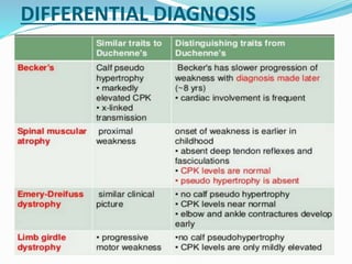
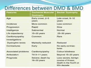
 Duchenne and Becker Muscular dystrophies.
 Emery-Dreifuss Muscular Dystrophy.
 Myotonic Muscular Dystrophy.
 Limb-Girdle Muscular Dystrophies.
 Facioscapulohumeral Muscular Dystrophy.
 Congenital Muscular Dystrophies.
TYPES:
FacioscapulohumeralMuscularDystrophy.
Duchenne muscular dystrophy (DMD)
It is the most common hereditary neuromuscular disease
affecting all races and ethnic groups.
 Its characteristic clinical features are progressive
weakness, intellectual impairment, hypertrophy of the
calves, and proliferation of connective tissue in muscle.
 This disease is inherited as an X-linked recessive trait. The
abnormal gene is at the Xp21 locus.
 The incidence is 1 in 3,600 live born infant boys.
 The disorder is caused by a mutation in the
gene, the largest gene located on the human X
chromosome which codes for the protein dystrophin.
 Without dystrophin, muscles are susceptible to
mechanical injury and undergo repeated cycles of
necrosis and regeneration.
 Ultimately, regenerative capabilities are exhausted or
inactivated.
Clinical Features
 •Clinical onset of muscular weakness usually occurs
between 2 and 3 years of age.
 •Histologic and laboratory evidence of a myopathy
may be observed from birth.
dystrophin
Posture changes during
progression of DMD
Stage 1
– Presymptomatic
Creatine kinase usually elevated
Positive family history
CLNICAL FEATURES
Stage 2- Early ambulatory
 Clumsy & Waddling gait, manifesting in children aged
2-6 years
 Progressive weakness in the proximal musculature,
initially in the lower extremities, but later involving
the neck flexors, shoulders, and arms.
 Possible toe-walking
 Can climb stairs
 Gower's sign -'climbing up legs' using the hands when
rising from the floor
Stage 3- Late ambulatory
 More difficulty walking Around age 8 years, most
patients notice difficulty with ascending stairs and
respiratory muscle strength begins a slow but steady
decline
 Cannot arise from the floor
 The forced vital capacity begins to gradually wane,
leading to symptoms of nocturnal hypoxemia such as
lethargy and early morning headaches
Stage 4 – Early nonambulatory
 Can self-propel for some time
 Able to maintain posture
 Possible development of scoliosis
Stage 5 – Late nonambulatory
 Scoliosis may progress, especially when more
wheelchair dependent
 If wheelchair bound and profoundly weak, patients
develop terminal respiratory or cardiac failure, usually
by the early 30s
 poor nutritional intake
 Contractures may develop
Complications
 Cardiomyopathy
 Decreased self-independence and mobility
 Lung failure (cause of death)
 Muscle tightness around joints
 Mental impairment
PHYSICAL EXAMINATION
 Generally, neck flexors, wrist extensors, quadriceps, tibialis
anterior, biceps, and triceps muscles are affected more.
 Deep tendon reflexes, slowly diminish and ultimately
disappear
 Calf muscle enlargement (pseudo hypertrophy)
contractures of the iliotibial bands, hip flexors, and heel
cords
 Equinovarus deformity of ankle is universal
 Asymmetric weakening of the paraspinal muscles leads to
kyphoscoliosis, which in turn further compromises
pulmonary and gastrointestinal function.
WORK UP
 Serum Creatine Phosphokinase (Elevated)
 Electromyography
 Nerve Conduction Velocity Study
 Molecular diagnosis
 Muscle biopsy
 Imaging Studies
 Electrocardiogram
 Echocardiogram
DIFFERENTIAL DIAGNOSIS
Differences between DMD & BMD
Carrier detection
 Carrier detection is an important aspect of the care
and evaluation of patients with DMD and their family
members
 For many years, CPK testing was the best method for
carrier detection; however, it is elevated in only two
thirds of female carriers
 If affected male in family has a known deletion or
duplication of the dystrophin gene, testing for carrier
status is performed accurately by testing possible
carriers for the same deletion or duplication
Start typing here..
TREATMENT
 There is no medical cure for this disease. Much can be
done to treat complications and to improve the quality
of life of affected children.
 Cardiac decompensation often responds initially well
to digoxin.
 Preservation of a good nutritional state is important.
GENETIC CONSELLING
Genetic counseling can offer many benefits for
families living with Duchenne or Becker
muscular dystrophy, including:
- Education about the disorder
- Coordination and explanation of genetic
testing
- Emotional counseling
EXON SKIPPING
In DMD, exon skipping is a potential treatment approach
that is under investigation to correct for specific genetic
mutations and restore production of dystrophin protein.
 As the name suggests, the principle of exon
skipping is to encourage the cellular machinery to
‘skip over’ an exon. Small pieces of DNA
called antisense oligonucleotides (AOs) or
‘molecular patches‘ are used to mask the exon
that you want to skip, so that it is ignored during
protein production
Drisapersen and eteplirsen are exon 51 skipping
antisense oligonucleotides that bind RNA and skip
(bridge) over the defective exon, thus producing a
shorter but potentially functional dystrophin protein.
71252786117d6aa1344a0f53179ba910 (1).pdf

71252786117d6aa1344a0f53179ba910 (1).pdf

  • 2.
     Duchenne andBecker Muscular dystrophies.  Emery-Dreifuss Muscular Dystrophy.  Myotonic Muscular Dystrophy.  Limb-Girdle Muscular Dystrophies.  Facioscapulohumeral Muscular Dystrophy.  Congenital Muscular Dystrophies. TYPES:
  • 3.
  • 4.
    Duchenne muscular dystrophy(DMD) It is the most common hereditary neuromuscular disease affecting all races and ethnic groups.  Its characteristic clinical features are progressive weakness, intellectual impairment, hypertrophy of the calves, and proliferation of connective tissue in muscle.  This disease is inherited as an X-linked recessive trait. The abnormal gene is at the Xp21 locus.  The incidence is 1 in 3,600 live born infant boys.
  • 5.
     The disorderis caused by a mutation in the gene, the largest gene located on the human X chromosome which codes for the protein dystrophin.  Without dystrophin, muscles are susceptible to mechanical injury and undergo repeated cycles of necrosis and regeneration.  Ultimately, regenerative capabilities are exhausted or inactivated.
  • 6.
    Clinical Features  •Clinicalonset of muscular weakness usually occurs between 2 and 3 years of age.  •Histologic and laboratory evidence of a myopathy may be observed from birth.
  • 7.
  • 8.
  • 9.
    Stage 1 – Presymptomatic Creatinekinase usually elevated Positive family history CLNICAL FEATURES
  • 10.
    Stage 2- Earlyambulatory  Clumsy & Waddling gait, manifesting in children aged 2-6 years  Progressive weakness in the proximal musculature, initially in the lower extremities, but later involving the neck flexors, shoulders, and arms.  Possible toe-walking  Can climb stairs
  • 11.
     Gower's sign-'climbing up legs' using the hands when rising from the floor
  • 12.
    Stage 3- Lateambulatory  More difficulty walking Around age 8 years, most patients notice difficulty with ascending stairs and respiratory muscle strength begins a slow but steady decline  Cannot arise from the floor  The forced vital capacity begins to gradually wane, leading to symptoms of nocturnal hypoxemia such as lethargy and early morning headaches
  • 13.
    Stage 4 –Early nonambulatory  Can self-propel for some time  Able to maintain posture  Possible development of scoliosis
  • 14.
    Stage 5 –Late nonambulatory  Scoliosis may progress, especially when more wheelchair dependent  If wheelchair bound and profoundly weak, patients develop terminal respiratory or cardiac failure, usually by the early 30s  poor nutritional intake  Contractures may develop
  • 15.
    Complications  Cardiomyopathy  Decreasedself-independence and mobility  Lung failure (cause of death)  Muscle tightness around joints  Mental impairment
  • 16.
    PHYSICAL EXAMINATION  Generally,neck flexors, wrist extensors, quadriceps, tibialis anterior, biceps, and triceps muscles are affected more.  Deep tendon reflexes, slowly diminish and ultimately disappear  Calf muscle enlargement (pseudo hypertrophy) contractures of the iliotibial bands, hip flexors, and heel cords  Equinovarus deformity of ankle is universal  Asymmetric weakening of the paraspinal muscles leads to kyphoscoliosis, which in turn further compromises pulmonary and gastrointestinal function.
  • 17.
    WORK UP  SerumCreatine Phosphokinase (Elevated)  Electromyography  Nerve Conduction Velocity Study  Molecular diagnosis  Muscle biopsy  Imaging Studies  Electrocardiogram  Echocardiogram
  • 18.
  • 19.
  • 20.
    Carrier detection  Carrierdetection is an important aspect of the care and evaluation of patients with DMD and their family members  For many years, CPK testing was the best method for carrier detection; however, it is elevated in only two thirds of female carriers  If affected male in family has a known deletion or duplication of the dystrophin gene, testing for carrier status is performed accurately by testing possible carriers for the same deletion or duplication Start typing here..
  • 21.
    TREATMENT  There isno medical cure for this disease. Much can be done to treat complications and to improve the quality of life of affected children.  Cardiac decompensation often responds initially well to digoxin.  Preservation of a good nutritional state is important.
  • 22.
    GENETIC CONSELLING Genetic counselingcan offer many benefits for families living with Duchenne or Becker muscular dystrophy, including: - Education about the disorder - Coordination and explanation of genetic testing - Emotional counseling
  • 23.
    EXON SKIPPING In DMD,exon skipping is a potential treatment approach that is under investigation to correct for specific genetic mutations and restore production of dystrophin protein.
  • 24.
     As thename suggests, the principle of exon skipping is to encourage the cellular machinery to ‘skip over’ an exon. Small pieces of DNA called antisense oligonucleotides (AOs) or ‘molecular patches‘ are used to mask the exon that you want to skip, so that it is ignored during protein production
  • 25.
    Drisapersen and eteplirsenare exon 51 skipping antisense oligonucleotides that bind RNA and skip (bridge) over the defective exon, thus producing a shorter but potentially functional dystrophin protein.