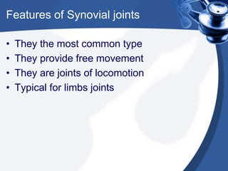
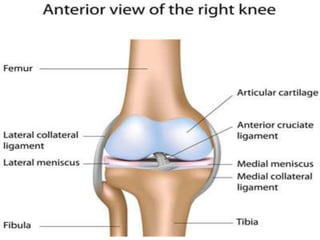
Introduction to Clinically
Oriented Anatomy
‫ــــــ‬ Joints
By: Hasan Arafat
Joints are unions between two or more
bones or rigid parts of the skeleton
• They exhibit a variety of forms and
functions
• They are classified according to the
type of the material by which the
articulating bones are united
• They can be also classified according
to mobility
Three general types of joints are recognized:
fibrous, cartilaginous and synovial
• These types differ in the manner of
material by which each joint is made
• Freely movable synovial joints are the
most common type
• Most of our movements happen at
synovial joints
• However, the other types perform a
number of very important functions
Synovial joints
• In Synovial joints, the articulating are
united by a joint capsule
• The joint capsule is composed of an
outer fibrous layer lined by serous
synovial membrane
• The joint capsule encloses an articular
cavity
Synovial joints (continued)
• The joint cavity is a potential space that
contains a small amount of lubricating
synovial fluid
• The lubricating synovial fluid is
secreted by the synovial membrane
that lines the fibrous layer of the joint
capsule
Synovial joints (continued)
• Inside the capsule, articular cartilage
covers the articulating surfaces of the
bones
• All other internal surfaces are covered
by synovial membranes
• The periosteum investing the
participating bones external to the joint
blends with the fibrous layer of the
capsule
Fibrous joints
• The articulating bones are united b
fibrous tissue
• The amount of movement at this joint
depends on the length of the fibers
uniting the articulating surfaces
• The sutures of the cranium are
examples
Fibrous joints (continued)
• Fibrous joints can be subdivided into 2
subtypes
• A syndesmosis type unites the bones
with a sheet of fibrous tissue, either a
ligament or fibrous membrane
• This type is partially movable
• The interosseous membrane between
the radius and ulna is an example
Dentatoalveolar syndesmosis: a fibrous joint in
which a peg-like process fits into a socket
articulation between the root of the tooth and the
alveolar process of the jaw
• It’s also called gomphosis or socket
• Mobility at this joint indicates a
pathological state, affecting the
supporting tissue of the tooth
• So, it’s normally immobile
• However, microscopic movements here
is related to proprioception
Cartilaginous joints
• They are united by hyaline cartilage or
fibrocartilage
• Can be divided into 2 types
• These types are primary cartilaginous
joints, or synchondroses, and
secondary cartilaginous joints, or
symphyses
Cartilaginous joints (continued)
• In synchondroses, the bones are joined
by hyaline cartilage
• Hyaline cartilage permits slight bending
during early life
• Primary cartilaginous joints are usually
temporary unions
• E.g. the growth plate
Cartilaginous joints (continued)
• Symphyses are strong, slightly movable
joints
• They are united by fibrocartilage
• E.g. intervertebral discs
• They provide strength and shock
absorption as well as considerable
flexibility to the vertebral column
Features of Synovial joints
• They the most common type
• They provide free movement
• They are joints of locomotion
• Typical for limbs joints
Features of Synovial joints (cont)
• They are reinforced by accessory
ligaments
• Accessory ligaments are either
separate (extrinsic) or thickening
portions of joint capsule (intrinsic)
Features of Synovial joints (cont)
• They also contain fibrocartilaginous
articular discs
• They are also called meniscus
• They are found where articulating
surfaces are incongruous
Classification of Synovial joints
• Six major types of synovial joints are
classified according to the shape of
articulating surfaces and the type of
movement they permit
• These types are plane joints, hinge
joints, saddle joints, condyloid joints
and ball-and-socket joints
1. Plane joints
• They are nonaxial
• They permit gliding or sliding
movements in the plane of the articular
surfaces
• The opposed surfaces are flat or almost
flat
• Movement is limited by their tight joint
capsules
1. Plane joints (cont)
• They are numerous and are nearly
always small
• E.g. the acromioclavicular joint
2. Hinge joints
• Permit flexion and extension only
• They are uniaxial
• Movements at these joints happen in
the sagittal plane around a transverse
axis
• Their joint capsules are thin
2. Hinge joints (cont)
• The joint capsule lax anteriorly and
posteriorly where movement occures
• The bones are joined by strong laterally
placed collateral ligaments
• The elbow is an example
Saddle joints
• Permit abduction, adduction, flexion
and extension
• Biaxial joints, movements occur in
sagittal and frontal planes
• Circumduction is also available
• E.g. carpometacarpal joint at the thumb
Condyloid joints
• Permit flexion, extension, adduction
and abduction
• Biaxial Joints
• The movement at the sagittal is greater
than the frontal
• More restricted circumduction
• E.g. metacarbophalangeal joints
Ball and socket joints
• Multiple axes and planes
• Allow flexion, extension, abduction,
adduction, medial and lateral rotation
and circumduction
• Spheroidal surface of one bone moves
within the socket of another
• E.g. hip joint
Pivot joints
• Permit rotation around a central axis
• Uniaxial joints
• The rounded process of bone rotates
within a sleeve or ring
• E.g. the median atlantoaxial joint
• The atlas rotates around the dens of
the axis during rotation of the head
Synovial joints

Introduction to Clinically Oriented Anatomy-Joints

  • 1.
    Introduction to Clinically OrientedAnatomy ‫ــــــ‬ Joints By: Hasan Arafat
  • 2.
    Joints are unionsbetween two or more bones or rigid parts of the skeleton • They exhibit a variety of forms and functions • They are classified according to the type of the material by which the articulating bones are united • They can be also classified according to mobility
  • 3.
    Three general typesof joints are recognized: fibrous, cartilaginous and synovial • These types differ in the manner of material by which each joint is made • Freely movable synovial joints are the most common type • Most of our movements happen at synovial joints • However, the other types perform a number of very important functions
  • 4.
    Synovial joints • InSynovial joints, the articulating are united by a joint capsule • The joint capsule is composed of an outer fibrous layer lined by serous synovial membrane • The joint capsule encloses an articular cavity
  • 5.
    Synovial joints (continued) •The joint cavity is a potential space that contains a small amount of lubricating synovial fluid • The lubricating synovial fluid is secreted by the synovial membrane that lines the fibrous layer of the joint capsule
  • 6.
    Synovial joints (continued) •Inside the capsule, articular cartilage covers the articulating surfaces of the bones • All other internal surfaces are covered by synovial membranes • The periosteum investing the participating bones external to the joint blends with the fibrous layer of the capsule
  • 8.
    Fibrous joints • Thearticulating bones are united b fibrous tissue • The amount of movement at this joint depends on the length of the fibers uniting the articulating surfaces • The sutures of the cranium are examples
  • 10.
    Fibrous joints (continued) •Fibrous joints can be subdivided into 2 subtypes • A syndesmosis type unites the bones with a sheet of fibrous tissue, either a ligament or fibrous membrane • This type is partially movable • The interosseous membrane between the radius and ulna is an example
  • 11.
    Dentatoalveolar syndesmosis: afibrous joint in which a peg-like process fits into a socket articulation between the root of the tooth and the alveolar process of the jaw • It’s also called gomphosis or socket • Mobility at this joint indicates a pathological state, affecting the supporting tissue of the tooth • So, it’s normally immobile • However, microscopic movements here is related to proprioception
  • 13.
    Cartilaginous joints • Theyare united by hyaline cartilage or fibrocartilage • Can be divided into 2 types • These types are primary cartilaginous joints, or synchondroses, and secondary cartilaginous joints, or symphyses
  • 14.
    Cartilaginous joints (continued) •In synchondroses, the bones are joined by hyaline cartilage • Hyaline cartilage permits slight bending during early life • Primary cartilaginous joints are usually temporary unions • E.g. the growth plate
  • 15.
    Cartilaginous joints (continued) •Symphyses are strong, slightly movable joints • They are united by fibrocartilage • E.g. intervertebral discs • They provide strength and shock absorption as well as considerable flexibility to the vertebral column
  • 18.
    Features of Synovialjoints • They the most common type • They provide free movement • They are joints of locomotion • Typical for limbs joints
  • 19.
    Features of Synovialjoints (cont) • They are reinforced by accessory ligaments • Accessory ligaments are either separate (extrinsic) or thickening portions of joint capsule (intrinsic)
  • 20.
    Features of Synovialjoints (cont) • They also contain fibrocartilaginous articular discs • They are also called meniscus • They are found where articulating surfaces are incongruous
  • 22.
    Classification of Synovialjoints • Six major types of synovial joints are classified according to the shape of articulating surfaces and the type of movement they permit • These types are plane joints, hinge joints, saddle joints, condyloid joints and ball-and-socket joints
  • 23.
    1. Plane joints •They are nonaxial • They permit gliding or sliding movements in the plane of the articular surfaces • The opposed surfaces are flat or almost flat • Movement is limited by their tight joint capsules
  • 24.
    1. Plane joints(cont) • They are numerous and are nearly always small • E.g. the acromioclavicular joint
  • 26.
    2. Hinge joints •Permit flexion and extension only • They are uniaxial • Movements at these joints happen in the sagittal plane around a transverse axis • Their joint capsules are thin
  • 27.
    2. Hinge joints(cont) • The joint capsule lax anteriorly and posteriorly where movement occures • The bones are joined by strong laterally placed collateral ligaments • The elbow is an example
  • 30.
    Saddle joints • Permitabduction, adduction, flexion and extension • Biaxial joints, movements occur in sagittal and frontal planes • Circumduction is also available • E.g. carpometacarpal joint at the thumb
  • 31.
    Condyloid joints • Permitflexion, extension, adduction and abduction • Biaxial Joints • The movement at the sagittal is greater than the frontal • More restricted circumduction • E.g. metacarbophalangeal joints
  • 32.
    Ball and socketjoints • Multiple axes and planes • Allow flexion, extension, abduction, adduction, medial and lateral rotation and circumduction • Spheroidal surface of one bone moves within the socket of another • E.g. hip joint
  • 33.
    Pivot joints • Permitrotation around a central axis • Uniaxial joints • The rounded process of bone rotates within a sleeve or ring • E.g. the median atlantoaxial joint • The atlas rotates around the dens of the axis during rotation of the head
  • 35.