Downloaded 42 times
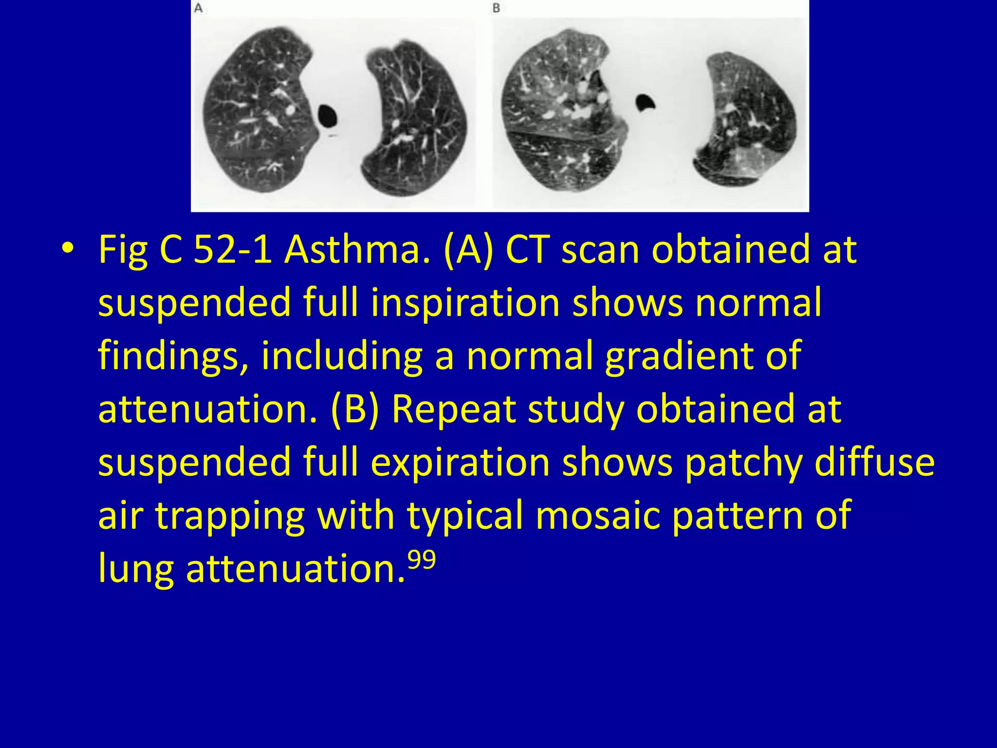
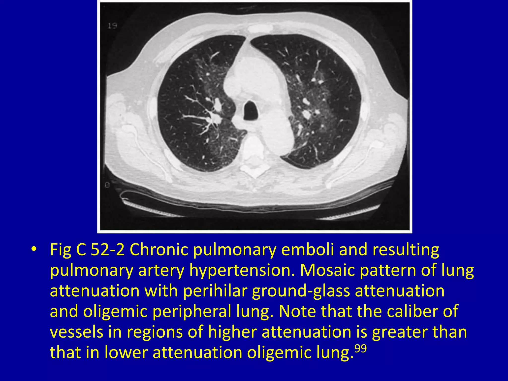
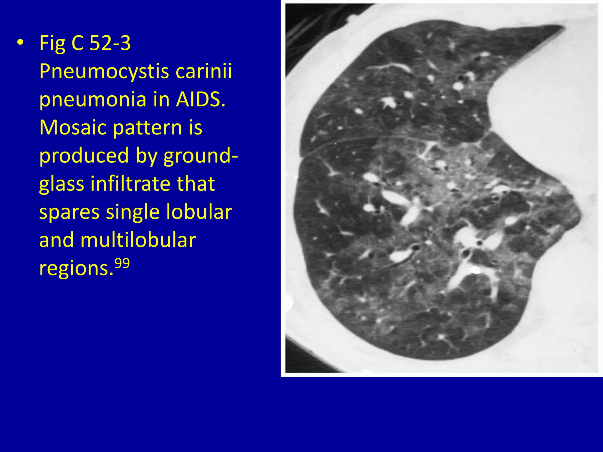


The document describes a mosaic pattern seen on chest CT scans, which appears as a patchy mixture of areas with different lung densities. This pattern is seen in conditions like asthma during expiration due to air trapping, chronic pulmonary embolism and pulmonary hypertension due to differing blood flow, and Pneumocystis pneumonia in AIDS patients where the ground glass infiltrate spares some lobular regions. Examples of each condition showing the mosaic pattern are provided.


