Karnataka University, Dharwad.
               B. Sc. Botany, Semester III Paper Anatomy and Embryology
                                           Paper:
       Unit 3. Microsporogenesis, Male Gametophyte, Male Ferm Unit and Pollen Morphology

Structure of Anther:
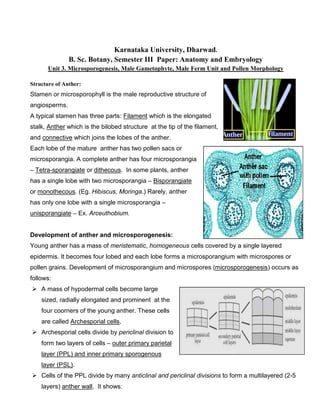
Stamen or microsporophyll is the male reproductive structure of
angiosperms.
A typical stamen has three parts: Filament which is the elongated
stalk, Anther which is the bilobed structure at the tip of the filament,
and connective which joins the lobes o the anther.
                                     of
Each lobe of the mature anther has two pollen sacs or
microsporangia. A complete anther has four microsporangia
– Tetra-sporangiate or dithecous In some plants, anther
                       dithecous.
has a single lobe with two microsporangia – Bisporangiate
or monothecous. (Eg. Hibiscus, Moringa Rarely, anther
                               Moringa.)
has only one lobe with a single microsporangia –
unisporangiate – Ex. Arceuthobium.


Development of anther and microsporogenesis:
Young anther has a mass of meristematic homogeneous cells covered by a single layered
                           meristematic,
epidermis. It becomes four lobed and each lobe forms a microsporangium with microspores or
pollen grains. Development of microsporangium and microspores (microsporogenesis) occurs as
                                                              (microsporogenesis
follows:
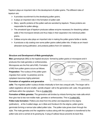
    A mass of hypodermal cells b
                               become large
    sized, radially elongated and prominent at the
    four coorners of the young anther. These cells
    are called Archesporial cells.
                                 .
    Archesporial cells divide by periclinal division to
    form two layers of cells – outer primary parietal
    layer (PPL) and inner primary sporogenous
                  d
    layer (PSL).
    Cells of the PPL divide by many anticlinal and periclinal divisions to form a multilayered (2
                                                                                               (2-5
    layers) anther wall. It shows:
                       .
o Epidermis-outermost layer, single layer of flattened cells.
                 outermost
     o Endothecium – Inner to epidermis,
        single layered, radially elongated cells,
        Has a thin layered area called
        stomium which helps in dehiscence of
        the anthers.
     o Middle layers: 2-3 layers of flattened
                        3
        cells inner to endothecium, and
     o Tapetum: Innermost layer, single laye
                                        layer
        of cells with dense cytoplasm and
        prominent nuclei, provides nutrition to
                        ,
        developing microspores. It is of two
        types – Amoeboid type (cell wall
        ruptures and protoplasm released,
        forms a protoplasmic network, provides nutrition to developing microspores Found
                                                                       microspores.
        mainly in monocots) and Secretory type (Cells remain intact, secret nutrition into the
        microsporangium).
   Cells of the PSL divide by mitosis and form a mass of diploid sporogenous tissue which then
   forms Microspore mother cells or Pollen mother cells (MMC or PMC). They are loosely
                                                                    .
   arranged when mature. PMC’s divide by meiosis to form haploid microspores or pollen grains
                                                                                       grains.
   They are arranged as tetrads (group of four).
   Tetrad formation is of two types:
     o Successive type: Each nuclear division
                      :
        followed by cell wall formation. Four cells
               d
        arranged in iso-bilateral manner.
                        bilateral
     o Simultaneous type: Nucleus of PMC
                        :
        divides twice to form four haploid nuclei.
        Cell wall forms simultaneously between
        four nuclei resulting in tetrads.
   Cells of the tetrad completely sep
                                  separate from
   each other and develop into mature pollen grains
   or microspores with characteristic structures.
   These microspores germinate and produce the male gametophyte.
Role of tapetum:
Tapetum plays an important role in the development of pollen grains. The different roles of
tapetum are:
      It provides nourishment to the developing pollen grains.
      It plays an important role in the formation of pollen wall.
      Many specific proteins of the pollen wall are secreted by tapetum. These proteins are
      responsible for pollen allergy.
      The amoeboid type of tapetum produces callase enzyme necessary for dissolving callose
      walls of the microspore tetrads and thus helps in their separation into individual pollen
      grains.
      Callase enzyme also plays an important role in making the pollen grains fertile or sterile.
      It produces a oily coating over some pollen grains called pollen kits. It helps as an insect-
      attractant during pollination, and protects pollens from UV radiations.


Structure and Development of Male gametophyte:
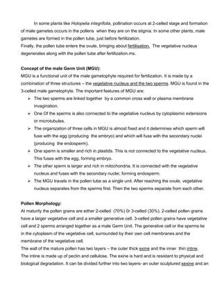
Male gametophyte (MG) is the haploid structure formed by pollen grains or microspore and it
produces the male gametes or antherozoides.
Microspores are the first cell of MG. Formation
of MG from pollen grains occurs as follows:
Preparation: The nucleus of the pollen grain
migrates from center to periphery and its
cytoplasm becomes highly polarized.
Formation of vegetative and generative
cells: The migrated pollen nucleus divides mitotically to form two unequal cells. The larger cell is
called vegetative cell and smaller, spindle shaped cell is the generative cell. Later, the generative
cell floats within the cytoplasm . This is 2-celled MG.
Formation of Male gametes: The generative cell divides by mitosis forming two male cells which
then give rise to two non-motile male gametes or antherozoides. This is 3-celled MG.
Pollen tube formation: Pollens are shed from the anther and deposited on the stigma
(pollination), at the 3-celled stage, as in Beta and Hordeum.On the stigma, pollen grains
germinate forming a narrow tube called pollen tube. The pollen tube grows down through the
style. The male gametes and the vegetative nucleus along with the cytoplasm is transferred to the
pollen tube and is carried at its growing tip. A plug of callose tissue prevents its back flow.
In some plants like Holopelia integrifolia, pollination occurs at 2-celled stage and formation
of male gametes occurs in the pollens when they are on the stigma. In some other plants, male
gametes are formed in the pollen tube, just before fertilization.
Finally, the pollen tube enters the ovule, bringing about fertilisation. The vegetative nucleus
degenerates along with the pollen tube after fertilization.ms.


Concept of the male Germ Unit (MGU):
MGU is a functional unit of the male gametophyte required for fertilization. It is made by a
combination of three structures – the vegetative nucleus and the two sperms. MGU is found in the
3-celled male gametophyte. The important features of MGU are:
       The two sperms are linked together by a common cross wall or plasma membrane
       invagination.
       One Of the sperms is also connected to the vegetative nucleus by cytoplasmic extensions
       or microtubules.
       The organization of three cells in MGU is almost fixed and it determines which sperm will
       fuse with the egg (producing the embryo) and which will fuse with the secondary nuclei
       (producing the endosperm).
       One sperm is smaller and rich in plastids. This is not connected to the vegetative nucleus.
       This fuses with the egg, forming embryo.
       The other sperm is larger and rich in mitochondria. It is connected with the vegetative
       nucleus and fuses with the secondary nuclei, forming endosperm.
       The MGU travels in the pollen tube as a single unit. After reaching the ovule, vegetative
       nucleus separates from the sperms first. Then the two sperms separate from each other.


Pollen Morphology:
At maturity the pollen grains are either 2-celled (70%) 0r 3-celled (30%). 2-celled pollen grains
have a larger vegetative cell and a smaller generative cell. 3-celled pollen grains have vegetative
cell and 2 sperms arranged together as a male Germ Unit. The generative cell or the sperms lie
in the cytoplasm of the vegetative cell, surrounded by their own cell membranes and the
membrane of the vegetative cell.
The wall of the mature pollen has two layers – the outer thick exine and the inner thin intine.
The intine is made up of pectin and cellulose. The exine is hard and is resistant to physical and
biological degradation. It can be divided further into two layers- an outer sculptured sexine and an
inner non-sculptured nexine. The sexine is further made up of three layers – tectum (roof layer),
columellae (bar like elements) and foot layer.
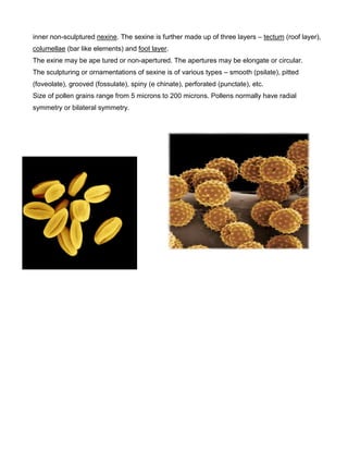
The exine may be ape tured or non-apertured. The apertures may be elongate or circular.
The sculpturing or ornamentations of sexine is of various types – smooth (psilate), pitted
(foveolate), grooved (fossulate), spiny (e chinate), perforated (punctate), etc.
Size of pollen grains range from 5 microns to 200 microns. Pollens normally have radial
symmetry or bilateral symmetry.