Anther wall and its
functions
Presentation by : Deepanshi patel
Botany honours
Acharya Narendra Dev college
University of Delhi
Anther : Introduction
● Stamens consist of an anther and a filament
● Anthers contain the pollen grains .
● Anther is the site of pollen development and dispersal.
● Filament transmits water and nutrients to the anther Also help in pollen
dispersal or positioning.
● Pollen grains are the carriers of male gametes (sperm cells)
● Male gametes play a central role in plant reproduction during the process of
double fertilization.
Structure:
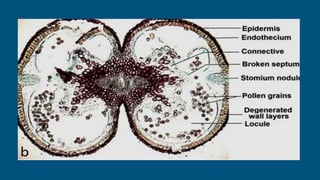
A typical anther is a bilobed, dithecous structure with two microsporangia in
each lobe. Therefore, an anther is a tetrasporangiate structure with four
microsporangia.
● Connective : the non-sporangial tissue (connects or joints the lobes)
● Intersporangial septum- strip of sterile tissue between the two
microsporangia.
● Monothecous-two microsporangia are fused at maturity to form a single
theca
Functions of various cells of an anther
Anther wall
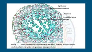
1. Epidermis
2. Endothecium
3. Middle layers
4. Tapetum
EPIDERMIS :
● Outermost layer of the anther
● Encloses archesporial cell and its products
● Stretched and Flattened in mature anther
● Protective in function.
● Prevents water loss from the anther
● Provides structural support to the anther alongwiith endothecium
● Plays a role in the anther dehiscence and release of pollen grains
● Helps in gaseous diffusion too
ENDOTHECIUM
● Hypodermal in origin ,Present beneath the epidermis
● Outermost layer derived from parietal cell , Persistent in the mature anther. Usually
single layered
● Generally uninucleate and highly vacuolated
● Few starch grains may be found in the cells. Cells are radially elongated •
Endothecial thickenings are of a-cellulose and little lignin
● Hygroscopic in nature
● Thickenings arise from the inner tangential walls and run outward and upward
● Outer tangential walls remain thin • Fibrous bands together with stomium provide
mechanical force for anther dehiscence
MIDDLE LAYERS :
● Usually ephemeral and become flattened by early meiosis in pollen mother cell
● 1-3 middle layers; sometimes more layers as in Lilium
● Absent in Wolffia and Vallisneria
● Cells are flattened, thin-walled, uninucleate and vacuolated
● May behave as endothecium by developing fibrous thickenings
● Store reserve food material such as starch, which gets mobilized during the
development of pollen.
● Get Crushed during meiosis in MMC
TAPETUM :
● The innermost and most conspicuous layer of the anther wall
● Completely surrounds the sporogenous tissue.
● Usually single layered and derived from parietal cells
● Primarily nutritive and secretory in function
● Cells contain prominent nucleus and dense cytoplasm , Mitochondria, plastids,
endoplasmic reticulum, dictyosomes, and ribosomes present in large numbers
● Degenerates completely by the time of anther dehiscence
● Mostly originates from parietal cell but can also arise from connective
Dimorphic in Alectra thomsonii
● Cells often become polyploid because of either mitotic divisions of nuclei
(multinucleate) ,formation of restitution nuclei, endomitosis or połyteny, all
indicating a high metabolic activity.
TYPES OF TAPETUM :
1. Amoeboid or invasive or periplasmodial or syncytial tapetum
Amoeboid tapetum is common in the monocots and in few dicots like
Asteraceae eg. Helianthus annuus,
e.g. Arum italicum, Tradescantia bracteata, Butomus umbellatus
1. Secretory or glandular or parietal or non-syncytial tapetum
Secretory tapetum is common in dicots Eg : Citrus limon, Capsicum annuum,
Helleborus foetidus and few monocots like Poaceae (Sorghum bicolor)
DIFFERENCE :
AMOEBOID TAPETUM
● Tapetum breaks down early .
● Cells fuse to form tapetal plasmodium
● Cells extrude in sporogenous cavity
● Cytoplasm engulfs microspores like
Amoeba
● Hydrolytic enzymes are secreted
● No Orbicules/Ubisch bodies are secreted
● Has no role in exine formation
● Moderate number of ribosomes
SECRETORY TAPETUM
● Tapetal cells remain intact
● No plasmodium is formed
● Cells remain in their place
● Cells do not engulf the microspores.
● No hydrolytic enzymes are secreted
● Orbicules/Ubisch bodies are secreted
● Involved in exine formation along with
sporopollenin
● High number of ribosomes.
ORBICULES :
● Orbicules or the Ubisch bodies are small sporopollenin particles
● Smaller than 1 un to a few micrometers in diameter
● Frequently fuse into larger compound aggregates - Originate as lipid
droplets in the tapetal cytoplasm (as pro orbicules or pro-ubisch bodies)
● Are extruded into the anther locule .
● Get coated with sporopollenin to form ubisch bodies
● Usually seen lining the inner tangential walls of the secretory tapetum.
● Develop simultaneously with the growing pollen exine
● Play a major role in pollen wall formation
TEM ubisch bodies at uninucleate stage of microspores (al, anther locule; pub,
pro-ubisch body, s, sporopollenin; ub, ubisch body; ve, vesicle)
FUNCTIONS OF TAPETUM :
● Tapetum is envolved in supply of nutrients to developing pollen (nurse
tissue).
● Tapetum plays role in breakdown of callose wall around microspore tetrads
by secreating enzyme called Callase (beta -1, 3 glucanase)
● It supplies sporopollinin precursors to pollen exine.
● It supplies pollen coat substance called as pollenkitt and tryphine
● Tapetum is involved in supply of pollen wall proteins.
Thankyou…!!!