2. MICROSPOROGENESIS
• Adult angiosperm plants are diploid sporophytes.
• They produce haploid spored by meiosis. This is
called sporogenesis.
• Spores are of two kinds namely microspores (male
spores or pollen grains) and megaspores (female
spores).
• The formation of microspores is called
microsporogenesis, and that of megaspores is
called megasporogenesis.
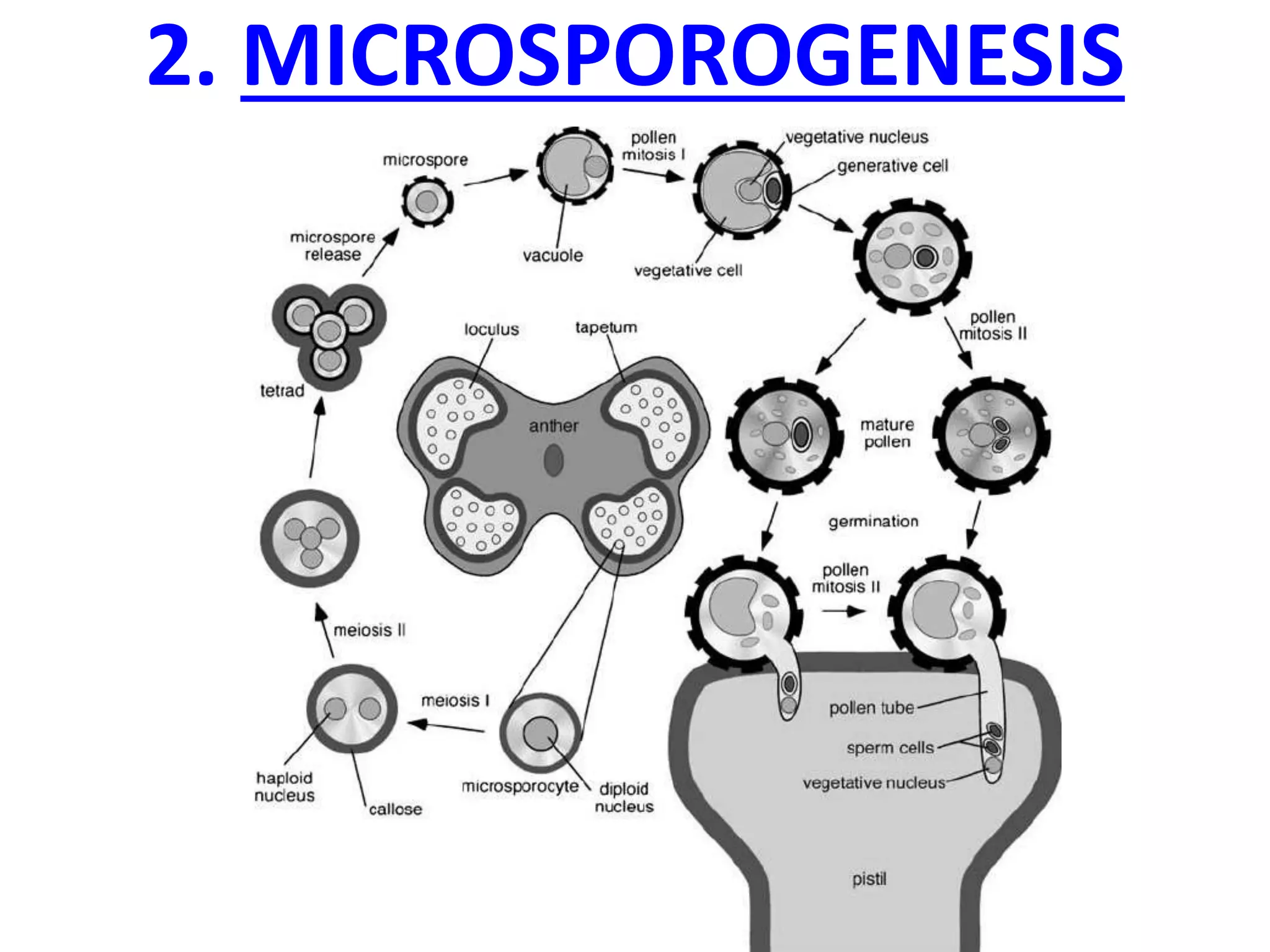
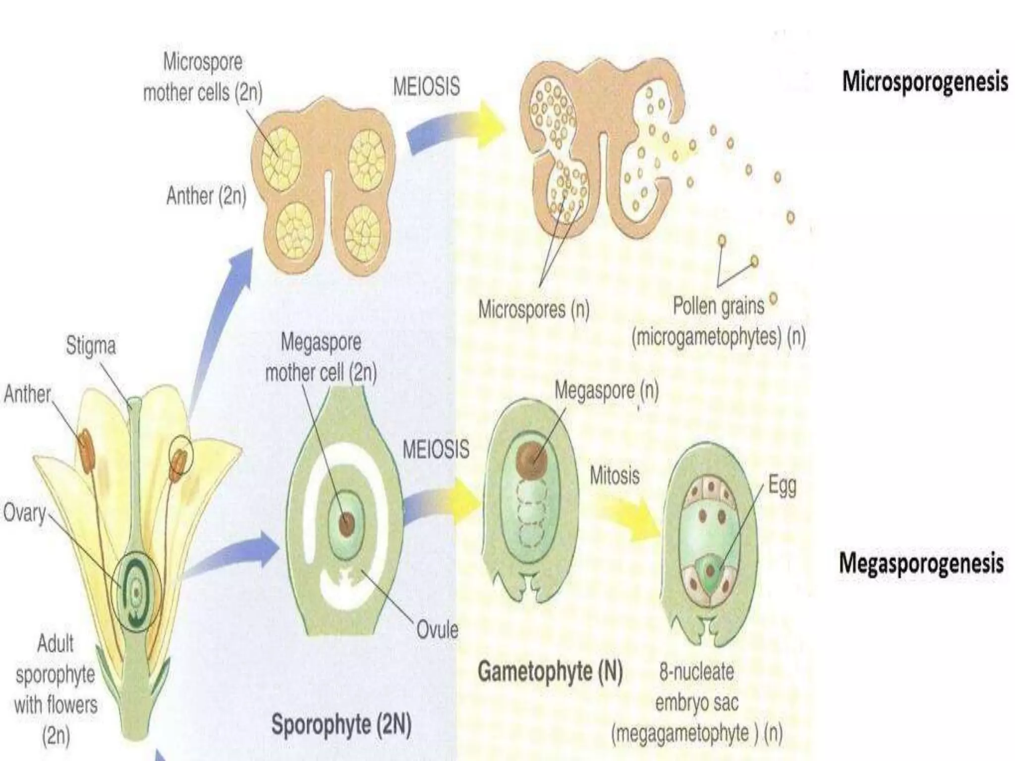
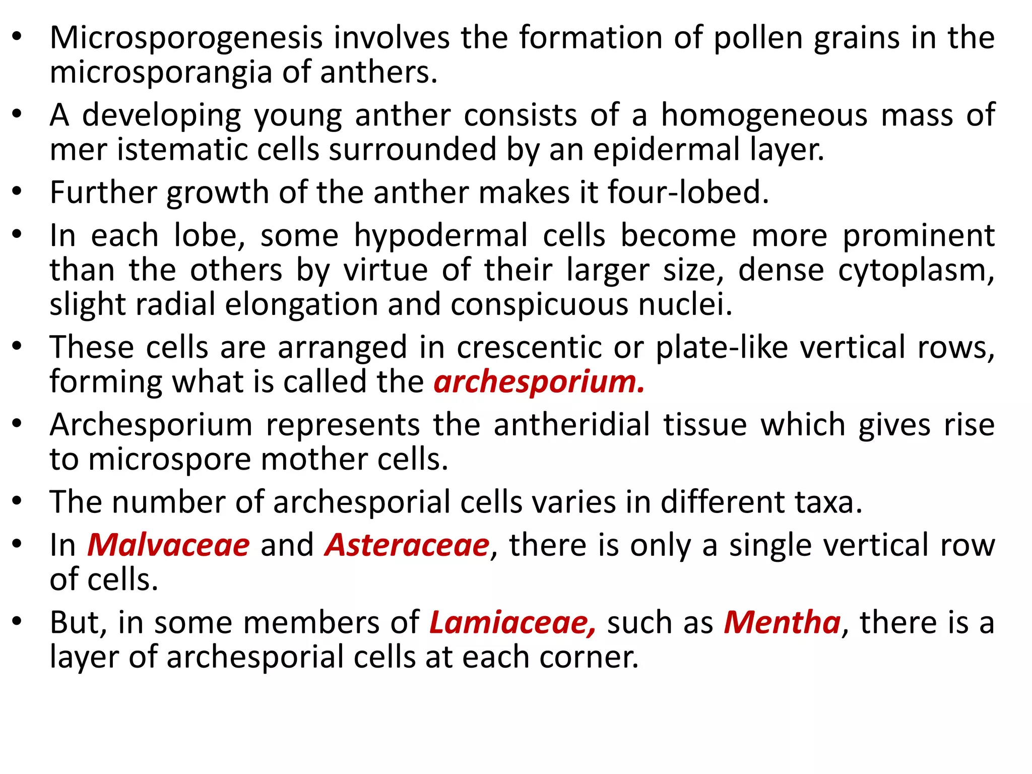
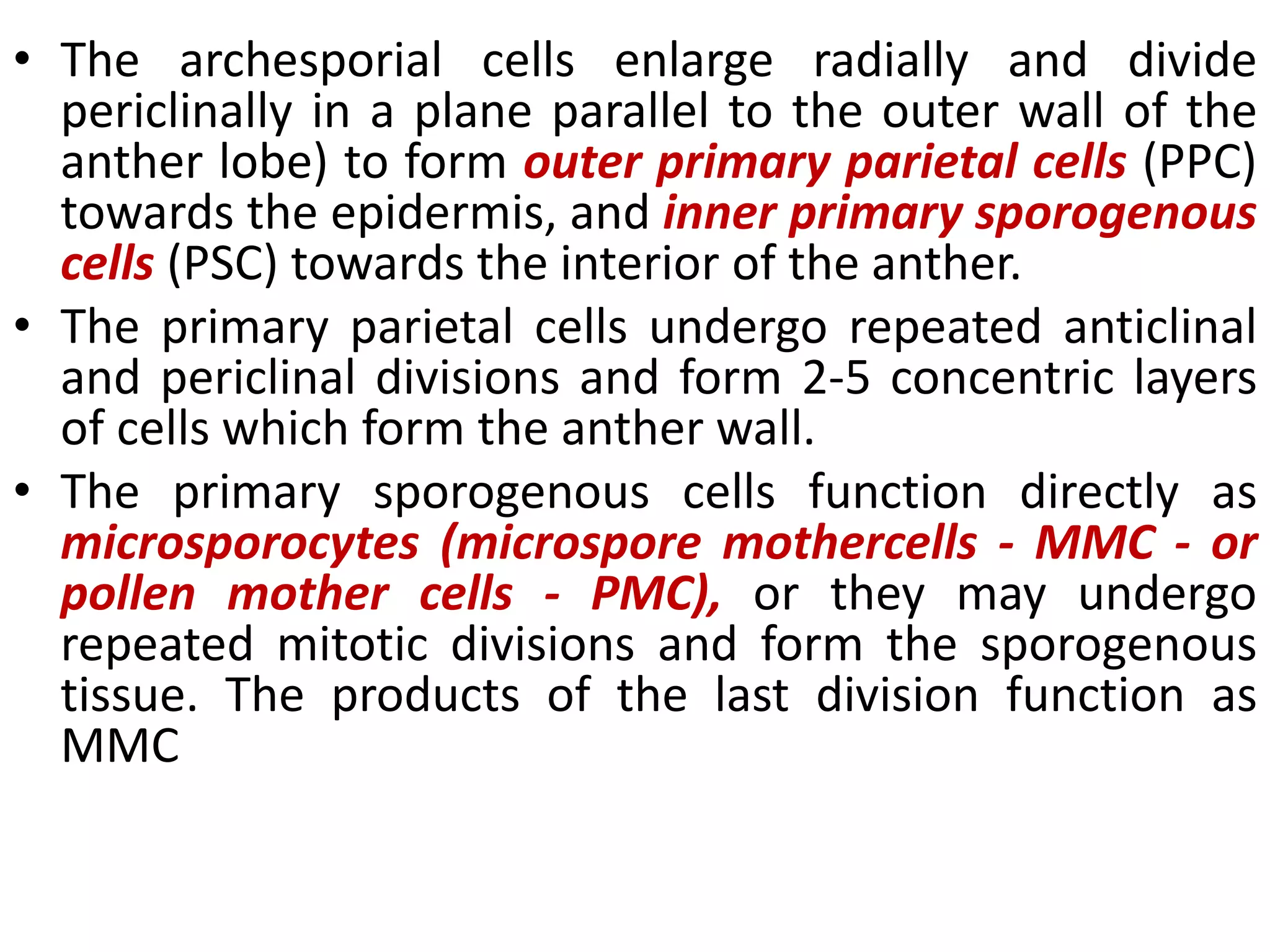
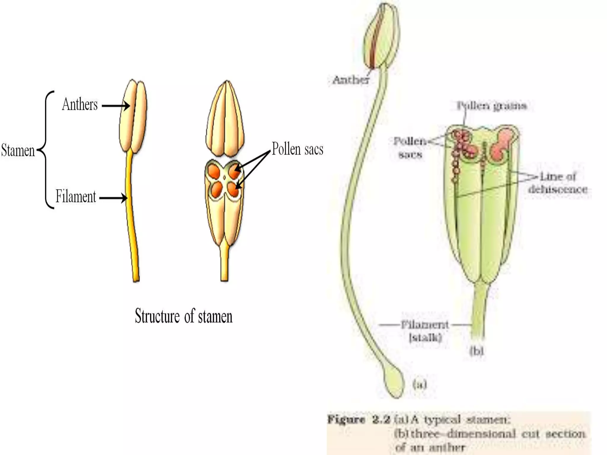
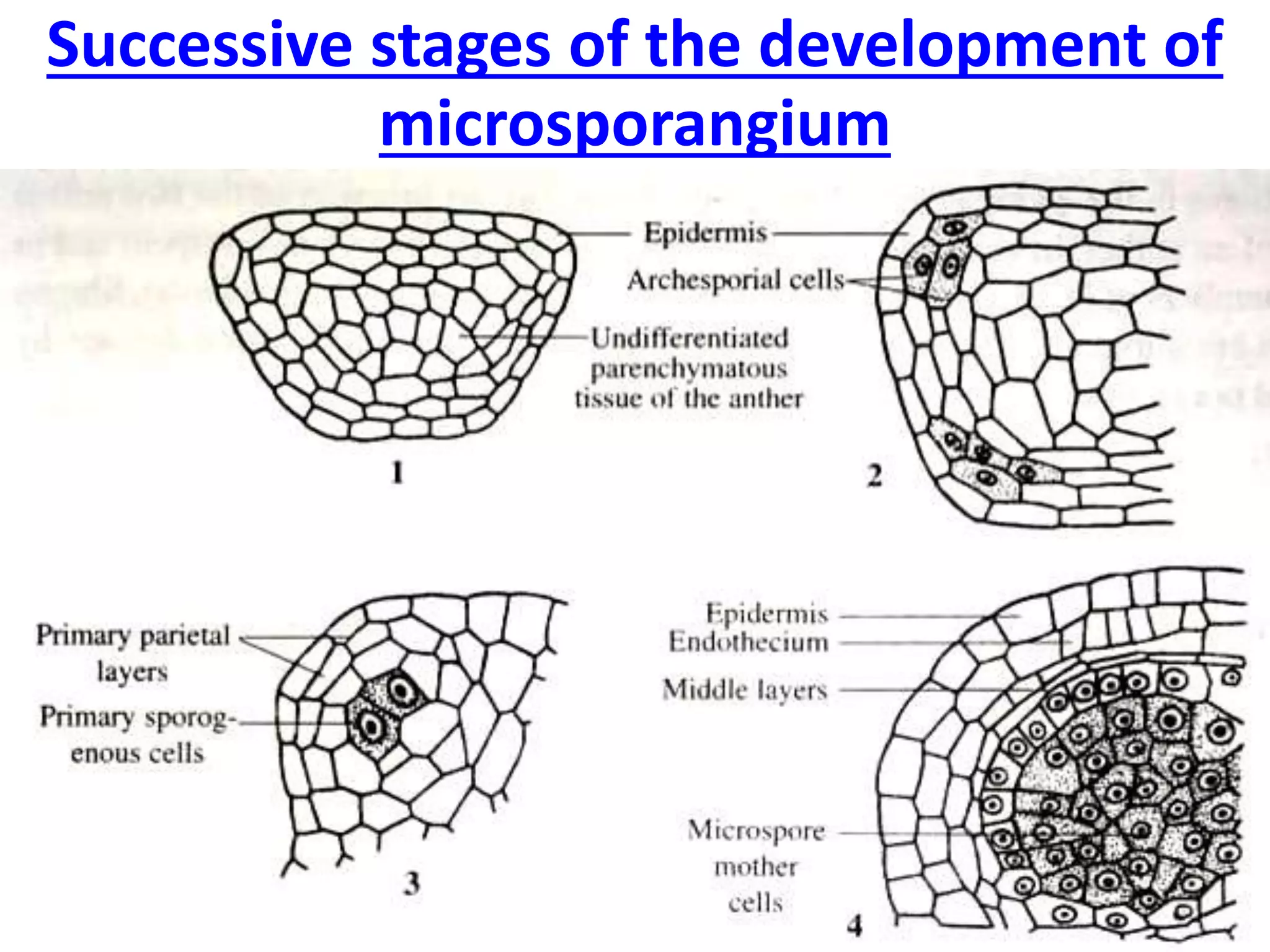
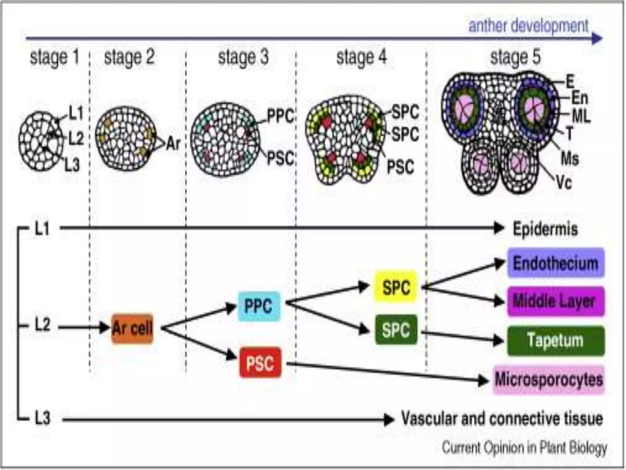
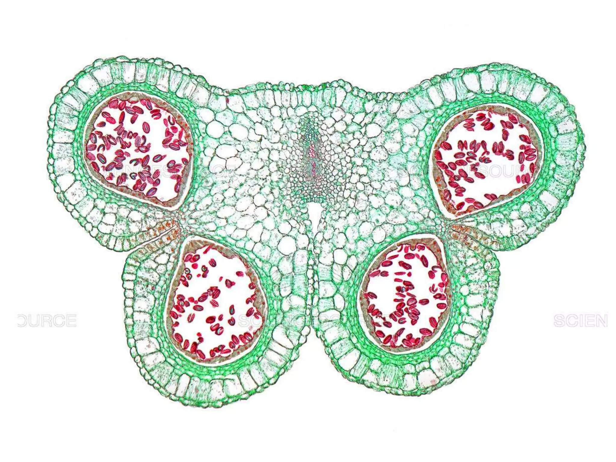
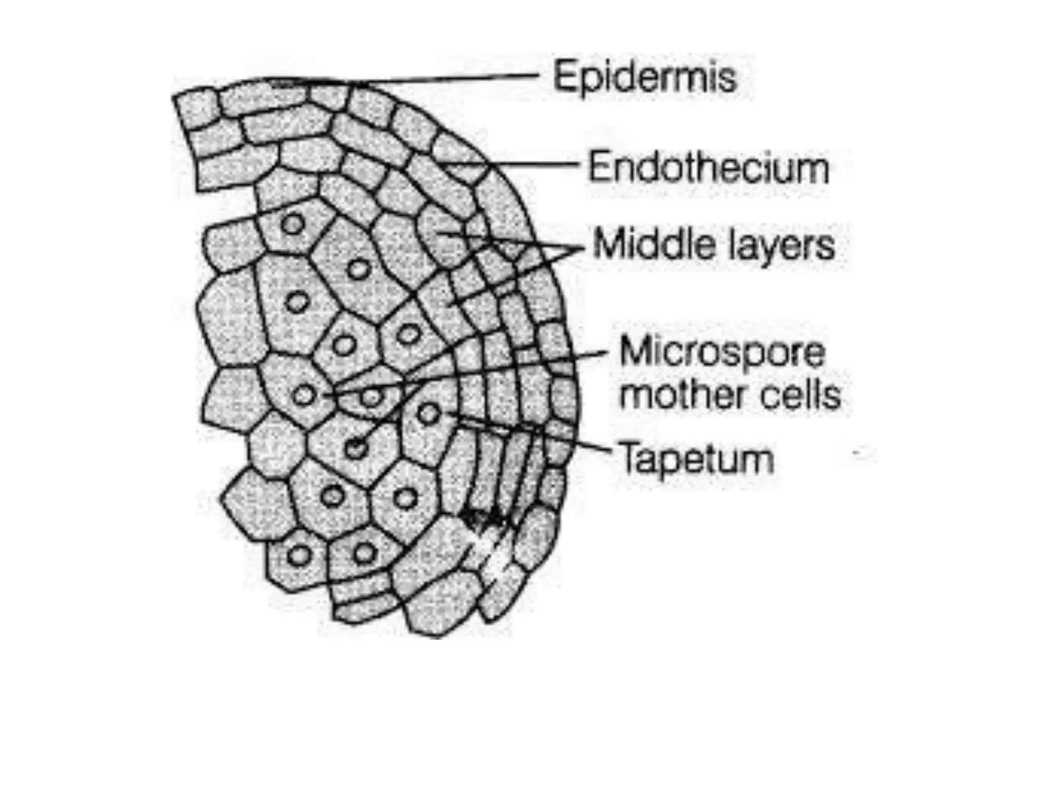
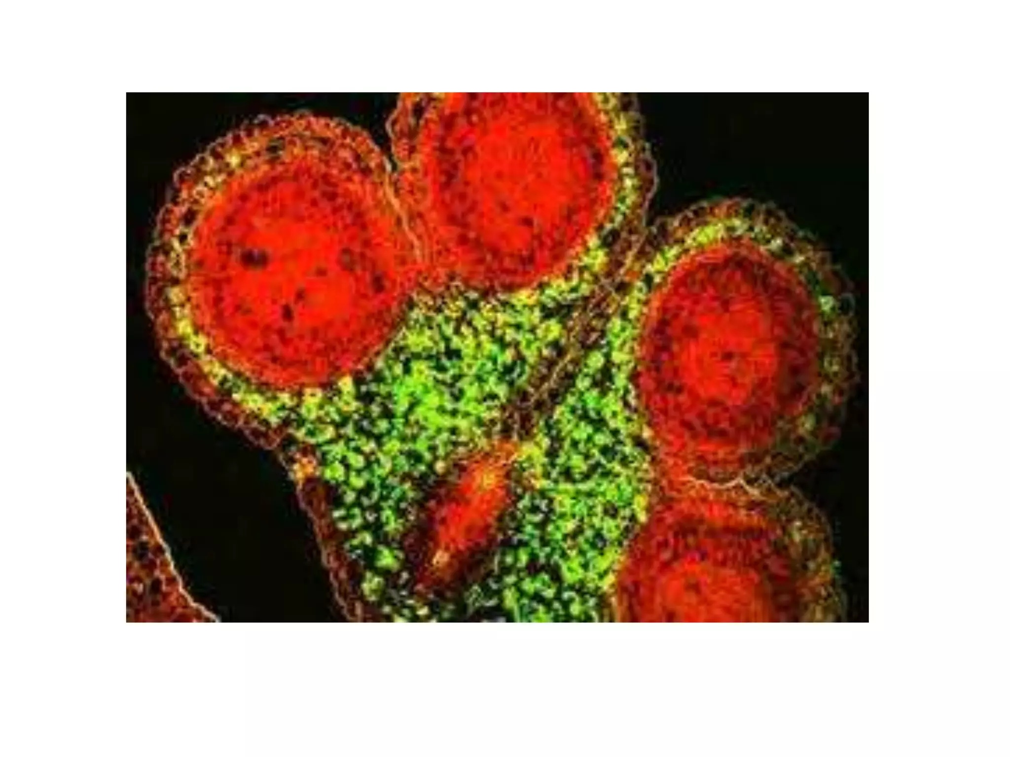
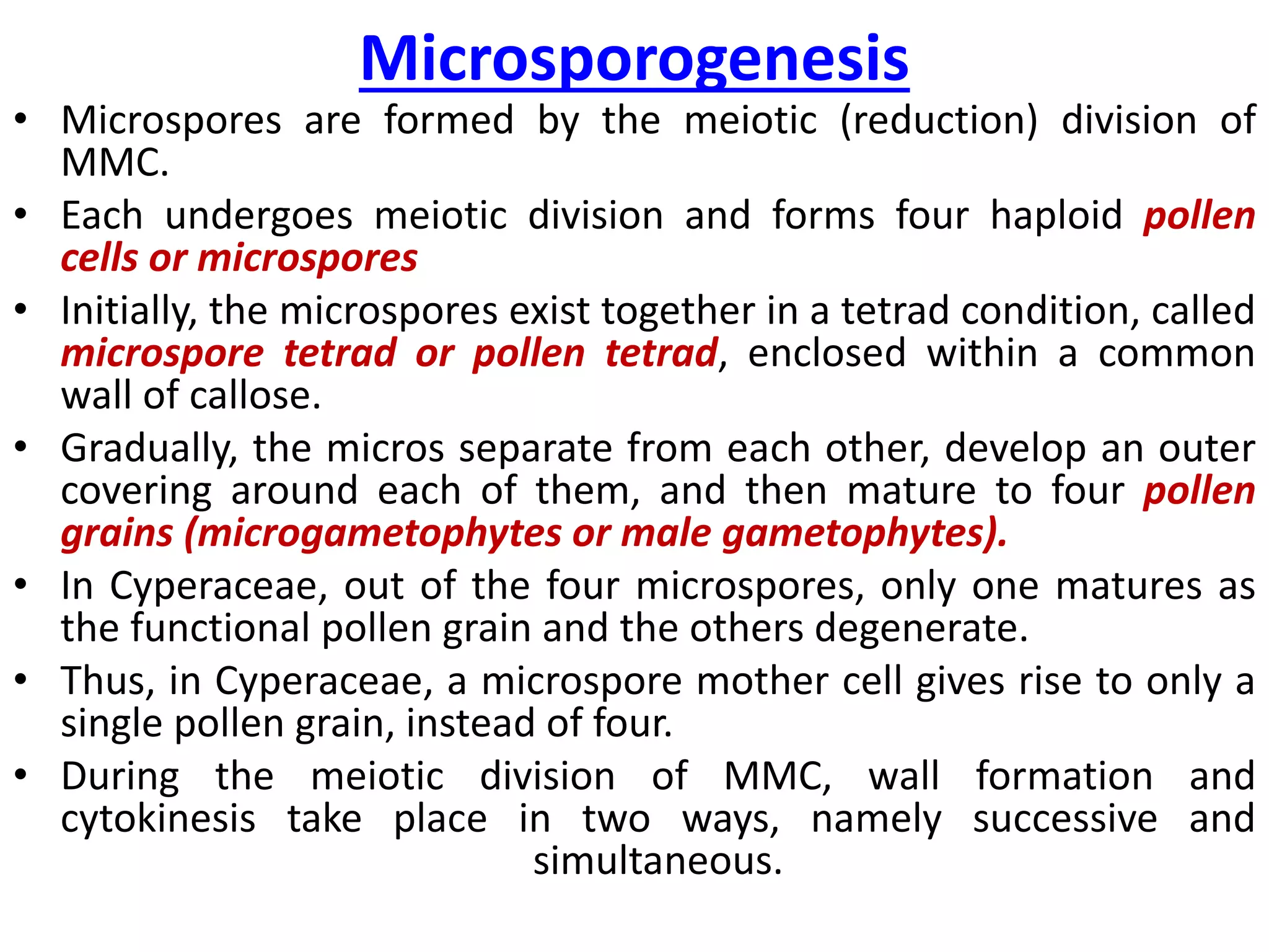
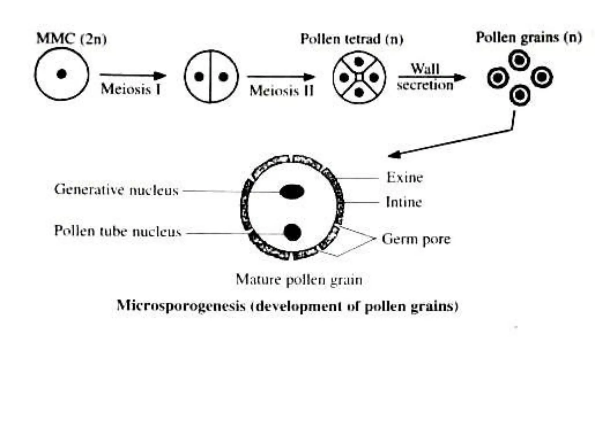
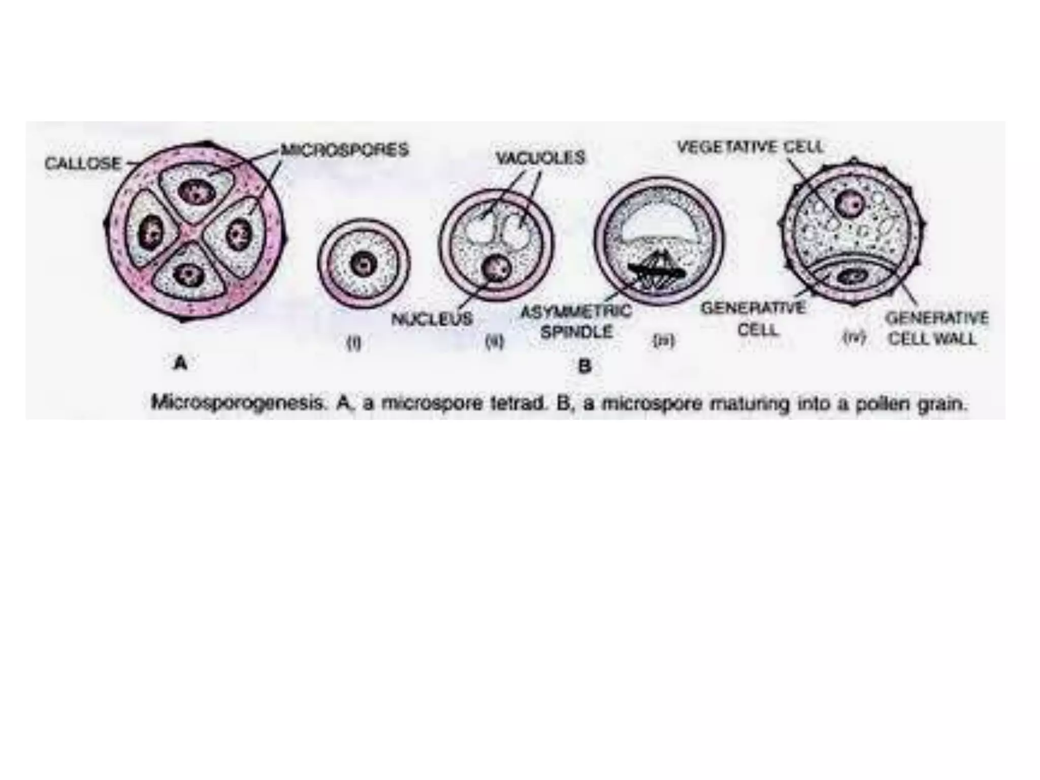
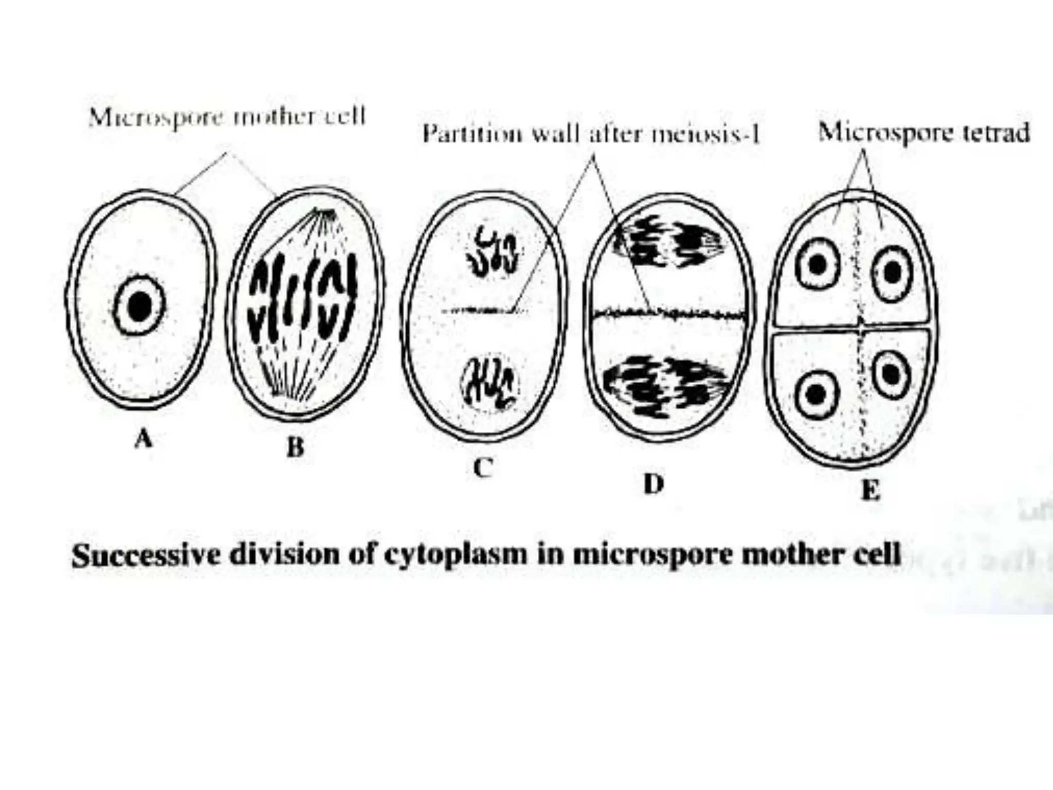
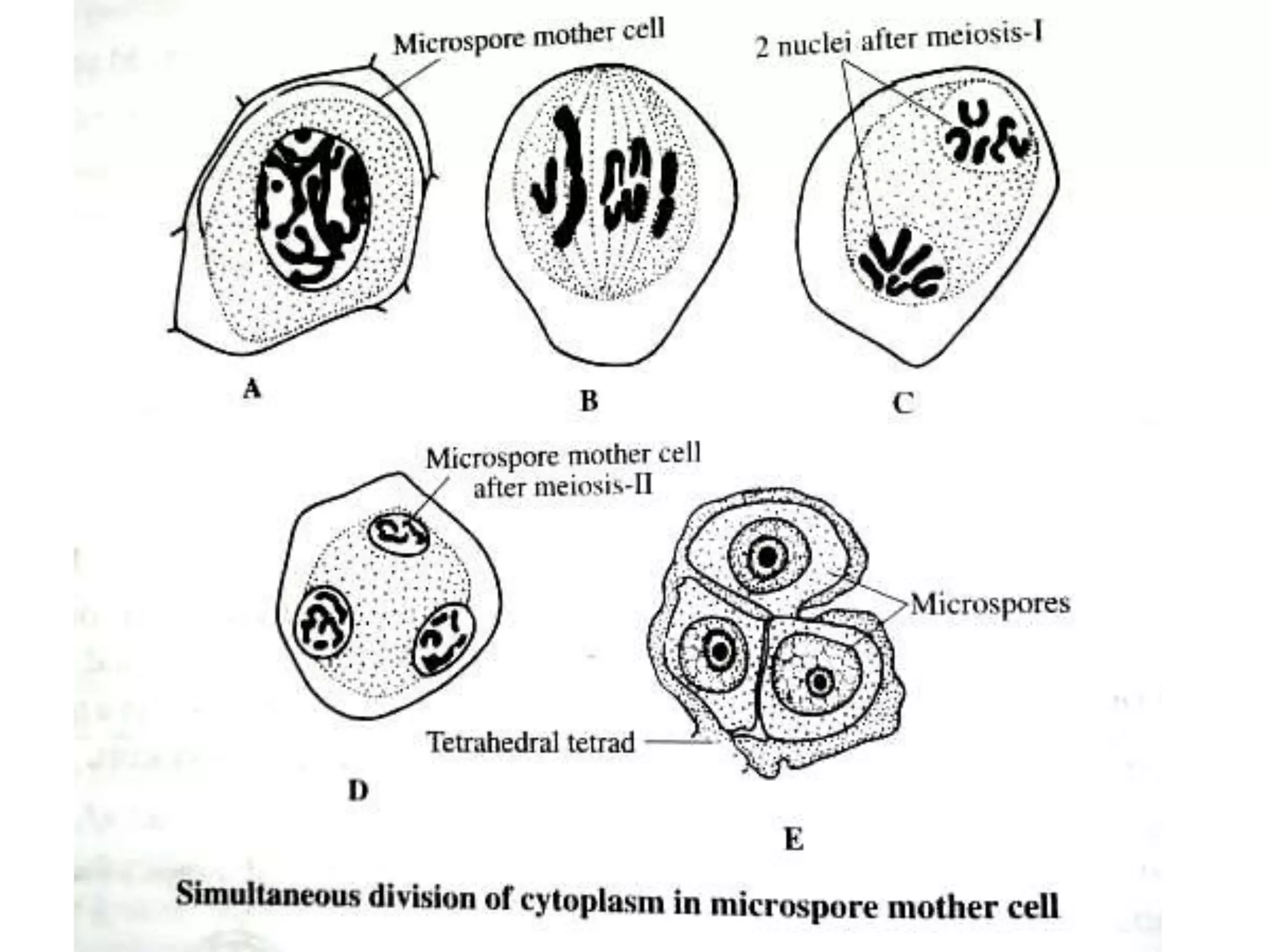
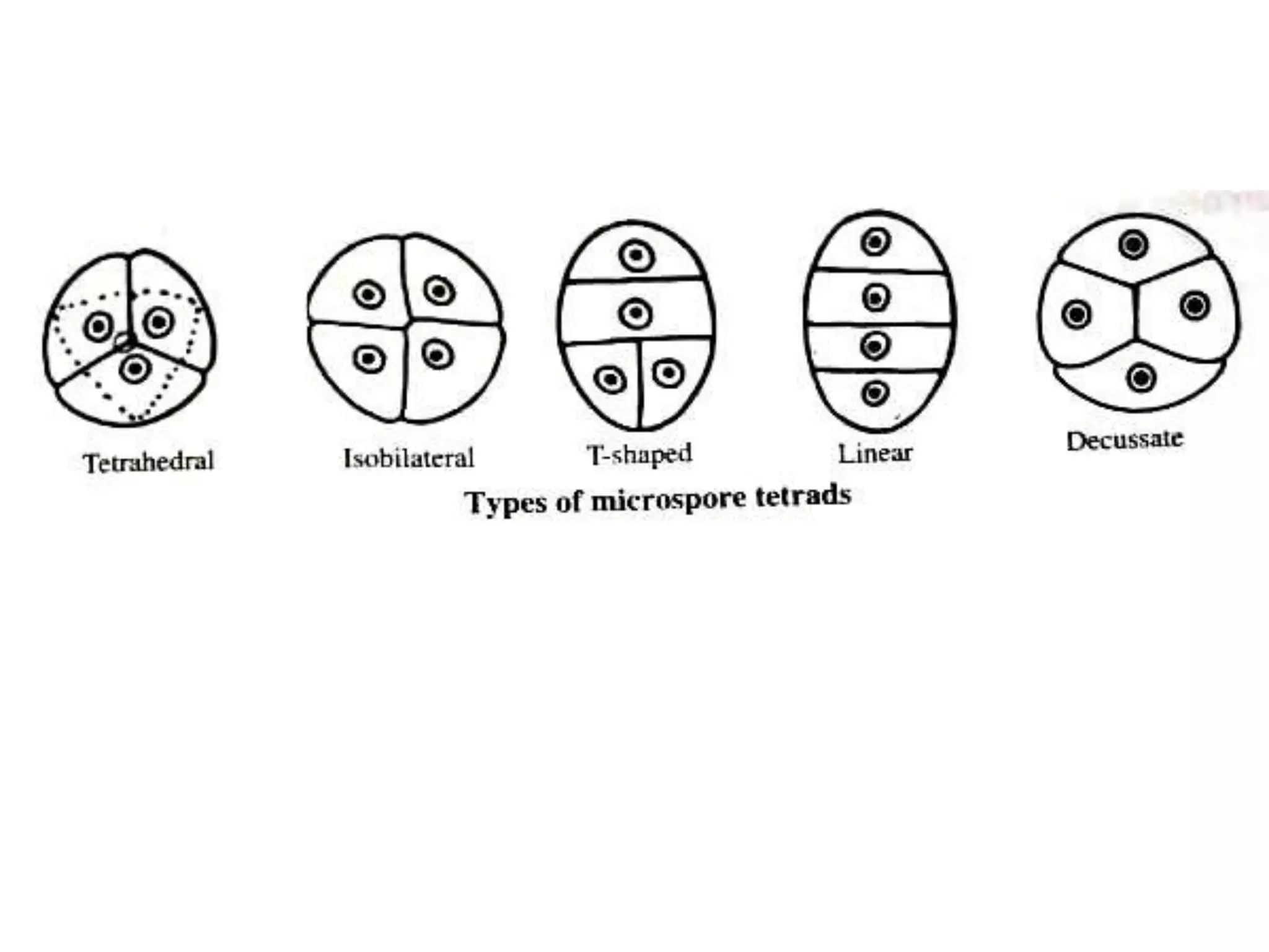
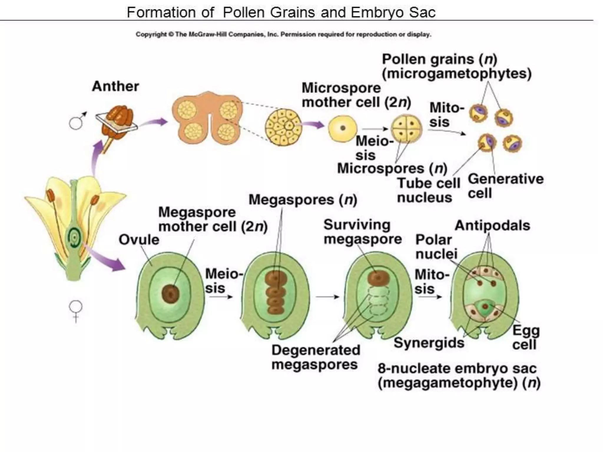
• Microsporogenesis involves the formation of pollen grains in the
microsporangia of anthers.
• A developing young anther consists of a homogeneous mass of
mer istematic cells surrounded by an epidermal layer.
• Further growth of the anther makes it four-lobed.
• In each lobe, some hypodermal cells become more prominent
than the others by virtue of their larger size, dense cytoplasm,
slight radial elongation and conspicuous nuclei.
• These cells are arranged in crescentic or plate-like vertical rows,
forming what is called the archesporium.
• Archesporium represents the antheridial tissue which gives rise
to microspore mother cells.
• The number of archesporial cells varies in different taxa.
• In Malvaceae and Asteraceae, there is only a single vertical row
of cells.
• But, in some members of Lamiaceae, such as Mentha, there is a
layer of archesporial cells at each corner.
• The archesporial cells enlarge radially and divide
periclinally in a plane parallel to the outer wall of the
anther lobe) to form outer primary parietal cells (PPC)
towards the epidermis, and inner primary sporogenous
cells (PSC) towards the interior of the anther.
• The primary parietal cells undergo repeated anticlinal
and periclinal divisions and form 2-5 concentric layers
of cells which form the anther wall.
• The primary sporogenous cells function directly as
microsporocytes (microspore mothercells - MMC - or
pollen mother cells - PMC), or they may undergo
repeated mitotic divisions and form the sporogenous
tissue. The products of the last division function as
MMC
Successive stages of the development of
microsporangium
Anther wall
• Mature anther wall has four main parts,
namely from
• outside epidermis,
• endothecium layer,
• 2 or 3 middle cell layers and
• a single-layered tapetum.
Epidermis
• Epidermis is the outermost protective layer, formed of
flattened and tangentially stretched cells.
• During the development of anther, the epidermal cells
divide anticlinally in order to cope up with the enlarging
internal tissues.
• In a mature anther, the cells are greatly stretched and
flattened.
• In plants of dry habitats, epidermal cells become so
much stretched that they lose contact with each other.
• So, at maturity only their withering remains.
• Epidermis is protective in function.
• An unusual feature is found in Arceuthobium, namely
the development of fibrous thickenings in the
epidermis, a feature characteristic of gymnosperms.
Endothecium (fibrous layer)
• Endothecium is the sub-epidermal fibrous layer.
• Generally, it is single-layered, but in some plants, it is multilayered.
• Endothecial cells attain maximum development when the anther is
ready to dehisce for releasing pollen.
• The walls of endothecial cells become radially elongated.
• From their inner tangential walls, fibrous bands develop upwards
and terminate near the outer wall.
• These bands contain high pro portions of a-cellulose.
• In those anthers which dehisce longitudinally, thickenings are
absent in the endothecial cells that are located at the junction of
the two pollen sacs of an anther.
• In some cleistogamous species (where flowers never open) and in
the members of Hydrocharitaceae (a family comprising only
aquatic plants), fibrous bands are altogether absent in the
endothecium.
• Also, in anthers which dehisce by apical pores, thickenings are
absent in the endothecium.
• The presence of fibrous bands, differential
expansion of the outer and inner tangential walls,
and the hygroscopic nature of the endothecial cells
play an important role in the dehiscence of anthers
at maturity.
• Endothecial cells are thin-walled along the line of
dehiscence of each anther lobe.
• The opening through which the pollen grains are
discharged from the pollen sac is called stomium.
• At maturity, a strain is exerted on the stomium due
to the loss of water from endothecial cells.
• As a result, stomium ruptures and the anther
dehisces.
Middle layers
• The cells of the middle layers are generally ephemeral and
they degenerate most completely even before the pollen
mother cells undergo meiosis.
• They & flattened and crushed during the meiosis of pollen
mother cells.
• In some plants, or more middle layers may persist indefinitely
(e.g., Ranunculus, Lilium), and one adjacent to the
endothecium may even develop fibrous thickenings.
• In Gloriosa, the outermost layer has fibrous bands, similar to
those of endothecium.
• Middle layers are storage centres for starch and other food
reserves which get mobilized during the later development of
pollen.
• They often participate in the formation of pollenkitt, a
viscous coat around pollen grains. Middle layers degenerate
at maturity.
Tapetum
• This is the innermost layer of the anther wall.
• Usually, it exists as a single layer around the sporogenous
tissue.
• But, in some cases, tapetal cells divide periclinally, giving rise
to supernumerary layers (e.g., Coccinia, Nicotiana, Costus).
• Tapetum attains maximum development at the tetrad stage
of microsporogenesis.
• Tapetal cells have dense cytoplasm and conspicuous nuclei.
• This layer is of great physiological significance in that it is
more nutritive in function.
• All the food materials entering into the sporogenous tissue
diffuse through this layer.
• Ultimately, the cells of this layer get disorganized.
• In most species, tapetum forms a homogeneous layer that
completely surrounds the sporogenous tissue.
• In some species, such as Alectra thomsonii, a dimorphic
tapetum is present.
• It consists of two kinds of morphologically different cells.
• The cells, lying close to the connective, form the connective
tapetum or C-tapetum, and those lying close to the periphery
form the parietal tapetum or P-tapetum.
• The cells of the C-tapetum are larger than those of the P-
tapetum.
• C-tapetal cells are derived from the connective tissue, and P-
tapetal cells from the primary parietal layer.
• In Triticales, tapetum is homogeneous and it originates
exclusively from the parietal layer.
• It is quite exceptional that in Antirrhinum majus tapetum is
formed from sporogenous tissue.
Classification of tapetum
• On the basis of behaviour, two types of tapetum
can be recognized, namely
• amoeboid tapetum and secretory tapetum.
(a) Amoeboid (invasive or periplasmodial)
tapetum
• This type is characterized by an early breakdown of
the inner and radial walls of its cells.
• The protoplast of these cells intrude between the
pollen mother cells and the developing pollen
grains.
• After intrusion, the protoplasts fuse with each other
and form a mass of periplasmodium.
• Amoeboid tapetum is found in Alisma,
Tradescantia, etc.
(b) Secretory (glandular or parietal)
tapetum
• In this type, the cells remain in their original
position throughout microspore development.
• The tapetal cells secrete nourishing substances into
the microsporangium and finally degenerate after
pollen maturity.
• This type is common among angiosperms.
• e.g., Mirabilis jalapa, Hellebores foetida.
• The characteristic feature of secretory tapetum is the presence of
a number spherical bodies, called pro-ubisch bodies, together with
mitochondria, plastids anddictyosomes.
• Pro-ubisch bodies are of lipid nature and they make their
appearance during microsporogenesis.
• Their number increases as cell division proceeds.
• Also there occurs a marked shift in their position towards the
anther cavity.
• At the tetrad stage of spores, they increase further in number and
get surrounded by a layer of nibosomes.
• Eventually, the pro-ubisch bodies are extruded from tapetal cells.
• They pass through the tapetal cell membrane into the space
between the membrane and the spore cell wall.
• Here, they get rapidly coated with sporopollenin and become
spherical bodies, called ubisch bodies.
• Ubisch bodies play a decisive role in organising the exine pattern.
Functions of tapetum
1. Tapetum transports the nutrients to the anther locule
where the microspore mothe cells undergo further
development.
2. Tapetum is involved in the synthesis of the enzyme
callase which is essential for the release of microspores
in a tetrad by degrading the callose wall.
3. Precocious release of callase by tapetum is probably
the reason for cytoplasmic male sterility in Petunia.
4. Proteins derived from tapetum occupy the cavities of
the exine and play a significant role in the recognition
of compatible pistils.
Sporogenous tissue
• The primary sporogenous layer forms the sporogenous tissue that
gives rise to the microspore mother cells (MMC), or pollen mother
cells (PMC).
• The sporogenous cells may function directly as the microspore
mother cells or undergo a series of mitotic divisions.
• The daughter cells of the last mitotic division function as
microspore mother cells.
• Pollen mother cells are polygonal in shape and are closely packed.
• As the anther increases in size, these cells become loosely
arranged and rounded in shape.
• All spore mother cells have the potential for forming microspores
or pollen grains.
• However, some of them disintegrate and act as a nourishing tissue
for the developing pollen grains.
• The formation of microspores from the MMC of sporogenous
tissue is called microsporogenesis.
Microsporogenesis
• Microspores are formed by the meiotic (reduction) division of
MMC.
• Each undergoes meiotic division and forms four haploid pollen
cells or microspores
• Initially, the microspores exist together in a tetrad condition, called
microspore tetrad or pollen tetrad, enclosed within a common
wall of callose.
• Gradually, the micros separate from each other, develop an outer
covering around each of them, and then mature to four pollen
grains (microgametophytes or male gametophytes).
• In Cyperaceae, out of the four microspores, only one matures as
the functional pollen grain and the others degenerate.
• Thus, in Cyperaceae, a microspore mother cell gives rise to only a
single pollen grain, instead of four.
• During the meiotic division of MMC, wall formation and
cytokinesis take place in two ways, namely successive and
simultaneous.
(i) Successive wall formation
• In this case, each nuclear division is followed by cell
wall formation.
• The first nuclear division is followed by cell wall
formation, resulting in the formation of two haploid
cells. i.e. a dyad.
• The two cells of the dyad undergo the second meiotic
division (meiosis II).
• It is also followed by wall formation and it results in a
tetrad of haploid cells.
• Successive wall formation is commonly found in
monocots.
(ii) Simultaneous wall formation
• In this case, meiosis I is not followed by cell wall formation.
• So, instead of a microspore dyad, a binucleate cell with two
haploid nuclei is formed.
• The two daughter nuclei then divide mitotically (meiosis II) to
form four haploid nuclei lying in a common mass of
cytoplasm.
• Now, cell wall formation occurs simultaneously in between
the 4 nuclei, resulting 4 haploid cells (microspores).
• Wall formation is centripetal; starts from the lateral walls and
meets in the centre of the cell.
• This kind of cytokinesis may lead to the arrangement of
microspore tetrads in various ways.
• Simultaneous wall formation is comon among dicots.
Arrangement of microspore tetrads
• The microspores of a tetrad are separated from each other by
callose walls.
• Practically, there is no connection between the microspores
of the different tetrads in same anther locule.
• The arrangement spores in a tetrads may show variations.
• The may be tetrahedral, isobilateral, decussate, linear, T-
shaped, etc.
• Of these, terahedral and isobilateral arrangements are the
commonest ones.
• In Aristalochia elegans all the five types of tetrad
arrangement are present.
• Usually microspores separate from one another shortly after
meiosis.
• However, in some plants, the spores tend to remain together
in tetrads for longer periods and develop into compound
pollen grains (e.g., Drimys, Annona, Acacia, Drosera etc.).
• In Asclepiadaceae and Orchidaceae, all the pollen grains in a
pollen sac are united into a single compact mass, known as
pollinium.
• Occurrence of more than four spores in a tetrad is called
polyspory.
• A number of plants exhibit this phenomenon as an
abnormality.
• Polyspory is often due to the division of tetrads.
• Tetrads with as many as eleven microspores have been
reported from Cuscuta reflexa.
• Pollinia
Mature pollen grains
• A mature pollen grain is a haploid spherical structure,
covered by two concentric walls.
• The outer wall is called exine, and the inner one intine.
• At one or more points, the pollen wall is extremely thin
or porous.
• They are called germpores.
• Exine is multilayered.
• It is formed of a highly resistant and durable biological
material, called sporopollenin, which is related to cutin
and suberin.
• Sporopollenin is believed to be the oxidative
polymer of carotenoids and carotenoid esters.
• It provides resistance against physical and biological
decomposition and thereby prevents the natural
decay of pollen grains.
• Structure of pollen grain Preservation of pollen
grains during fossilization is due to the presence of
sporopollenin.
• In most cases, exine itself is two layered, consisting
of an outer layer, called exo-exine, and an inner
layer, called endo-exine
• The surface of the exine may be sculptured, spiny or warty.
• Exine has one or more thin and circular germ pores.
• Through these, the intine protrudes out as the pollen tube
during germination.
• In insect-pollinated microspores, exine has a viscous and
sticky outer coat, called pollenkitt.
• Intine is single-layered, and formed mostly of pectocellulose.
• It encloses the cytoplasm of the pollen grain.
• Pollen grains are packed with dense cytoplasm so long as they
are in the tetrad stage.
• The cytoplasm of a mature pollen grain contains ER,
mitochondria, dictyosomes, a generative nucleus and a
vegetative nucleus.
• The study of pollen grains is called palynology.
Dehiscence of anther
• Mature anther consists of a mass of haploid pollen
tetrads enclosed within the sporangial wall.
• On maturation, the sporongial wall consists of only
epidermis and endothecium: middle layers and the
tapetum disintegrate.
• Once the microspores attain maturity, the anther wall
splits open and liberates them. This is called dehiscence
of anther.
• During this, the partition wall between the two
microsporangia or pollen sacs of each side of the
anther disintegrates, forming a common pollen sac.
• A pressure is exerted on the anther wall by the
mass of pollen grains, rupturing the anther wall and
liberating the pollen grains from the common
pollen sac.
• The point of this rupture is called stomium.
• It is an area of thin-walled cells.
• The fibrous thickening of the endothecium is
believed to help the dehiscence of the anther and
the dispersal of pollen grains.
• Dehiscence of the anther may take place by
transverse slit, longitudinal slit, pores, valves, etc.
Structure of mature dehisced anther
(a) Dehiscence by transverse slit (transverse dehiscence):
The anther lobe dehisces by transverse slits along the
breadth. e.g., Ocimum sanctum.
(b) Dehiscence by longitudinal slit (longitudinal
dehiscence): The anther lobe splits by longitudinal slits
along the length. This is the commonest type of
anther dehiscence e.g., Gossypium, Hibiscus,
Helianthus, etc.
(c) Dehiscence by pores (apical or porous dehiscence):
Pollen grains are liberated through special apical pores
present in the anther lobes. e.g., Solanum.
(d) Dehiscence by valves (valvular dehiscence):
Dehiscence takes place by shutterlike valves, which
open out at the top of the anther. e.g., Berberis.
Development of
male gametophyte
• Microspores represent the first cell or the beginning of the
male gametophytic generation.
• Its maturation to a pollen grain occurs within the anther.
• Mature microspores, after their release from the tetrads, are
termed pollen grains. During maturation, the microspore
becomes free from the tetrad.
• Then, it undergoes unequal mitosis to form a small lenticular
cell or generative cell and a large vegetative cell or tube cell.
• In due course, the vegetative cell develops to a germ tube and
the generative cell undergoes division and gives rise to two
male gametes (microgametes or sperms).
• Initially, the generative cell remains attached to the intine of
the pollen wall. But, later on, it detaches from the pollen wall
and lies free in the cytoplasm.
• In most angiosperms, pollen grains are shed at this two-celled
stage.
• So, in such cases, the division of the generative cell takes place
within the pollen tube after pollen germination.
• While these changes are in progress, the microspores synthesizes
its wall.
• After release, pollen grains undergo rapid enlargement by the
uptake of locular fluid.
• At the initial stage, the pollen grains are non-vacuolated.
• But later on, vacuoles appear and the cytoplasm forms a thin lining
of the wall.
• The first division of a pollen grain results into two unequal cells.
• The larger one is the vegetative cell, which later on forms the
pollen tube.
• The smaller cell is the generative cell, which produces the male
gametes by another mitosis.
• Initially, the generative cell is attached to the wall of the pollen
grain.
• But later on, it comes to lie in the cytoplasm of the vegetative cell.
Vegetative cell
• After pollen mitosis, the vegetative cell continues to
grow.
• Cell organelles increase in size and number.
• Vacuole gradually disappears and the RNA and
protein contents of cell increase very much.
• Nuclear envelope becomes highly convoluted. At a
later stage, nucleus lacks nucleolus. But, in certain
orchids, nucleolus is present.
Generative cell
• The generative cell is small and spherical in shape at the time of
detachment from the pollen wall.
• During pollen development, it changes its shape and becomes
elongated and cylindrical.
• The elongated nature of the cell helps its movement into the
pollen tube.
• Eventually, the generative cell loses contact with the pollen wall
and gets into the vegetative cell.
• Thus, the pollen grain becomes two-celled.
• The function of the generative cell is to give rise to the male
gametes or sperms.
• The nucleus of the generative cell divides by mitosis, forming two
cells.
• The partition wall and the surrounding wall of the sperms
disappear and naked sperms are released.
• Nuclear division in the generative cell may take
place either when pollen grains are present within
the anther (e.g., Beta, Hordeum) or after the pollen
has been shed from the anther.
• In the former, pollen grains are shed at the 3-celled
stage, and in the latter at the 2-celled stage.
• More over, in the latter, the generative cell may
divide after the pollen has reached the stigma or
just before it reaches the embryo sac.
• In rare cases, the generative cell divides after the
pollen tube has reached the embryo sac.
• Each male gamete consists of a large nucleus
surrounded by a thin sheath of cytoplasm, externally
limited by a cell membrane.
• The cytoplasm of the male cells contains mitochondria,
dictyosomes, ribosomes and microtubules.
• The microtubules probably help in maintaining or
altering the shape of male gametes while they pass
through the pollen tube.
• Functional plastids are generally absent in male cells.
• The two male cells of a gametophyte do have physical
contact with each other.
• Such a contact is important for the coordinated
development of both the cells.
Microsporogenesis
Microsporogenesis

Microsporogenesis

  • 1.
  • 2.
    • Adult angiospermplants are diploid sporophytes. • They produce haploid spored by meiosis. This is called sporogenesis. • Spores are of two kinds namely microspores (male spores or pollen grains) and megaspores (female spores). • The formation of microspores is called microsporogenesis, and that of megaspores is called megasporogenesis.
  • 4.
    • Microsporogenesis involvesthe formation of pollen grains in the microsporangia of anthers. • A developing young anther consists of a homogeneous mass of mer istematic cells surrounded by an epidermal layer. • Further growth of the anther makes it four-lobed. • In each lobe, some hypodermal cells become more prominent than the others by virtue of their larger size, dense cytoplasm, slight radial elongation and conspicuous nuclei. • These cells are arranged in crescentic or plate-like vertical rows, forming what is called the archesporium. • Archesporium represents the antheridial tissue which gives rise to microspore mother cells. • The number of archesporial cells varies in different taxa. • In Malvaceae and Asteraceae, there is only a single vertical row of cells. • But, in some members of Lamiaceae, such as Mentha, there is a layer of archesporial cells at each corner.
  • 5.
    • The archesporialcells enlarge radially and divide periclinally in a plane parallel to the outer wall of the anther lobe) to form outer primary parietal cells (PPC) towards the epidermis, and inner primary sporogenous cells (PSC) towards the interior of the anther. • The primary parietal cells undergo repeated anticlinal and periclinal divisions and form 2-5 concentric layers of cells which form the anther wall. • The primary sporogenous cells function directly as microsporocytes (microspore mothercells - MMC - or pollen mother cells - PMC), or they may undergo repeated mitotic divisions and form the sporogenous tissue. The products of the last division function as MMC
  • 8.
    Successive stages ofthe development of microsporangium
  • 10.
    Anther wall • Matureanther wall has four main parts, namely from • outside epidermis, • endothecium layer, • 2 or 3 middle cell layers and • a single-layered tapetum.
  • 12.
    Epidermis • Epidermis isthe outermost protective layer, formed of flattened and tangentially stretched cells. • During the development of anther, the epidermal cells divide anticlinally in order to cope up with the enlarging internal tissues. • In a mature anther, the cells are greatly stretched and flattened. • In plants of dry habitats, epidermal cells become so much stretched that they lose contact with each other. • So, at maturity only their withering remains. • Epidermis is protective in function. • An unusual feature is found in Arceuthobium, namely the development of fibrous thickenings in the epidermis, a feature characteristic of gymnosperms.
  • 13.
    Endothecium (fibrous layer) •Endothecium is the sub-epidermal fibrous layer. • Generally, it is single-layered, but in some plants, it is multilayered. • Endothecial cells attain maximum development when the anther is ready to dehisce for releasing pollen. • The walls of endothecial cells become radially elongated. • From their inner tangential walls, fibrous bands develop upwards and terminate near the outer wall. • These bands contain high pro portions of a-cellulose. • In those anthers which dehisce longitudinally, thickenings are absent in the endothecial cells that are located at the junction of the two pollen sacs of an anther. • In some cleistogamous species (where flowers never open) and in the members of Hydrocharitaceae (a family comprising only aquatic plants), fibrous bands are altogether absent in the endothecium. • Also, in anthers which dehisce by apical pores, thickenings are absent in the endothecium.
  • 15.
    • The presenceof fibrous bands, differential expansion of the outer and inner tangential walls, and the hygroscopic nature of the endothecial cells play an important role in the dehiscence of anthers at maturity. • Endothecial cells are thin-walled along the line of dehiscence of each anther lobe. • The opening through which the pollen grains are discharged from the pollen sac is called stomium. • At maturity, a strain is exerted on the stomium due to the loss of water from endothecial cells. • As a result, stomium ruptures and the anther dehisces.
  • 17.
    Middle layers • Thecells of the middle layers are generally ephemeral and they degenerate most completely even before the pollen mother cells undergo meiosis. • They & flattened and crushed during the meiosis of pollen mother cells. • In some plants, or more middle layers may persist indefinitely (e.g., Ranunculus, Lilium), and one adjacent to the endothecium may even develop fibrous thickenings. • In Gloriosa, the outermost layer has fibrous bands, similar to those of endothecium. • Middle layers are storage centres for starch and other food reserves which get mobilized during the later development of pollen. • They often participate in the formation of pollenkitt, a viscous coat around pollen grains. Middle layers degenerate at maturity.
  • 19.
    Tapetum • This isthe innermost layer of the anther wall. • Usually, it exists as a single layer around the sporogenous tissue. • But, in some cases, tapetal cells divide periclinally, giving rise to supernumerary layers (e.g., Coccinia, Nicotiana, Costus). • Tapetum attains maximum development at the tetrad stage of microsporogenesis. • Tapetal cells have dense cytoplasm and conspicuous nuclei. • This layer is of great physiological significance in that it is more nutritive in function. • All the food materials entering into the sporogenous tissue diffuse through this layer. • Ultimately, the cells of this layer get disorganized.
  • 20.
    • In mostspecies, tapetum forms a homogeneous layer that completely surrounds the sporogenous tissue. • In some species, such as Alectra thomsonii, a dimorphic tapetum is present. • It consists of two kinds of morphologically different cells. • The cells, lying close to the connective, form the connective tapetum or C-tapetum, and those lying close to the periphery form the parietal tapetum or P-tapetum. • The cells of the C-tapetum are larger than those of the P- tapetum. • C-tapetal cells are derived from the connective tissue, and P- tapetal cells from the primary parietal layer. • In Triticales, tapetum is homogeneous and it originates exclusively from the parietal layer. • It is quite exceptional that in Antirrhinum majus tapetum is formed from sporogenous tissue.
  • 21.
    Classification of tapetum •On the basis of behaviour, two types of tapetum can be recognized, namely • amoeboid tapetum and secretory tapetum.
  • 23.
    (a) Amoeboid (invasiveor periplasmodial) tapetum • This type is characterized by an early breakdown of the inner and radial walls of its cells. • The protoplast of these cells intrude between the pollen mother cells and the developing pollen grains. • After intrusion, the protoplasts fuse with each other and form a mass of periplasmodium. • Amoeboid tapetum is found in Alisma, Tradescantia, etc.
  • 24.
    (b) Secretory (glandularor parietal) tapetum • In this type, the cells remain in their original position throughout microspore development. • The tapetal cells secrete nourishing substances into the microsporangium and finally degenerate after pollen maturity. • This type is common among angiosperms. • e.g., Mirabilis jalapa, Hellebores foetida.
  • 25.
    • The characteristicfeature of secretory tapetum is the presence of a number spherical bodies, called pro-ubisch bodies, together with mitochondria, plastids anddictyosomes. • Pro-ubisch bodies are of lipid nature and they make their appearance during microsporogenesis. • Their number increases as cell division proceeds. • Also there occurs a marked shift in their position towards the anther cavity. • At the tetrad stage of spores, they increase further in number and get surrounded by a layer of nibosomes. • Eventually, the pro-ubisch bodies are extruded from tapetal cells. • They pass through the tapetal cell membrane into the space between the membrane and the spore cell wall. • Here, they get rapidly coated with sporopollenin and become spherical bodies, called ubisch bodies. • Ubisch bodies play a decisive role in organising the exine pattern.
  • 26.
    Functions of tapetum 1.Tapetum transports the nutrients to the anther locule where the microspore mothe cells undergo further development. 2. Tapetum is involved in the synthesis of the enzyme callase which is essential for the release of microspores in a tetrad by degrading the callose wall. 3. Precocious release of callase by tapetum is probably the reason for cytoplasmic male sterility in Petunia. 4. Proteins derived from tapetum occupy the cavities of the exine and play a significant role in the recognition of compatible pistils.
  • 27.
    Sporogenous tissue • Theprimary sporogenous layer forms the sporogenous tissue that gives rise to the microspore mother cells (MMC), or pollen mother cells (PMC). • The sporogenous cells may function directly as the microspore mother cells or undergo a series of mitotic divisions. • The daughter cells of the last mitotic division function as microspore mother cells. • Pollen mother cells are polygonal in shape and are closely packed. • As the anther increases in size, these cells become loosely arranged and rounded in shape. • All spore mother cells have the potential for forming microspores or pollen grains. • However, some of them disintegrate and act as a nourishing tissue for the developing pollen grains. • The formation of microspores from the MMC of sporogenous tissue is called microsporogenesis.
  • 29.
    Microsporogenesis • Microspores areformed by the meiotic (reduction) division of MMC. • Each undergoes meiotic division and forms four haploid pollen cells or microspores • Initially, the microspores exist together in a tetrad condition, called microspore tetrad or pollen tetrad, enclosed within a common wall of callose. • Gradually, the micros separate from each other, develop an outer covering around each of them, and then mature to four pollen grains (microgametophytes or male gametophytes). • In Cyperaceae, out of the four microspores, only one matures as the functional pollen grain and the others degenerate. • Thus, in Cyperaceae, a microspore mother cell gives rise to only a single pollen grain, instead of four. • During the meiotic division of MMC, wall formation and cytokinesis take place in two ways, namely successive and simultaneous.
  • 33.
    (i) Successive wallformation • In this case, each nuclear division is followed by cell wall formation. • The first nuclear division is followed by cell wall formation, resulting in the formation of two haploid cells. i.e. a dyad. • The two cells of the dyad undergo the second meiotic division (meiosis II). • It is also followed by wall formation and it results in a tetrad of haploid cells. • Successive wall formation is commonly found in monocots.
  • 35.
    (ii) Simultaneous wallformation • In this case, meiosis I is not followed by cell wall formation. • So, instead of a microspore dyad, a binucleate cell with two haploid nuclei is formed. • The two daughter nuclei then divide mitotically (meiosis II) to form four haploid nuclei lying in a common mass of cytoplasm. • Now, cell wall formation occurs simultaneously in between the 4 nuclei, resulting 4 haploid cells (microspores). • Wall formation is centripetal; starts from the lateral walls and meets in the centre of the cell. • This kind of cytokinesis may lead to the arrangement of microspore tetrads in various ways. • Simultaneous wall formation is comon among dicots.
  • 37.
    Arrangement of microsporetetrads • The microspores of a tetrad are separated from each other by callose walls. • Practically, there is no connection between the microspores of the different tetrads in same anther locule. • The arrangement spores in a tetrads may show variations. • The may be tetrahedral, isobilateral, decussate, linear, T- shaped, etc. • Of these, terahedral and isobilateral arrangements are the commonest ones. • In Aristalochia elegans all the five types of tetrad arrangement are present.
  • 39.
    • Usually microsporesseparate from one another shortly after meiosis. • However, in some plants, the spores tend to remain together in tetrads for longer periods and develop into compound pollen grains (e.g., Drimys, Annona, Acacia, Drosera etc.). • In Asclepiadaceae and Orchidaceae, all the pollen grains in a pollen sac are united into a single compact mass, known as pollinium. • Occurrence of more than four spores in a tetrad is called polyspory. • A number of plants exhibit this phenomenon as an abnormality. • Polyspory is often due to the division of tetrads. • Tetrads with as many as eleven microspores have been reported from Cuscuta reflexa.
  • 41.
  • 42.
    Mature pollen grains •A mature pollen grain is a haploid spherical structure, covered by two concentric walls. • The outer wall is called exine, and the inner one intine. • At one or more points, the pollen wall is extremely thin or porous. • They are called germpores. • Exine is multilayered. • It is formed of a highly resistant and durable biological material, called sporopollenin, which is related to cutin and suberin.
  • 44.
    • Sporopollenin isbelieved to be the oxidative polymer of carotenoids and carotenoid esters. • It provides resistance against physical and biological decomposition and thereby prevents the natural decay of pollen grains. • Structure of pollen grain Preservation of pollen grains during fossilization is due to the presence of sporopollenin. • In most cases, exine itself is two layered, consisting of an outer layer, called exo-exine, and an inner layer, called endo-exine
  • 47.
    • The surfaceof the exine may be sculptured, spiny or warty. • Exine has one or more thin and circular germ pores. • Through these, the intine protrudes out as the pollen tube during germination. • In insect-pollinated microspores, exine has a viscous and sticky outer coat, called pollenkitt. • Intine is single-layered, and formed mostly of pectocellulose. • It encloses the cytoplasm of the pollen grain. • Pollen grains are packed with dense cytoplasm so long as they are in the tetrad stage. • The cytoplasm of a mature pollen grain contains ER, mitochondria, dictyosomes, a generative nucleus and a vegetative nucleus. • The study of pollen grains is called palynology.
  • 48.
    Dehiscence of anther •Mature anther consists of a mass of haploid pollen tetrads enclosed within the sporangial wall. • On maturation, the sporongial wall consists of only epidermis and endothecium: middle layers and the tapetum disintegrate. • Once the microspores attain maturity, the anther wall splits open and liberates them. This is called dehiscence of anther. • During this, the partition wall between the two microsporangia or pollen sacs of each side of the anther disintegrates, forming a common pollen sac.
  • 49.
    • A pressureis exerted on the anther wall by the mass of pollen grains, rupturing the anther wall and liberating the pollen grains from the common pollen sac. • The point of this rupture is called stomium. • It is an area of thin-walled cells. • The fibrous thickening of the endothecium is believed to help the dehiscence of the anther and the dispersal of pollen grains. • Dehiscence of the anther may take place by transverse slit, longitudinal slit, pores, valves, etc.
  • 50.
    Structure of maturedehisced anther
  • 51.
    (a) Dehiscence bytransverse slit (transverse dehiscence): The anther lobe dehisces by transverse slits along the breadth. e.g., Ocimum sanctum. (b) Dehiscence by longitudinal slit (longitudinal dehiscence): The anther lobe splits by longitudinal slits along the length. This is the commonest type of anther dehiscence e.g., Gossypium, Hibiscus, Helianthus, etc. (c) Dehiscence by pores (apical or porous dehiscence): Pollen grains are liberated through special apical pores present in the anther lobes. e.g., Solanum. (d) Dehiscence by valves (valvular dehiscence): Dehiscence takes place by shutterlike valves, which open out at the top of the anther. e.g., Berberis.
  • 52.
  • 54.
    • Microspores representthe first cell or the beginning of the male gametophytic generation. • Its maturation to a pollen grain occurs within the anther. • Mature microspores, after their release from the tetrads, are termed pollen grains. During maturation, the microspore becomes free from the tetrad. • Then, it undergoes unequal mitosis to form a small lenticular cell or generative cell and a large vegetative cell or tube cell. • In due course, the vegetative cell develops to a germ tube and the generative cell undergoes division and gives rise to two male gametes (microgametes or sperms). • Initially, the generative cell remains attached to the intine of the pollen wall. But, later on, it detaches from the pollen wall and lies free in the cytoplasm.
  • 59.
    • In mostangiosperms, pollen grains are shed at this two-celled stage. • So, in such cases, the division of the generative cell takes place within the pollen tube after pollen germination. • While these changes are in progress, the microspores synthesizes its wall. • After release, pollen grains undergo rapid enlargement by the uptake of locular fluid. • At the initial stage, the pollen grains are non-vacuolated. • But later on, vacuoles appear and the cytoplasm forms a thin lining of the wall. • The first division of a pollen grain results into two unequal cells. • The larger one is the vegetative cell, which later on forms the pollen tube. • The smaller cell is the generative cell, which produces the male gametes by another mitosis. • Initially, the generative cell is attached to the wall of the pollen grain. • But later on, it comes to lie in the cytoplasm of the vegetative cell.
  • 61.
    Vegetative cell • Afterpollen mitosis, the vegetative cell continues to grow. • Cell organelles increase in size and number. • Vacuole gradually disappears and the RNA and protein contents of cell increase very much. • Nuclear envelope becomes highly convoluted. At a later stage, nucleus lacks nucleolus. But, in certain orchids, nucleolus is present.
  • 63.
    Generative cell • Thegenerative cell is small and spherical in shape at the time of detachment from the pollen wall. • During pollen development, it changes its shape and becomes elongated and cylindrical. • The elongated nature of the cell helps its movement into the pollen tube. • Eventually, the generative cell loses contact with the pollen wall and gets into the vegetative cell. • Thus, the pollen grain becomes two-celled. • The function of the generative cell is to give rise to the male gametes or sperms. • The nucleus of the generative cell divides by mitosis, forming two cells. • The partition wall and the surrounding wall of the sperms disappear and naked sperms are released.
  • 64.
    • Nuclear divisionin the generative cell may take place either when pollen grains are present within the anther (e.g., Beta, Hordeum) or after the pollen has been shed from the anther. • In the former, pollen grains are shed at the 3-celled stage, and in the latter at the 2-celled stage. • More over, in the latter, the generative cell may divide after the pollen has reached the stigma or just before it reaches the embryo sac. • In rare cases, the generative cell divides after the pollen tube has reached the embryo sac.
  • 66.
    • Each malegamete consists of a large nucleus surrounded by a thin sheath of cytoplasm, externally limited by a cell membrane. • The cytoplasm of the male cells contains mitochondria, dictyosomes, ribosomes and microtubules. • The microtubules probably help in maintaining or altering the shape of male gametes while they pass through the pollen tube. • Functional plastids are generally absent in male cells. • The two male cells of a gametophyte do have physical contact with each other. • Such a contact is important for the coordinated development of both the cells.