*
FLOWERING PLANTS

1
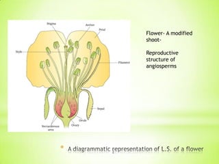
Flower- A modified
shootReproductive
structure of
angiosperms

*

2
* FLORICULTURE : the cultivation of
flowering plants.
* Several hormonal and structural
changes are initiated which lead
to the differentiation and
development of the floral
PRIMORDIUM
* primordium- an organ in its
earliest stage of development;
the foundation for subsequent
development
* Inflorescences are formed which
bear the floral buds and then the
flowers.
* In the flower the male and
female reproductive structures,
the androecium (MALE) and the
gynoecium ( FEMALE )
differentiate and develop.

*

3
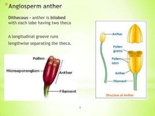
A typical stamen• the long and slender stalk
called the filament
• the terminal bilobed structure
called the anther.
• Monothecous
Hibiscus, Dithecous brinjal

The proximal end of the filament
is attached to the thalamus or the
petal of the flower. Check
terminology.
The number and length of
stamens vary.

*

4
*
Dithecous - anther is bilobed
with each lobe having two theca

A longitudinal groove runs
lengthwise separating the theca.

5
*

6
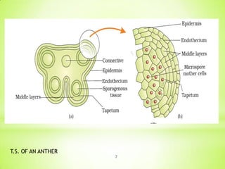
T.S. OF AN ANTHER
7
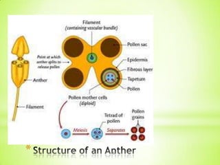
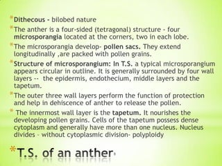
* Dithecous - bilobed nature
* The anther is a four-sided (tetragonal) structure - four

microsporangia located at the corners, two in each lobe.
* The microsporangia develop- pollen sacs. They extend
longitudinally ,are packed with pollen grains.
* Structure of microsporangium: In T.S. a typical microsporangium
appears circular in outline. It is generally surrounded by four wall
layers -- the epidermis, endothecium, middle layers and the
tapetum.
* The outer three wall layers perform the function of protection
and help in dehiscence of anther to release the pollen.
* The innermost wall layer is the tapetum. It nourishes the
developing pollen grains. Cells of the tapetum possess dense
cytoplasm and generally have more than one nucleus. Nucleus
divides – without cytoplasmic division- polyploidy

*

8
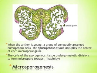
* When the anther is young, a group of compactly arranged

homogenous cells -the sporogenous tissue occupies the centre
of each microsporangium.
* The cells of the sporogenous tissue undergo meiotic divisions
to form microspore tetrads. ( haploidy)

*

9
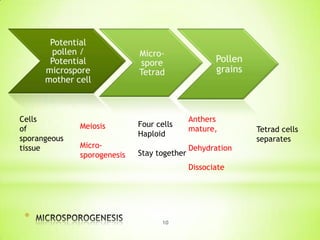
Potential
pollen /
Potential
microspore
mother cell

Cells
of
sporangeous
tissue

Microspore
Tetrad

Meiosis

Four cells
Haploid

Microsporogenesis

Stay together

Pollen
grains

Anthers
mature,
Dehydration
Dissociate

*

10

Tetrad cells
separates
*

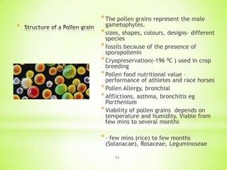
* The pollen grains represent the male

gametophytes.
* sizes, shapes, colours, designs- different
species
* fossils because of the presence of
sporopollenin
* Cryopreservation(-196 0C ) used in crop
breeding
* Pollen food nutritional value –
performance of athletes and race horses
* Pollen Allergy, bronchial
* Afflictions, asthma, bronchitis eg
Parthenium
* Viability of pollen grains depends on
temperature and humidity. Viable from
few mins to several months

* – few mins (rice) to few months

(Solanacae), Rosaceae, Leguminoseae
11
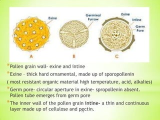
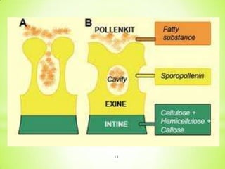
* Pollen grain wall- exine and intine
* Exine – thick hard ornamental, made up of sporopollenin
( most resistant organic material high temperature, acid, alkalies)

* Germ pore- circular aperture in exine- spropollenin absent.
Pollen tube emerges from germ pore

* The inner wall of the pollen grain intine- a thin and continuous
layer made up of cellulose and pectin.
12
13
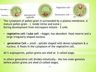
The cytoplasm of pollen grain is surrounded by a plasma membrane. A
mature pollen grain – ( inside intine and exine )
During development from microspore mitosis – 2 cells
• vegetative cell / tube cell --bigger, has abundant -food reserve and a
large irregularly shaped nucleus.
•

generative Cell -- small , spindle shaped with dense cytoplasm & a
nucleus. It floats in the cytoplasm of the vegetative cell.

60 % angiosperms, pollen grains are shed at 2-celled stage.
In others generative cell divides mitotically - the two male gametes
before pollen grains are shed (3-celled stage).
14
* Pistil/gynoecium innermost whorl
* Carpel – stigma , style , Ovary
* Ovary many ovule ( megasporangia) attached to a tissue
inside ovary – placenta – ovarian cavity ( locules) –

* Placentation – recap
* number of ovules in an ovary may be one (wheat,
* paddy, mango) to many (papaya, water melon, orchids).
* Monocarpellary / multicarpellary
* Syncarpous ( pistils fused together) , aopcarpous ( free)

*

15
*

(1)

Orthotropous : The micropyle, chalaza
and funicle are in a straight line. This is the
most primitive type of ovule
e.g., Piper, Polygonum, Cycas. - A

* (2) Anatropous : The ovule turns at

180 angle. Thus it is inverted ovule.
Micropyle lies close to hilum or at
side of hilum e.g, found in 90% of
angiosperm families. B

* (3) Campylotropous : Ovule is curved more

or less at right angle to funicle. Micropylar
end is bend down slightly e.g., in members
of Leguminosae, Cruciferae. D
* (4) Hemianatropous : Ovule turns at 90
angle upon the funicle or body of ovule and
is at right angle to the funicle
e.g., Ranunculus. C
* (5) Amphitropous : Ovule as well as
embryo sac is curved like horse shoe
e.g, Lemna, Poppy, Alisma. E
* (6) Circinotropous : The ovule turns at
more than 360 angle, so funicle becomes
coiled around the ovule e.g., Opuntia
(Cactaceae), Plumbaginaceae. F
16
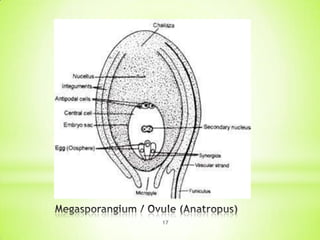
17
1.

Funicle

The ovule is attached to the placenta by means of a stalk called
funicle.

2.

Hilum

The body of the ovule fuses with funicle in the region called
hilum.(hilum - the junction between ovule and funicle)

3.

Integuments

Each ovule has one or two protective envelopes called
integuments.(inner and outer )

4.

Micropyle

The integuments leave small opening at one end called micropyle.this end is micropylar end.

5.

Chalaza

Opposite the micropylar end, is the chalaza, representing the basal
part of the ovule.

6.

Nucellus

Tissue encloses embryo sac

7.

Embryo sac
(female
gametophyte)

An ovule generally has a single embryo sac formed from a megaspore
through reduction division.
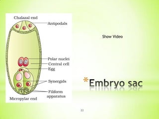
* Nucleate and 7 celled.
1. Egg apparatus- at micropylar end. It has 2 synergids and an egg.
2. Fili form apparatus- synegids – special thickening at micropylar
end. It guides the pollen tube.
3. Antipodals- 3 in number twds chalazal end.
4. Two Haploid nuclei at polar end below egg appataus

*

18
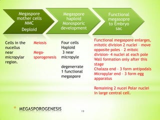
Megaspore
mother cells
MMC
Deploid
Cells in the
nucellus
near
micropylar
region.

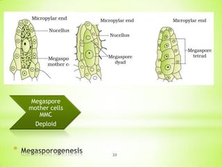
Meiosis
Megasporogenesis

Megaspore
haploid
Monosporic
development
Four cells
Haploid
3 near
micropyle
degenerrate
1 functional
megaspore

Functional
megasopre
to Embryo
sac
Functional megaspore enlarges,
mitotic division 2 nuclei – move
opposite poles – 2 mitoic
division- 4 nuclei at each pole
Wall formation only after this
stage
Chalaza end – 3 form antipodals
Micropylar end – 3 form egg
apparatus
Remaining 2 nucei Polar nuclei
in large central cell.

*

19
Megaspore
mother cells
MMC
Deploid

*

20
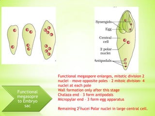
Functional
megasopre
to Embryo
sac

Functional megaspore enlarges, mitotic division 2
nuclei – move opposite poles – 2 mitoic division- 4
nuclei at each pole
Wall formation only after this stage
Chalaza end – 3 form antipodals
Micropylar end – 3 form egg apparatus
Remaining 221
nucei Polar nuclei in large central cell.
Show Video

*
22
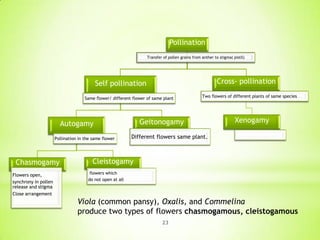
Pollination
Transfer of pollen grains from anther to stigma( pistil)

Cross- pollination

Self pollination
Same flower/ different flower of same plant

Geitonogamy

Autogamy
Pollination in the same flower

Chasmogamy
Flowers open,
synchrony in pollen
release and stigma
Close arrangement

Two flowers of different plants of same species

Xenogamy

Different flowers same plant.

Cleistogamy
flowers which
do not open at all

Viola (common pansy), Oxalis, and Commelina
produce two types of flowers chasmogamous, cleistogamous
23
Advantages of cleistogamy
Assured seed-set even in the absence of pollinators.
But no variation in offspring.
Compare self and cross pollination.
Agents of pollination – discussion book
Self study / discussion by students
Show other ppt

24
*All events

from deposition of pollen grains on stigma – entry
of pollen tube into the ovule -Pollen pistil interaction

*Identification of compatible (same species) or incompatible(
other species) pollen grains by stigma

*a dynamic process involving pollen recognition followed by
promotion or inhibition of the pollen.

*Reason – chemical interaction between pollen and stigma
*Compatible pollen accepted by stigma, initiates post
pollination events

*

25
*
*Germination- formation of pollen tube through germ

pore- content move to pollen tube- through stigma, style
reaches ovary
*pollen grains - two-celled condition (a vegetative cell
and a generate cell)- the generative cell divides and
forms the two male gametes during the growth of pollen
tube in the stigma.
*three-celled condition, pollen tubes carry the two male
gametes from the beginning.
*Pollen tube, after reaching the ovary, enters the ovule
through the micropyle and then enters one of the
synergids through the filiform apparatus
*Interaction is important in plant breeding- for superior
plant development
26
* Emasculation

* When the stigma of bagged

flower attains receptivity,
mature pollen grains collected
from anthers of the male
parent are dusted on the
stigma, and the flowers are
rebagged, and the fruits
allowed to develop.

female parent bears bisexual
flowers, removal of anthers
from the flower bud before
the anther dehisces using a
pair of forceps.

* Bagging
Emasculated flowers have to
be covered with a bag of
suitable size, generally made
up of butter paper, to prevent
contamination of its stigma
with unwanted pollen.

* unisexual flowers,

no
emasculation. The female
flower buds are bagged before
the flowers open.
* When the stigma becomes
receptive, pollination is carried
out using the desired pollen
and the flower rebagged.
27
*
Pollen tube into
synergid

Syngamy

Triple fusion
3 haploid nucleus
fuse

• the pollen tube releases the two male gametes into the
cytoplasm of the synergid.

• the pollen tube releases the two male gametes into the
cytoplasm of the synergid.
• Diploid Zygote formation - zygote develops into an embryo.

• other male gamete moves towards the two polar nuclei located
in the central cell and fuses with them to produce a triploid
PEN This central cell forms
• primary endosperm cell (PEC) and develops into the endosperm
28
*
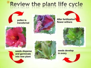
After fertilization
flower withers

pollen is
transferred

2

3

1
seeds disperse
and germinate
into new plant

4

seeds develop
in ovary
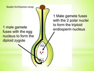
Double fertilization recap

1 male gamete
fuses with the egg
nucleus to form the
diploid zygote

1 Male gamete fuses
with the 2 polar nuclei
to form the triploid
endosperm nucleus
*
* After double fertilization events of endosperm and embryo

development, maturation of ovule(s) into seed(s) and ovary
into fruit, are collectively termed post-fertilisation events.

Endosperm development
Embryogeny
Seed formation
* Endosperm development :

Primary Endosperm Cell (Triple

fusion) – Endosperm (3n)

* Endosperm – cells filled with reserved food material -main
source of nutrition for the embryo

31
*
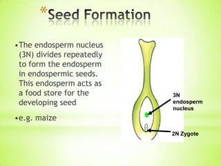
•The endosperm nucleus
(3N) divides repeatedly
to form the endosperm
in endospermic seeds.
This endosperm acts as
a food store for the
developing seed

3N
endosperm
nucleus

•e.g. maize
2N Zygote
*
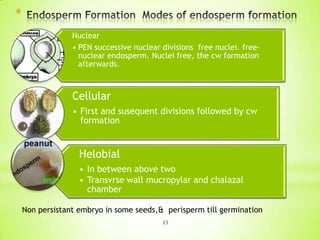
Nuclear
• PEN successive nuclear divisions free nuclei. freenuclear endosperm. Nuclei free, the cw formation
afterwards.

Cellular
• First and susequent divisions followed by cw
formation

Helobial
• In between above two
• Transvrse wall mucropylar and chalazal
chamber
Non persistant embryo in some seeds,& perisperm till germination
33
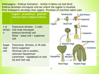
Embryogeny - Embryo formation – similar in Mono cot and dicot
Embryo develops micropylar end sac where the zygote is situated.
First endosperm develops then zygote. Provision of nutrition taken care
Zygote – proembryo – globular
mature heart shaped embryo

1 st
Transverse division – 2 cells
divisio Cell twds micropyle
n
embryo/terminal cell
Other – basal cell / suspensor
cell
Suspe
nsor
cell

Transverse division, 6-10 cells
forms suspensor
Terminal cell is swollen,
vesicular Haustorium
Lowermost – hypophysis to root
tip and root cap

34
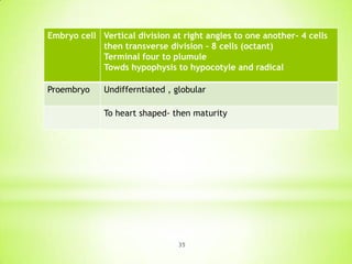
Embryo cell Vertical division at right angles to one another- 4 cells
then transverse division – 8 cells (octant)
Terminal four to plumule
Towds hypophysis to hypocotyle and radical

Proembryo

Undifferntiated , globular
To heart shaped- then maturity

35
Dicot embryo consists of an
embryonal axis and two
cotyledons.
The portion of embryonal axis above
the level of cotyledons is the
epicotyl, which terminates with the
plumule or stem tip.
The cylindrical portion below the
level of cotyledons is hypocotyl that

* terminates at its lower end in the

radical or root tip. The root tip is
covered with a root cap.

*

36
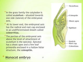
* In the grass family the cotyledon is

called scutellum - situated towards
one side (lateral) of the embryonal
axis.

* At its lower end, the embryonal axis

has the radical and root cap enclosed
in an undifferentiated sheath called
coleorrhiza.

* The portion of the embryonal axis

above the level of attachment of
scutellum is the epicotyl. Epicotyl
has a shoot apex and a few leaf
primordia enclosed in a hollow foliar
structure, the coleoptile.

*

37
*
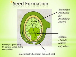
Endosperm
Food store
for
developing
embryo

Micropyle – pore-entry
Of oxygen, water during
germination

Integuments, becomes the seed coat

Embryo
Plumule,
radicle,
cotyledons
*
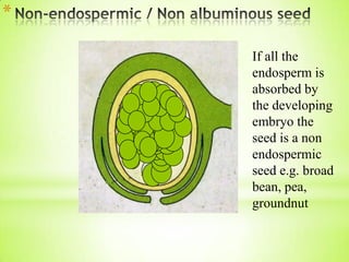
If all the
endosperm is
absorbed by
the developing
embryo the
seed is a non
endospermic
seed e.g. broad
bean, pea,
groundnut
*
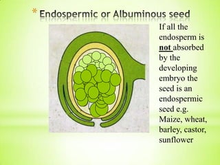
If all the
endosperm is
not absorbed
by the
developing
embryo the
seed is an
endospermic
seed e.g.
Maize, wheat,
barley, castor,
sunflower
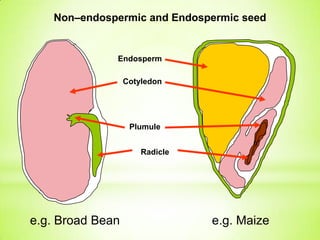
Non–endospermic and Endospermic seed

Endosperm

Cotyledon

Plumule
Radicle

e.g. Broad Bean

e.g. Maize
*

42
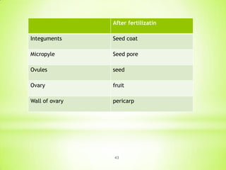
After fertilizatin

Integuments

Seed coat

Micropyle

Seed pore

Ovules

seed

Ovary

fruit

Wall of ovary

pericarp

43
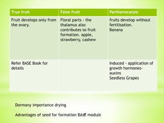
True fruit

False fruit

Parthenocarpic

Fruit develops only from
the ovary.

Floral parts - the
thalamus also
contributes to fruit
formation. apple,
strawberry, cashew

fruits develop without
fertilisation.
Banana

Refer BASE Book for
details

Dormany importance drying
44
Advantages of seed for formation BAsE module

Induced - application of
growth hormonesauxins
Seedless Grapes
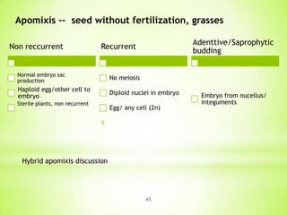
Apomixis -- seed without fertilization, grasses
Non reccurrent

Adenttive/Saprophytic
budding

Recurrent

Normal embryo sac
production

No meiosis

Haploid egg/other cell to
embryo

Diploid nuclei in embryo

Sterile plants, non recurrent

Egg/ any cell (2n)

Hybrid apomixis discussion

45

Embryo from nucellus/
integuments
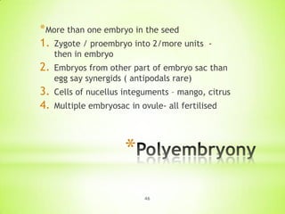
* More than one embryo in the seed
1. Zygote / proembryo into 2/more units

-

then in embryo

2.

Embryos from other part of embryo sac than
egg say synergids ( antipodals rare)

3.
4.

Cells of nucellus integuments – mango, citrus
Multiple embryosac in ovule- all fertilised

*
46